Liver Lesions on Imaging: A Radiology Perspective
- Dr. Surabhi karthik , Assistant Professor, Department of Radiology, Bhaskara Medical College, Hyderabad, Telangana, India
- Dr. Sunitha gattigorla , Professor, Department of Pathology, Nova institute of medical sciences and research centre, Telangana, India
- Dr. Kavitha K , Assistant Professor, Department of Ophthalmology, RVM Medical college, Siddipet, Telangana, India
- Dr. Mood Narayan , Associate Professor, Department of Respiratory Medicine, ESIC Medical College, Hyderabad, Telangana, India
- Dr. Vishal Parekar , Professor, Department of Pathology, Kamineni Institute of Medical Sciences, Narketpally, Telangana, India
Article Information:
Abstract:
Background: Chronic liver disease (CLD) is a major global public health problem due to its diverse spectrum of causes and symptoms. Accurate diagnosis, staging, and prognosis are essential for managing a disease. In this study, we aimed to investigate the clinicopathological profile and etiological pattern of chronic liver problems in cases where a biopsy had been conducted. Liver biopsy remains the preferred method for precise staging and grading utilizing established scoring systems such as Ishak, METAVIR, and Batts-Ludwig classifications. It makes it possible to evaluate architectural distortion, necroinflammatory activity, and hepatic fibrosis. Among the major etiologies that histopathological evaluation is crucial for differentiating are non-alcoholic steatohepatitis (NASH), chronic viral hepatitis (HCV and HBV), metabolic dysfunction-associated fatty liver disease (MAFLD), alcohol-related liver disease (ARLD), hereditary hemochromatosis, hepatocellular carcinoma, and non-alcoholic fatty liver disease (NAFLD). Despite the increasing use of non-invasive techniques, liver biopsy remains the gold standard for definitive tissue diagnosis, aids in diagnosing problems with many or overlapping causes, and guides therapy decisions. The study highlights the variety of histological patterns seen in CLD and demonstrates that, despite its limitations, liver biopsy may still be helpful in some therapeutic settings.
Keywords:
Article :
INTRODUCTION:
Chronic liver disease (CLD) is a condition that affects millions of individuals yearly and is considered to be one of the leading causes of liver failure as well as a significant contributor to mortality and morbidity around the world. According to the findings of the clinicopathological study, prompt treatment or removal of the agent that caused the condition may result in a regression of the fibrosis and the repair of the parenchyma. This demonstrates the importance of making an accurate diagnosis as soon as possible [1].
When identifying chronic liver disease in individuals, a liver biopsy is a crucial component. It is often used to assess the severity of the disease by identifying the grading (level of necroinflammatory activity) and staging (extent of fibrosis). In addition to establishing a definitive diagnosis, it assists in determining the severity of the sickness. This purpose has resulted in the creation of a great deal of grading systems that have been confirmed. Grading and staging systems have recently been focusing their attention on chronic viral hepatitis in particular as a result of recent advancements in therapy. Although chronic cholestatic disorders, allograft rejection, and fatty liver disease each have their own scoring systems, these scoring systems are not used nearly as often in clinical practice as other scoring systems [2].
The coexistence of many etiologies is common and has the potential to significantly influence the progression of illness, despite the fact that many individuals who suffer from liver disease have a single diseased process that predominates. In spite of the fact that the histological features of many liver ailments may sometimes be comparable to one another, a comprehensive histopathological examination may frequently differentiate between them, resulting in a more accurate explanation of the underlying cause [3,4]. When a biopsy is performed to confirm a case, the significance of this clinicopathological relationship becomes even more evident. This is because it becomes essential to differentiate between overlapping illnesses such as liver damage caused by alcohol, viral hepatitis, fatty liver disease, and other chronic hepatopathies.
The diagnosis and treatment of hepatic diseases have been significantly influenced by developments in medical technology, improvements in medicine, and an increase in the sophistication of surgical operations. As a consequence of this, non-invasive diagnostic tools are turning into more significant, and liver biopsy is becoming an increasingly insignificant component of this process [5]. In spite of this, there are some situations in which a liver biopsy is still the most effective method for determining the underlying etiology, the amount of inflammation, and the disease activity via histological examination. The current research set out to determine the potential etiological diagnosis by correlating clinical, histological, biochemical, and serological results; to evaluate the severity of hepatic injury and the stage of fibrosis; and to examine the clinicopathological profile of chronic liver diseases in cases where the biopsy had confirmed the diagnosis.
OBJECTIVES
1. To assess the clinicopathological characteristics of chronic liver disorders in instances with biopsy confirmation.
2. To use histological analysis to identify the etiological pattern and evaluate the degree of liver damage and fibrosis stage.
CLINICAL IMPORTANCE OF LIVER BIOPSY IN THE EVALUATION OF CHRONIC LIVER DISEASE (CLD):
A liver biopsy shows hepato-pathology, or its absence. As far as anybody knows, it's the sole technique to distinguish CLD's multiple pathological stages and identify its causes. A liver biopsy is needed to evaluate NAFLD and NASH fibrosis [6]. The difference between bland and necroinflammation-related fibrosis. This helps diagnose and assess illness severity [7,8]. Therefore, fibrosis is largely examined histologically, and only a liver biopsy can do so. The fibrosis stage is crucial for illness therapy since it precedes the more severe cirrhosis stage and has a better chance of disease reversal following intervention. As fibrosis progresses in NAFLD, transplant-free survival falls. Unlike non-invasive procedures, which may be affected by underlying diseases, liver biopsy scoring systems provide trustworthy findings independent of prior problems. This is helpful for distinguishing autoimmune hepatitis from non-alcoholic steatohepatitis (NASH) in obese individuals with abnormal liver function tests and positive autoimmune serology [9]. Liver biopsy specimens may aid pathological diagnosis of autoimmune hepatitis [10] and chronic hepatitis B and C infections [11]. Even though non-invasive procedures are more common, liver biopsy results have been utilized to validate them. A liver biopsy may assist diagnose, grade, stage, forecast, and treat liver disease. Table 1 summarizes liver biopsy processes.
Table 1: Methods of Performing Liver Biopsy
|
Method |
Characteristics |
|
Percutaneous biopsy |
It's the most frequent liver biopsy. A tiny tissue sample is taken by inserting a needle through the skin into the liver. An ultrasound or CT scan guides the surgery for precise localization. |
|
Transvenous biopsy |
Often used in patients with severe ascites, morbid obesity, thrombocytopenia, or coagulopathy who cannot undergo percutaneous biopsy. The transjugular or transfemoral veins are used to introduce a catheter into the hepatic vein. A biopsy needle is inserted through the catheter to collect the sample. |
|
Laparoscopy liver biopsy |
Used when transvenous biopsy fails or when severe coagulopathies need focused lesion biopsy. Traditional abdominal laparoscopy is used. |
|
Plugged biopsy |
Modified percutaneous biopsy. In individuals with poor coagulation, ascites, or unsuccessful transvenous biopsy. A coaxial introducer needle is put into the liver, a cutting needle is used to take tissue samples, and a gelatin sponge is used to fill the biopsy tract to avoid bleeding. |
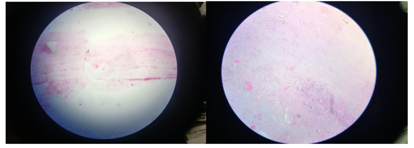
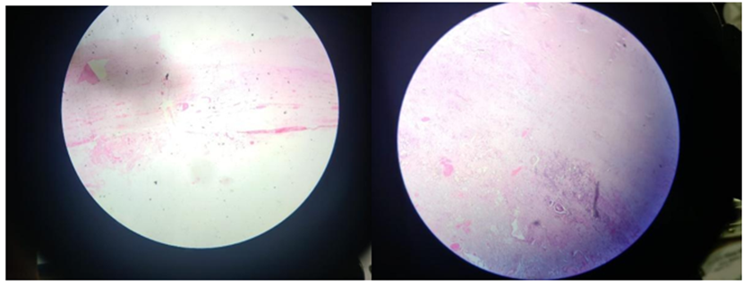
Figure 1. Histopathology of liver lesions
HISTOLOGICAL ASSESSMENT OF CHRONIC LIVER DISEASES: APPLICATION OF ISHAK, METAVIR, AND BATTS–LUDWIG SCORING SYSTEMS:
Many grading methods have been developed to classify and rank liver biopsies' histological samples. Employable regardless of illness etiology. In this case, liver fibrosis indicates disease progression. Grades of necroinflammation and hepatocellular damage indicate disease severity and progression [12]. Therefore, these scoring methods mostly use inflammation (hepatitis) and fibrosis to determine severity. Staging and grading together may enhance CLD diagnosis, prognosis, and therapy. Figures 1, 2, and 3 describe Batts-Ludwig, METAVIR, and Ishak scoring systems. Figure 4 compares these systems. Complexity makes the Ishak technique less applicable to clinical practice and less reproducible than the METAVIR and Batts-Ludwig score systems. The American Association for the Study of Liver Disease (AASLD) recommends using Batts-Ludwig and METAVIR for daily clinical practice with individual patients and more complicated scoring systems for large-scale clinical trials. A study compared these systems using the kappa-(k)-coefficient. The k-coefficient is a typical statistical tool for assessing observer agreement on qualitative or category items. It may also be used between two qualitative rating systems since its values range from 0 to 1. The usual interpretation of these statistics shows no agreement from 0 to 0.2, moderate agreement from 0.21 to 0.4, 0.41 to 0.6, 0.61 to 0.8, and practically perfect agreement from 0.81 to 1 [13]. A study comparing the METAVIR and Ishak systems demonstrated a substantial association (k=0.627) between necroinflammatory scores and a near-perfect correlation (k=0.998) with fibrosis in histological samples from 82 serologically confirmed chronic viral hepatitis patients [14]. The Ishak score is more complete than the METAVIR activity score, which only incorporates piecemeal and lobular necrosis (Figs. 2 and 3), which may explain the poorer agreement in necroinflammatory ratings. It considers confluent, localized, apoptosis, and portal inflammation.
Figure. 1: Batts–Ludwig scoring system for grading necroinflammatory activity and staging hepatic fibrosis in chronic hepatitis liver biopsy specimens.
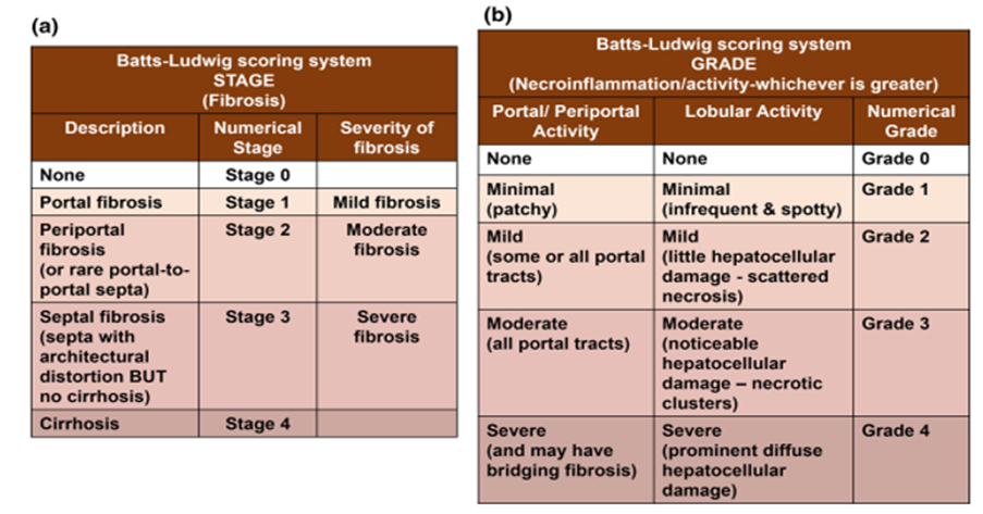
Figure. 1: Batts–Ludwig scoring system for grading necroinflammatory activity and staging hepatic fibrosis in chronic hepatitis liver biopsy specimens.
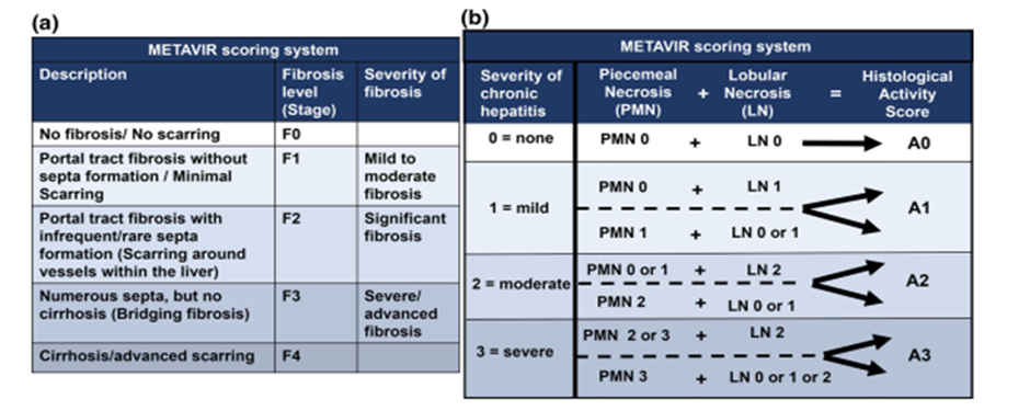
Figure. 2: METAVIR scoring system for evaluating fibrosis stage and histological activity grade in liver biopsy specimens.
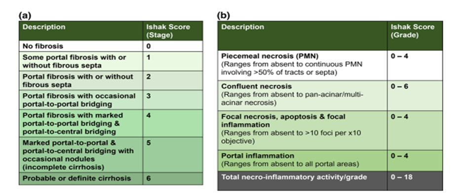
Figure. 3: Ishak (modified HAI) scoring system for grading necroinflammatory activity and staging fibrosis from 0 (no fibrosis) to 6 (cirrhosis) in liver biopsy specimens.
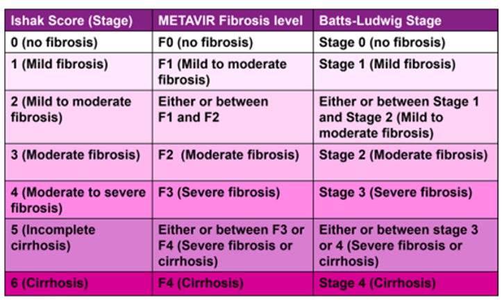
Figure. 4: Comparative correlation of Ishak, METAVIR, and Batts–Ludwig scoring systems for grading and staging chronic liver disease.
ROLE OF LIVER BIOPSY IN THE DIAGNOSIS OF VARIOUS HEPATIC DISORDERS
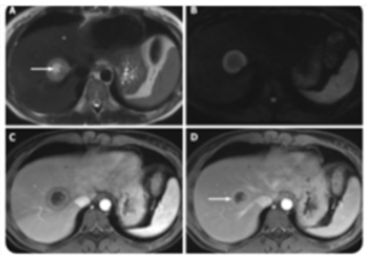
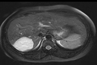
Figure 5. “Axial T2-weighted imaging of the metastatic lesion shows a target sign – hyperintense center (necrosis) with rim of viable tumor.”
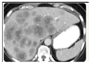
Figure 6. Multiple Heterogeneously Enhancing Liver Mets
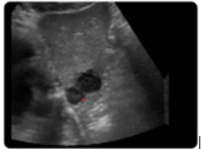
Figure 7. Two hypoechoic lesions on USG (patient with lung cancer)
ROLE OF LIVER BIOPSY IN THE DIAGNOSIS OF VARIOUS HEPATIC DISORDERS Alcohol-Associated Liver Disease:
Alcohol-induced ARLD pathology includes hepatic steatosis, alcoholic hepatitis, alcohol-associated cirrhosis, liver failure, and hepatocellular carcinoma. ARLD causes liver disease and accelerates the development of NAFLD, viral hepatitis, haemochromatosis, and others. ARLD will be treated with "screening, brief intervention and referral to treatment". Alcohol indicators for ARLD include liver enzymes, bilirubin, gamma-glutamyl transferase, carbohydrate-deficient transferrin, ethyl glucuronide, and phosphatidylethanol [15]. Biopsies for ARLD (alcoholic steatosis) are rare. It is utilized in rare cases with ARLD when the clinical presentation and serological tests are inconclusive or when other liver illnesses exacerbate and confuse the clinical presentation [16, 17]. Alcoholic hepatitis and cirrhosis are characterized by neutrophilic lobule inflammation, hepatocyte degeneration (including Mallory-Denk bodies and ballooning), steatosis, and fibrosis. Due to the dearth of well-validated non-invasive diagnostic methods for alcoholic hepatitis, a biopsy-based histology score (AHHS) was established and verified for prognostic categorization. Figure 5 shows the severity-rated four histological aspects. These include fibrosis (bridging or cirrhosis), billirubinostasis, polymorphonuclear neutrophil infiltration, and megamitochondria. Note that AHHS cannot stage or grade ARLD. It improves mortality prediction [18]. However, Dubois et al. showed that AHHS did not predict 1, 3, or 6-month survival in severe alcoholic hepatitis [19].
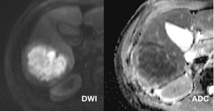
Figure. 5: there is an abscess in the right lobe of the liver showing Restriction on DWI and corresponding suppression on ADC
Non-Alcoholic Fatty Liver Disease (NAFLD) and Non-Alcoholic Steatohepatitis (NASH)
Hepatic steatosis, defined as >5% liver fat accumulation by histology or imaging, occurs without alcohol, steatogenic medicine, or monogenic genetic diseases. The ailment is NAFLD. Unlike ARLD, liver biopsies are the best way to detect NASH and fibrosis in NAFLD patients [20]. NAFLD patients at high risk of steatohepatitis and/or severe fibrosis (e.g., metabolic syndrome, increased aminotransferases, or age 60) or when other liver disorders cannot be ruled out should get liver biopsies, according to the AASLD [21]. NAFLD patients with high blood ferritin and iron saturation may need liver biopsies. This indicates severe hepatic iron buildup. Among NAFLD patients, 70-75% have non-alcoholic fatty liver (NAFL), whereas 25-30% have progressive non-alcoholic steatohepatitis (NASH). NAFL, or basic steatosis, is 5% or more, moderate non-specific inflammation, and minimal hepatocyte ballooning. NASH has 5% steatosis, lobular inflammation, hepatocyte ballooning, fibrosis, or both. Unlike NASH patients, NAFL patients have cirrhosis less than 4% of the time. A biopsy is now needed to diagnose, differentiate, and stage NAFLD. Brunt et al described NASH-specific stages and grades [22].
We created and validated a semiquantitative NAFLD-suspected histological grading technique. The system dominates NAFLD, including NAFL and NASH. This study is by the NASH Clinical Research Network Pathology Committee. Clinically, this is the NAFLD Activity Score (NAS) [23] (Fig. 6). A study ties NAS-5 to NASH diagnosis. Several pathologists diagnosed NASH using this instead of histology [24]. NAS grades and stages NAFLD to aid histological diagnosis, not NASH. Instead, it intended to examine therapy-induced changes. Brunt et al. studied NAS misuse. In pathologists' histological assessment, 7% of patients exhibited NAS≥5. Replacing NASH diagnosis with NAS≥5 may result in false positives. NASH and ARLD are characterized by steatosis, hepatitis, steatohepatitis, hepatocyte enlargement, apoptosis, necrosis, and fibrosis. A liver biopsy cannot differentiate NASH from alcohol-related steatohepatitis. Clinically, a history of alcohol consumption and the right clinical indications may distinguish the two. Some differences exist between NASH and ARLD. Satellitosis—polymorphonuclear cell clusters—is a hallmark of ARLD lobular inflammation. ARLD patients have thickened, fibrosed terminal hepatic venules and venoocclusive lesions, although not invariably steatosis. Only alcohol-induced hepatitis induces sclerosing hyaline necrosis.
Metabolic Dysfunction–Associated Fatty Liver Disease (MAFLD)
Rise in metabolic disease [25]. Recently, metabolic dysregulation may induce NAFLD [26, 27]. Therefore, metabolic dysfunction-associated fatty liver disease (MAFLD) encompasses the whole condition [28]. Hepatic steatosis (proven by imaging, serum fatty liver biomarkers, or histology), type 2 diabetes, overweight or obesity (BMI>25 kg/m2 in whites and>23 kg/m2 in Asians), or metabolic dysregulation with lean or normal weight describe MAFLD. Metabolic dysregulation is characterized by two or more metabolic risk factors, including waist circumference (≥102/88 cm in whites or ≥ 90/80 cm in Asians), prediabetes (5.6-6.9 mmol/L fasting plasma glucose), inflammation (hs-CRP >2 mg/L), high blood pressure (≥130/85 mmHg) or drug treatment, and low HDL-cholesterol levels. MAFLD is affected by age, sex, ethnicity, nutrition, metabolism, gut microbiota, and genetics [29]. MALFD diagnosis criteria are broader than NAFLD.
The latter does not tolerate high CLDs or alcohol, even with steatosis>5%. Fatty liver disease may increase CLDs. Liver disease is possible with MAFLD [30]. MAFLD improves clinical suspicion and lowers missed diagnosis, improving investigations. Metabolic illnesses including obesity and type 2 diabetes cause low-grade systemic inflammation. Clinical practice requires adding hs-CRP as an inflammatory marker to MAFLD diagnostic criteria. Lean, non-diabetic adults with normal BMI are seldom examined for hepatic steatosis with hs-CRP and HOMA-IR. These criteria for MAFLD diagnosis may screen this group. Researchers are researching this new phrase's practical applications. MAFLD and NAFLD were compared in fatty liver disease patients. Advanced fibrosis, age, BMI, HOMA-IR, lipid and liver enzyme levels, and metabolic comorbidities such diabetes and hypertension were greater in alcoholics. The data suggest the MAFLD criteria may identify high-risk hepatic steatosis individuals [31]. Importantly, MAFLD diagnostic criteria exist, but data limitations dispute their practical application. A steatosis in the liver indicates MAFLD. Hepatic fat deposits and blood lipid indicators may be detected by ultrasound. Hepatic steatosis may be assessed by liver biopsies and non-invasive methods. This method evaluates illness better. Its invasiveness and risks make this method only suitable for severe situations like many CLDs. MAFLD may be graded and staged like other CLDs to determine severity and progression. However, staging and grading depend on inflammatory-informed fibrosis assessments. CLD liver biopsies only indicate the illness at a given time. MAFLD, like other CLDs, cannot detect chronic-relapsing or intermittent inflammation.
Chronic Viral Hepatitis (Hepatitis B and C Infections)
For viral hepatitis, viruses A, B, C, D, or E infect the liver. Most hepatitis B and C infections cause liver inflammation, which may cause cirrhosis and HCC. Curing the worst virus, hepatitis C, has advanced. The incidence of hepatitis B is a public health issue. Although therapy has improved, cirrhosis and hepatocellular carcinoma are the main reasons of its high mortality [32, 33]. The liver biopsy is not advised for chronic hepatitis B and C diagnosis. It is still used for prognosis, staging/grading, and therapeutic management when biochemical or serological indications are ambiguous. Blood biomarkers are challenging to use to measure disease activity and fibrosis, especially in chronic hepatitis B [34]. Another application of orcein staining is to detect hepatitis B surface antigen in liver samples. CccDNA hepatitis B testing requires liver samples. This finding is crucial because viral cccDNA is the most critical genomic type for infection persistence. EASL advises non-invasive chronic hepatitis C severity assessments. If non-invasive treatments fail, mixed aetiologies are suspected or known, fibrosis is uncertain, or other causes are suspected, a liver biopsy may be indicated.
Histologically, chronic viral hepatitis has inflammatory fibrosis, cirrhosis, portal tract inflammation with mononuclear cells, interface hepatitis, localized lobular necrosis, and bile duct damage. Liver histology may reveal liver cell dysplasia, steatosis, hemosiderosis, necroinflammation, and fibrosis [35]. Chronic Hepatitis C may cause steatosis and bile duct damage. Necroinflammatory activity and fibrosis influence treatment and prognosis. Clinical studies show that fibrosis predicts disease progression better than necroinflammation. Besides elastography, Ishak, METAVIR, and Batts-Ludwig are used to evaluate fibrosis. We recommend liver biopsies before antiviral medication. Chronic hepatitis B patients with HBV DNA>20,000 IU/mL and alanine aminotransferase levels twice normal may start antiviral treatment without a liver biopsy. Because the drug helps all fibrosis phases.
Hereditary Hemochromatosis
Many northern Europeans inherit haemochromatosis, an iron overload illness. Dietary iron absorption rises in hemochromatosis (HFE), leading organ iron accumulation. HFE gene mutations (C282Y or H63D) cause end-stage liver, diabetes, and cardiac disease [36]. Tissue iron deposits were previously detected only by liver biopsy. Advances in genetic testing for HFE gene mutations, early identification, and non-invasive technologies like MRI that monitor liver iron levels have rendered liver biopsies obsolete for hereditary haemochromatosis diagnosis. However, it can still identify fibrosis or cirrhosis (which can be used for prognosis; see references [37, 38]), manage fibrosis in C282Y homozygotes with serum ferritin levels greater than 1000 ng/mL, distinguish C282Y from compound homozygotes by determining hepatic iron concentration, and rule out other chronic liver diseases (CLDs) like alcoholic and non- C282Y homozygotes with ferritin levels below 1000 ng/mL seldom need liver biopsies unless they have other cirrhosis risk factors.
Liver biopsies may be used if other testing suggest advanced fibrosis or cirrhosis [39]. Hepatocellular carcinoma risk rises with cirrhosis, therefore detection is crucial [40]. Iron values that don't match HFE genotypes or non-HFE hemochromatosis need a liver biopsy [41]. A liver biopsy and histology frequently detect hereditary hemochromatosis. This test grades fibrosis with Masson's trichrome and haematoxylin-eosin and evaluates hepatic iron cell distribution with Perls' Prussian blue to rule out other liver illnesses. For "the proportion of hepatocytes that stain for iron," Batts-Ludwig scores from 1 to 4, with 4 indicating pan-lobular iron deposition. The historical hepatic iron index may help improve iron evaluation. Iron distribution patterns in liver biopsies may detect viral hepatitis, NAFLD, and ARLD. About 60% had secondary iron loading. The reticuloendothelial system (macrophages and specialized endothelial cells) deposits iron most often in ferroportin disease, which is caused by a mutation in the cellular iron exporter gene. Most hepatocytes and Kupffer cells have modest iron distribution.
Hepatocellular Carcinoma (HCC)
HCC, the major cause of liver cancer, is becoming increasingly common. It often develops from advanced cirrhosis. In unclear imaging data, a liver biopsy may prevent a misdiagnosis of HCC in non-cirrhotic patients. Using biomarkers of hepatocellular differentiation like Arginase or bile salt export pump (BSEP) may help diagnose HCC beyond morphology and separate it from other primary and secondary hepatic malignancies. The hepatic lesion may be better described and early HCC separated from high-grade dysplastic nodule using tissue markers such glypican-3, glutamine synthetase, and heat shock protein-70. Liver biopsies may enable more accurate and customized treatment for hepatocellular carcinoma (HCC) patients, including prognostic categorization and therapeutic targets [42]. The American Association for the Study of Liver Diseases does not recommend a liver biopsy if the lesion is greater than 1 cm and two imaging scans indicate malignancy. Available screening methods for HCC in CLD patients, reliable ultrasonography, and HCC serum indicators such alpha-fetoprotein are making liver biopsies less required. Thus, biopsy for HCC diagnosis and assessment remains controversial. Needless to add, 2.7% needle tract tumor seeding after HCC biopsy is a major reason [43].
CONCLUSION:
When it comes to diagnosing, staging, grading, and predicting the prognosis of chronic liver illnesses, liver biopsies continue to be the gold standard. This is particularly true in circumstances when the biopsies have already been obtained. It differentiates between a broad variety of causal factors, such as hepatocellular carcinoma, chronic viral hepatitis, hereditary hemochromatosis, and alcoholic liver disease, as well as nonalcoholic fatty liver disease (NAFLD), major alcohol flush disease (MAFLD), and major alcohol flush disease. Despite the fact that non-invasive diagnostic tools have evolved, the biopsy is still considered the gold standard for fibrosis assessment as well as for resolving clinical circumstances that are difficult or overlap with other scenarios. It is necessary to use it with caution because of its invasive nature, the potential hazards involved, and the unpredictability of the samples. When it comes to individuals who have chronic liver disease, the connection of clinical, biochemical, radiographic, and histopathological data is what guides the optimal disease characterization and effective management strategies.
REFERENCES:
1. Quaglia A, Alves VA, Balabaud C, Bhathal PS, Bioulac-Sage P, Crawford JM et al. Role of aetiology in the progression, regression and parenchymal remodelling of liver disease: implications for liver biopsy interpretation. Histopathology. 2016;68(7):953-67.
2. Goodman ZD. Grading and staging systems for inflammation and fibrosis in chronic liver diseases. J Hepatol. 2007;47(4):598-607.
3. Bateman AC. Patterns of histological change in liver disease: my approach to ‘medical’ liver biopsy reporting. Histopathology 2007;51(5):585-96.
4. Brunt EM, Ramrakhaini S, Cordes BG, Neuschwander-Tetri BA, Janney CG, Bacon BR et al. Concurrence of histologic features of steatohepatitis with other forms of chronic liver diseases. Mod Pathol. 2003;16(1):49-56.
5. Rockey DC, Caldwell SH, Goodman ZD, Nelson RC, Smith AD. Liver Biopsy. Hepatology. 2009;49(3):1017-44.
6. Berger D, Desai V, Janardhan S. Con: Liver Biopsy Remains the Gold Standard to Evaluate Fibrosis in Patients With Nonalcoholic Fatty Liver Disease. Clinical Liver Disease [Internet]. 2019 [cited 2021 Jul 31];13:114–6. Available from: https://aasldpubs.onlin elibrary.wiley.com/doi/abs/10.1002/cld.740.
7. Goodman ZD. Grading and staging systems for inflammation and fibrosis in chronic liver diseases. J Hepatol. 2007;47:598–607.
8. Rockey DC, Caldwell SH, Goodman ZD, et al. Liver biopsy. Hepatology. 2009;49:1017–44.
9. Pandey N, Hoilat GJ, John S. Liver Biopsy. StatPearls [Internet]. Treasure Island (FL): StatPearls Publishing; 2020 [cited 2021 Jan 23]. Available from: http://www.ncbi.nlm.nih.gov/books/NBK47 0567/.
10. Washington K, Manns MP. 8 - Autoimmune Hepatitis. In: Burt AD, Ferrell LD, Hübscher SG, editors. Macsween’s Pathology of the Liver (Seventh Edition) [Internet]. Elsevier; 2018 [cited 2021 Sep 9]. p. 491–514. Available from: https://www.sciencedirect. com/science/article/pii/B978070206697900008X.
11. Mani H, Kleiner DE. Liver biopsy findings in chronic hepatitis B. Hepatology [Internet]. 2009 [cited 2021 Sep 9];49:S61–71. Available from: https://aasldpubs.onlinelibrary.wiley.com/doi/abs/ 10.1002/hep.22930
12. Boyd A, Cain O, Chauhan A, et al. Medical liver biopsy: background, indications, procedure and histopathology. Frontline Gastroenterology [Internet]. British Medical Journal Publishing Group; 2020 [cited 2021 Sep 9];11:40–7. Available from: https:// fg.bmj.com/content/11/1/40.
13. McHugh ML. Interrater reliability: the kappa statistic. Biochem Med (Zagreb). 2012;22:276–82.
14. Rozario R, Ramakrishna B. Histopathological study of chronic hepatitis B and C: a comparison of two scoring systems. J Hepatol. 2003;38:223–9.
15. Crabb DW, Im GY, Szabo G, et al. Diagnosis and Treatment of Alcohol-Associated Liver Diseases: 2019 Practice Guidance From the American Association for the Study of Liver Diseases. Hepatology [Internet]. 2020 [cited 2021 Aug 6];71:306–33. Available from: https://aasldpubs.onlinelibrary.wiley.com/doi/abs/10.1002/ hep.30866.
16. Torruellas C, French SW, Medici V. Diagnosis of alcoholic liver disease. World J Gastroenterol. 2014;20:11684–99. 28.
17. Kobyliak N, Dynnyk O, Abenavoli L. The role of liver biopsy to assess alcoholic liver disease. Rev Recent Clin Trials. 2016;11:175–9.
18. Altamirano J, Miquel R, Katoonizadeh A, et al. A histologic scoring system for prognosis of patients with alcoholic hepatitis. Gastroenterology. 2014;146:1231-1239.e6.
19. Dubois M, Sciarra A, Trépo E, et al. Histologic parameter score does not predict short-term survival in severe alcoholic hepatitis. United Eur Gastroenterol J. 2020;8:1003–12.
20. Chalasani N, Younossi Z, Lavine JE, et al. The diagnosis and management of nonalcoholic fatty liver disease: practice guidance from the American association for the study of liver diseases. Hepatology. 2018;67:328–57.
21. Sheka AC, Adeyi O, Thompson J, et al. Nonalcoholic steatohepatitis: a review. JAMA. 2020;323:1175–83.
22. Brunt EM, Janney CG, Di Bisceglie AM, et al. Nonalcoholic steatohepatitis: a proposal for grading and staging the histological lesions. Am J Gastroenterol. 1999;94:2467–74.
23. Kleiner DE, Brunt EM, Van Natta M, et al. Design and validation of a histological scoring system for nonalcoholic fatty liver disease. Hepatology. 2005;41:1313–21.
24. Brunt EM, Kleiner DE, Wilson LA, et al. The NAS and The histopathologic diagnosis in NAFLD: distinct clinicopathologic meanings. Hepatology. 2011;53:810–20.
25. Younossi Z, Anstee QM, Marietti M, et al. Global burden of NAFLD and NASH: trends, predictions, risk factors and prevention. Nat Rev Gastroenterol Hepatol. 2018;15:11–20.
26. Byrne CD, Targher G. NAFLD: a multisystem disease. J Hepatol. 2015;62:S47-64.
27. Miele L, Targher G. Understanding the association between developing a fatty liver and subsequent cardio-metabolic complications. Expert Rev Gastroenterol Hepatol. 2015;9:1243–5.
28. Eslam M, Newsome PN, Sarin SK, et al. A new definition for metabolic dysfunction-associated fatty liver disease: AN international expert consensus statement. J Hepatol Elsevier. 2020;73:202–9.
29. Tilg H, Effenberger M. From NAFLD to MAFLD: when pathophysiology succeeds. Nat Rev Gastroenterol Hepatol [Internet]. 2020 [cited 2022 Jan 15];17:387–8. Available from: https://www. nature.com/articles/s41575-020-0316-6.
30. De A, Ahmad N, Mehta M, et al. NAFLD vs. MAFLD – It is not the name but the disease that decides the outcome in fatty liver. Journal of Hepatology [Internet]. Elsevier; 2022 [cited 2022 Jan 15];76:475–7. Available from: https://www.journal-of-hepatology. eu/article/S0168-8278(21)02032-8/fulltext.
31. Lin S, Huang J, Wang M, et al. Comparison of MAFLD and NAFLD diagnostic criteria in real world. Liver International [Internet]. 2020 [cited 2022 Jan 15];40:2082–9. Available from: https://onlinelibrary.wiley.com/doi/abs/10.1111/liv.14548.
32. European Association for the Study of the Liver. Electronic address: easloffice@easloffice.eu, European Association for the Study of the Liver. EASL Recommendations on Treatment of Hepatitis C 2018. J Hepatol. 2018;69:461–511.
33. European Association for the Study of the Liver. Electronic address: easloffice@easloffice.eu, European Association for the Study of the Liver. EASL 2017 Clinical Practice Guidelines on the management of hepatitis B virus infection. J Hepatol. 2017;67:370–98.
34. Misdraji J. Changing indications for liver biopsy: viral hepatitis. Diagnostic Histopathology [Internet]. Elsevier; 2014 [cited 2021 Sep 21];20:119–24. Available from: https://www.diagnostichisto pathology.co.uk/article/S1756-2317(14)00033-4/abstract.
35. Pokorska-Śpiewak M, Kowalik-Mikołajewska B, Aniszewska M, et al. Is liver biopsy still needed in children with chronic viral hepatitis? World J Gastroenterol [Internet]. 2015 [cited 2021 Sep 21];21:12141–9. Available from: https://www.ncbi.nlm.nih.gov/ pmc/articles/PMC4641131/.
36. Pietrangelo A. Genetics, genetic testing, and management of hemochromatosis: 15 years since hepcidin. Gastroenterology. 2015;149:1240-1251.e4.
37. Bacon BR, Adams PC, Kowdley KV, et al. Diagnosis and management of hemochromatosis: 2011 practice guideline by the American association for the study of liver diseases. Hepatology. 2011;54:328–43.
38. Fitzsimons EJ, Cullis JO, Thomas DW, et al. Diagnosis and therapy of genetic haemochromatosis (review and 2017 update). Br J Haematol. 2018;181:293–303.
39. Kowdley KV, Brown KE, Ahn J, et al. ACG Clinical Guideline: Hereditary Hemochromatosis. Official journal of the American College of Gastroenterology | ACG [Internet]. 2019 [cited 2021 Sep 12];114:1202–18. Available from: https://journals.lww.com/ ajg/Fulltext/2019/08000/ACG_Clinical_Guideline__Hereditary_ Hemochromatosis.11.aspx.
40. Tarao K, Nozaki A, Ikeda T, et al. Real impact of liver cirrhosis on the development of hepatocellular carcinoma in various liver diseases—meta‐analytic assessment. Cancer Med [Internet]. 2019 [cited 2021 Sep 12];8:1054–65. Available from: https://www.ncbi. nlm.nih.gov/pmc/articles/PMC6434205/.
41. Bassett ML, Hickman PE, Dahlstrom JE. The changing role of liver biopsy in diagnosis and management of haemochromatosis. Pathology. 2011;43:433–9.
42. Di Tommaso L, Spadaccini M, Donadon M, et al. Role of liver biopsy in hepatocellular carcinoma. World J Gastroenterol [Internet]. 2019 [cited 2021 Sep 12];25:6041–52. Available from: https://www.ncbi.nlm.nih.gov/pmc/articles/PMC6824282/.
43. Silva MA, Hegab B, Hyde C, et al. Needle track seeding following biopsy of liver lesions in the diagnosis of hepatocellular cancer: a systematic review and meta-analysis. Gut. 2008;57:1592–6.