Morphologic Diversity of Liver Lesions: A Histopathology-Based Analysis
- Dr Sharath Chandra kashapaga , Assistant professor, department of pathology, Kamineni institute of medical sciences
- Dr Surabhi Karthik , Assistant Professor, Department of Radiology, Bhaskara Medical College Hyderabad
- Dr Sunitha gattigorla , Professor, Department of pathology, Nova institute of medical sciences
- Dr Mood Narayan , Professor, Department of respiratory medicine ESIC Medical college, Hyderabad
- Dr Vishal Parekar , Professor, Department of Pathology, Kamineni Institute of Medical Sciences, Narketpally
Article Information:
Abstract:
Background: Chronic liver disease (CLD) encompasses a wide range of conditions, including and not limited to inflammatory and benign hepatic lesions, primary and metastatic malignancies, and more. It is impossible to overstate the importance of a liver biopsy when it comes to determining the final diagnosis and determining the severity of a disease. The objective of this research is to investigate the clinicopathological relationship of liver lesions that have been established by biopsy and to evaluate the histological spectrum of these lesions. In this descriptive retrospective study, there were a total of 137 liver biopsy specimens that were included. These specimens were judged suitable for the diagnosis of chronic liver disease and associated hepatic abnormalities. All of the hospital's medical records were combed through in order to obtain information on health and demography. Histopathological methods that were considered standard comprised staining with hematoxylin and eosin, as well as the application of particular stains and immunohistochemistry when their use was required. With the use of descriptive statistics and a chi-square test, the data was analyzed to see whether or not there were any relationships between age, gender, and histological kinds. Individuals who were between the ages of 51 and 60 made up the majority of the cases. Men made up 42.3% of the population, whereas women made up 57.7% of the population. In terms of histological diagnosis, the most common ones were hepatocellular carcinoma, chronic hepatitis with fibrosis, and metastatic carcinoma. The link between being older and malignant liver lesions was found to be substantial (p < 0.001), but there was no significant correlation between sex and malignancy (p > 0.05). A significant amount of reliance is placed on liver biopsies for the diagnosis and categorization of hepatic abnormalities. Due to the high link that exists between increasing age and malignant liver pathology, patients who are above the age of 60 and have chronic liver disease should undergo further examinations.
Keywords:
Article :
INTRODUCTION:
The prevalence of liver diseases is continuously increasing all across the globe, and this trend is occurring regardless of factors such as age, gender, geography, or ethnicity. Chronic liver disease (CLD) is a primary cause of mortality and disability around the world. This is due to the fact that it may get more severe over time and could potentially lead to the development of cancer. There have been a number of variables that have contributed to the steady increase in the prevalence of hepatic diseases. These factors include metabolic risk factors, chronic infections, shifting lifestyles, and overall life expectancy. Tertiary care centers often deal with cases of these illnesses that are considered to be more advanced and complex.
Role of Liver Biopsy in Diagnosis
In the context of the ultimate clinical treatment for hepatic anomalies, liver biopsy continues to be a procedure that is widely used, well-established, and provides a high level of sensitivity [1,2]. Even though non-invasive diagnostic tools have evolved, liver biopsy and its histological examination are still regarded the gold standard when it comes to diagnosing and treating numerous hepatic problems. These disorders include autoimmune hepatitis, cirrhosis, and hepatocellular carcinoma, among others. Histopathological interpretation becomes very significant in situations when non-invasive methods of diagnosis, treatment planning, prognosis, or disease monitoring (such as clinical evaluation, radiographic imaging, or serological testing) are unable to meet the requirements. Given these circumstances, a liver biopsy is able to reliably categorize hepatic lesions as either benign, inflammatory, or malignant due to the direct morphological evidence that it provides.
Techniques of Liver Biopsy
Biopsies of the liver may be performed using a number of different techniques. A transthoracic percussion-guided liver biopsy is the method that is considered to be the gold standard. This operation is performed after a comprehensive history and physical examination, as well as imaging examinations. Additional techniques include the subcostal biopsy, real-time image-guided procedures, and the transthoracic image-guided biopsy that are also available. Patients with coagulopathy, ascites, or other conditions that prevent percutaneous biopsy are excellent candidates for advanced procedures such as transvenous or transjugular liver biopsy. These procedures involve inserting the biopsy needle into the jugular or femoral veins while being guided by fluoroscopic imaging. Patients who have these conditions are prime candidates for these advanced procedures. Due to the advancements in technology, liver biopsies that are carried out at hospitals that provide tertiary care are now considered to be diagnostic instruments that are both safer and more effective.
Indications and Clinical Utility
A liver biopsy is usually done for one of three reasons: to diagnose, to predict the outcome, or to check up on the patient.
1. Diagnostic Indications: When alternative diagnostic options have been exhausted, such as in the instances of overlap syndrome, nonalcoholic steatohepatitis (NASH), autoimmune hepatitis, or when discriminating between cholangiocarcinoma, hepatocellular carcinoma, and localized nodular hyperplasia, a liver biopsy is advised [3,4].
2. Prognostic Assessment: [5] Histopathological evaluation is an essential diagnostic technique that plays a significant role in identifying the extent and severity of chronic liver diseases such as advanced fibrosis, cirrhosis, non-alcoholic fatty liver disease (NASH), and other similar conditions. It is possible to gain significant information on the progression of the disease as well as its long-term effects by analyzing the degree of fibrosis and architectural distortion.
3. Follow-Up and Treatment Monitoring: In some conditions, such as autoimmune hepatitis, liver biopsies are recommended as a follow-up procedure. This is done with the purpose of evaluating the disease activity, predicting the recurrence of the illness, and monitoring the response to therapy [6]. As a result, it continues to be an essential instrument for assessing patients both before and after therapy.
Because of the great range of hepatic lesions that are observed in tertiary care institutions and the fact that histopathology continues to be the gold standard for final diagnosis, it is very important to evaluate the clinicopathological patterns of liver diseases that have been verified by biopsy in our setting. Although imaging and laboratory tests provide significant contributions to clinical assessment, there is a possibility that there may be differences between the findings obtained from clinical, radiographic, and histopathological examinations. This highlights the critical importance of tissue diagnosis.
OBJECTIVES
1. To investigate the histological range of liver lesions at a tertiary care hospital that have been biopsy-proven.
2. To establish a relationship between malignant and non-malignant liver lesions and clinicodemographic characteristics (sex and age).
RESEARCH METHODOLOGY:
The retrospective, descriptive, and observational study that served as the foundation for this research was carried out by the pathology department of a teaching hospital that provides tertiary care patients. Blood samples taken from liver biopsy specimens that were histopathologically determined to be chronic liver disease or related hepatic lesions were included in the research. These samples were collected during the course of the specified study period. Cases that had been verified by biopsy and had adequate tissue for evaluation were included in the research. Tissues that were either insufficient, autolyzed, or poorly preserved were not included in the study.
A search was conducted using the hospital records and the pathology requisition forms to look for relevant demographic and clinical information. These characteristics included age, gender, clinical diagnosis, and probability of cause. For the purpose of analyzing liver biopsy samples, typically used histopathology methods were used. Standard processing was performed on the tissues, which included embedding them in paraffin, sectioning them at a thickness of 3-5 μm, and staining them with hematoxylin and eosin. This was done after the tissues were preserved in 10% neutral buffered formalin. To assist in arriving at a definitive diagnosis, immunohistochemistry and specialized stains were used wherever they were deemed required [7].
On the basis of the histological findings, malignant and non-cancerous lesions were separated into several categories. Following the importation of data into Microsoft Excel, statistical software was used for the purpose of analysis. Descriptive statistics were used in order to provide a summary of the clinicopathological characteristics. When assessing connections between age group, sex, and histological category, which are all categorical variables, a p-value of less than 0.05 was used to define statistical significance. This allows for the evaluation of the relationships between these factors. Before the beginning of the investigation, the Institutional Ethics Committee gave its consent for the study to be conducted ethically.
DATA ANALYSIS
Table 1. Age Distribution of Patients with Chronic Liver Disease (CLD)
|
Age Group (Years) |
Number of Cases |
|
≤ 20 |
7 |
|
21–30 |
11 |
|
31–40 |
27 |
|
41–50 |
29 |
|
51–60 |
31 |
|
61–70 |
22 |
|
> 70 |
10 |
|
Total |
137 |
Table 1 displays the age-specific distribution of people with chronic liver disease confirmed by biopsy. There were 31 cases reported among those aged 51–60, 29 among those aged 41–50, and 27 among those aged 31–40. These age categories together accounted for the majority of the cases, indicating that middle-aged and older persons are the most often affected by chronic liver disease. A considerable proportion of patients were 60 years of age or older; 22 cases were between the ages of 61 and 70, and 10 cases were above 70, suggesting that the burden of sickness rises with age. However, there were only seven cases among individuals under the age of twenty, which is in line with the fact that chronic liver disease is less common in this age range [8]. The general pattern of the age distribution indicates that the incidence of chronic liver disease rises with age, which is in line with the combined effects of long-term hepatic damage, chronic inflammation, and metabolic factors.
Table 2. Sex Distribution of CLD Patients
|
Sex |
Number of Cases |
Percentage (%) |
|
Female (F) |
79 |
57.7 |
|
Male (M) |
59 |
42.3 |
|
Total |
137 |
100 |
Table 2 presents the gender distribution of those who have been diagnosed with chronic liver disease. The population under study consisted of 79 female cases, which accounted for 57.7% of the total, and 59 male cases, which accounted for 42.3% of the total. The fact that this research was conducted with a majority of female participants increases the potential that hepatic lesions and chronic liver disease are more prevalent among this particular group of women. The incidence of breast cancer is higher in females, and it is possible that autoimmune liver illnesses, malignancies linked to the gall bladder, and metastatic involvement from breast cancer might all play a part in explaining the observed increase [9]. According to a statistical analysis, the likelihood of developing liver lesions, whether they are malignant or non-malignant, is the same for both sexes. The study also discovered that there is no significant connection between sex and malignant liver illness, despite the fact that the majority of people with liver disease are male.
Table 3. Etiological Pattern Based on Clinical Diagnosis
|
Etiology / Clinical Diagnosis |
Number of Cases |
|
Hepatocellular carcinoma (HCC) |
11 |
|
?HCC (suspected) |
8 |
|
Cholangiocarcinoma |
3 |
|
Chronic liver disease (CLD) |
1 |
|
Autoimmune hepatitis |
3 |
|
Hydatid cyst (? / confirmed) |
11 |
|
Hemangioma |
5 |
|
Metastatic liver disease |
13 |
|
CA gall bladder |
9 |
|
CA breast |
10 |
|
CA pancreas |
7 |
|
Other malignancies* |
46 |
|
Total |
137 |
*Includes colon, stomach, cervix, ovary, esophagus, lymphoma, neuroendocrine tumors, etc.
The clinical diagnosis-based etiology pattern of chronic liver disease is shown in Table 3. A significant percentage of patients were identified as having cancerous conditions. To be more specific, there were thirteen cases of metastatic liver illness, eleven cases of hepatocellular carcinoma, eight cases of probable hepatocellular carcinoma, and carcinomas of the gall bladder, breast, and pancreas. The liver is a common place for secondary deposits that originate from other primary tumors, which is the reason why a substantial number of cases (46 instances) were categorized as other types of cancer [10]. In the category of benign liver illnesses, there were eleven cases of hydatid cysts, five cases of hemangiomas, three cases of autoimmune hepatitis, and one case of chronic liver disease with an unknown etiology. All of these cases were reported. The distribution of these cases demonstrates that the majority of patients who had liver lesions that were confirmed by biopsy had malignant or metastatic liver lesions. This highlights the need of histological confirmation in order to arrive at an accurate etiological diagnosis and to plan therapy accordingly.
Table 4. Histopathological Spectrum in Liver Biopsies
|
Histopathological Diagnosis |
Number of Cases |
|
Hepatocellular carcinoma (HCC) |
10 |
|
Metastatic carcinoma (all types) |
27 |
|
Hydatid cyst |
6 |
|
Cavernous hemangioma |
5 |
|
Chronic hepatitis with fibrosis / cirrhosis |
9 |
|
Cholangiocarcinoma |
4 |
|
Hepatic adenoma |
1 |
|
Simple / hepatic cyst |
2 |
|
Other benign & rare lesions* |
73 |
|
Total |
137 |
*Includes xanthogranulomatous reaction, neonatal cholestasis, vascular lesions, inflammatory lesions, etc.
As may be observed in Table 4, the histological spectrum associated with liver samples is shown here. As a result of the liver's extensive blood supply, it is susceptible to secondary tumor involvement. This is shown by the fact that the most common diagnosis of metastatic carcinoma (27 occurrences) is occurring in the liver. A total of ten cases of hepatocellular carcinoma and four cases of cholangiocarcinoma were identified as the primary hepatic cancers that were discovered. In the category of benign and inflammatory illnesses, there were a total of nine cases of chronic hepatitis with fibrosis or cirrhosis, six cases of hydatid cysts, five cases of cavernous hemangiomas, and two cases of simple or hepatic cysts. In addition, 73 individuals were found to have a wide range of benign and unusual abnormalities, which is a reflection of the fact that liver disease might vary from patient to patient [11]. Despite the fact that many of these diseases overlap radiological and clinical features, our findings demonstrate that liver biopsy is necessary for distinguishing between inflammatory diseases, benign diseases, and malignant diseases.
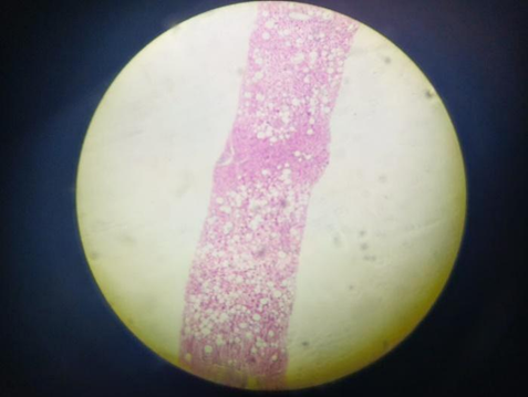
Figure 1. Low-Power View of Liver Showing Diffuse Macrovesicular Steatosis (H&E stain, 10×)
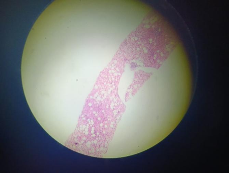
Figure 2. Liver Section Demonstrating Fatty Change with Large Clear Vacuoles
(H&E stain, 10×)
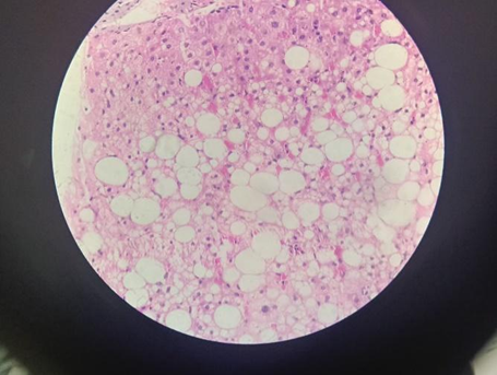
Figure 3. High-Power View of Hepatocytes with Macrovesicular Fat Accumulation
(H&E stain, 40×)
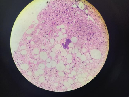
Figure 4. Hepatic Parenchyma Showing Extensive Steatosis with Nuclear Displacement
(H&E stain, 40×)
Table 5. Correlation of Clinical Diagnosis with Histopathology
|
Clinical Category |
Histopathology Concordant |
Discordant / Revised Diagnosis |
|
Suspected HCC |
Majority confirmed as HCC |
Few revised to metastasis |
|
Suspected cystic lesions |
Hydatid / simple cyst confirmed |
Minimal |
|
Suspected CLD |
Chronic hepatitis / fibrosis |
Occasional malignancy |
|
Suspected metastasis |
Metastatic carcinoma |
Rare primary liver tumors |
There is a correlation between the clinical diagnostic results and the histological findings, as shown in Table 5. Although a few patients were reclassified as having metastatic carcinoma, histology indicated that the great majority of cases were hepatocellular carcinoma. This indicates that there is a good agreement between the clinicopathological findings and the clinical findings. There was a high degree of concordance between clinical suspicions of cystic lesions and radiological and clinical examinations in these individuals [12]. This was due to the fact that the majority of cases were confirmed to be hydatid or simple cysts. Histopathology often revealed chronic hepatitis with fibrosis in patients who had been clinically diagnosed with chronic liver disease. However, in a few cases, an unexpected malignancy was discovered, which demonstrates the importance of biopsy in the diagnostic process. The histological confirmation of suspected metastases was rather frequent, with only a few instances of liver tumors that were the initial cause of the disease. When seen as a whole, the facts shown in this table demonstrate how important histological testing is for confirming clinical diagnoses, identifying malignancies that were not anticipated, and ensuring that patients get the best possible therapy.
Table 6. Association Between Sex and Histopathological Category
|
Sex |
Malignant |
Non-Malignant |
Total |
|
Female |
39 |
40 |
79 |
|
Male |
33 |
26 |
59 |
In this particular research, the chi-square (χ2) test was used to evaluate the connection between gender and histological category, primarily focusing on the distinction between malignant and non-malignant liver lesions. 39 of the 137 cases that were verified by biopsy were found to have malignant lesions, whereas 33 of the cases were found to have non-malignant lesions. A statistical analysis was conducted, and the findings revealed that the p-value was 0.554, while the χ2 value was found to be 0.35. Considering that the p-value was more than 0.05, it may be concluded that there was no statistically significant link between sex and liver cancer [13]. Based on these findings, it can be deduced that gender did not play a part in predicting the risk of developing malignant liver disease among the participants in the present study cohort. In cases of chronic liver disease that were verified by biopsy, sex was not shown to be an independent risk factor for malignancy. This was due to the fact that the distribution of malignant and non-malignant lesions was comparable in both males and females.
Table 7. Association Between Age Group and Histopathological Category
|
Age Group |
Malignant |
Non-Malignant |
|
≤20 |
1 |
6 |
|
21–30 |
1 |
10 |
|
31–40 |
11 |
16 |
|
41–50 |
14 |
15 |
|
51–60 |
23 |
8 |
|
61–70 |
15 |
7 |
|
>70 |
7 |
3 |
The chi-square test was used in order to investigate the relationship between age group and histological category in order to conduct an investigation into the occurrence of malignant liver lesions. The study showed a χ² value of 23.37 and a p-value of 0.00068, which is extremely significant. According to the findings, there is a significant relationship between becoming older and the development of malignant liver disease by a significant margin. The majority of malignant lesions were discovered in those who were between the ages of 51 and 60 and 61 and 70, but non-malignant lesions were more prevalent in younger age groups, particularly those who were under the age of 30 [14]. As a consequence of the findings, the likelihood of developing cancer continuously rises with increasing age. This age-related trend may have been caused by a number of factors, including chronic liver damage, cumulative hepatotoxic insults, and long-term inflammatory processes that raise the risk of cancer. Therefore, age becomes a significant determinant in the development of malignant liver disease in cases of chronic liver diseases that have been established clinically by biopsy.
In spite of the fact that age is a significant clinicopathological factor, the findings suggest that there is no significant link between malignant liver lesions and gender. In view of the fact that the likelihood of developing cancer increases dramatically with age, the findings of this study underline the need of conducting thorough screenings and making a fast diagnosis in older patients who arrive with chronic liver sickness. It is reasonable to presume that patients of either gender should get the same level of clinical care when they are suspected of having malignant liver lesions. This is because there is no significant gender bias in the medical field.
RESULTS:
One hundred thirty-seven cases of chronic liver disease and accompanying hepatic lesions that were verified by liver biopsy were included in this study. The majority of the cases were discovered in the medium and older age groups, which included individuals between the ages of 51 and 60 (31 cases), 41 to 50 (29 cases), and 31 to 40 (27 cases). There was a significant proportion of patients who were over the age of sixty, which is indicative of the fact that the incidence of liver disease rises with increasing age [15]. Both male and female patients were affected, with 59 male patients accounting for 42.3% of the total and 79 female patients making up 57.7% of the total.
An examination of the etiological pattern that was based on clinical diagnosis revealed that malignant illnesses were responsible for a significant number of the cases. The number of cases of metastatic liver disease was thirteen, the number of cases of hepatocellular carcinoma was eleven, and the number of cases of suspected HCC was eight. Primary malignancies of the gall bladder, breast, and pancreas were often the root cause of secondary liver involvement [16]. Among the disorders that were not malignant were hemangiomas, hydatid cysts, autoimmune hepatitis, and chronic liver disease for which there was no previously identified etiology.
There were 27 instances that were determined to be metastatic carcinoma, 10 cases that were found to be hepatocellular carcinoma, and 9 cases that were found to be chronic hepatitis with fibrosis or cirrhosis after the histological examination. Representative photomicrographs of fatty liver changes are shown in Figures 1–4. The diversity of liver disease was brought into even greater focus. According to the findings of the research, there was no statistically significant link between the presence of malignant liver disease and the gender of the individual (χ2 = 0.35, p = 0.554). The prevalence of malignant lesions was found to be higher in those who were above the age of 50, and there was a very significant association between age group and malignancy (χ2 = 23.37, p < 0.001).
DISCUSSION:
Chronic liver disease is a significant problem for the health of people all over the globe because of the progressive nature of the illness and the chance that it might develop into cancer. Due to the fact that the majority of cases were discovered in patients who were middle-aged or older, the present inquiry places an emphasis on the cumulative impact of metabolic stress, chronic inflammation, long-standing liver injury, and exposure to hepatotoxic chemicals over the course of time. The concept that age is a significant risk factor for hepatic cancers has been well-established [17], and the fact that our research found a robust link between increasing age and malignant liver pathology gives validity to the hypothesis that age is a risk factor. One possible explanation for the higher incidence of autoimmune liver illnesses, malignancies linked to the gall bladder, and breast cancer with metastasis among women in the study group is that women make up a disproportionately large portion of the population. When additional risk factors are present, males and females are equally prone to developing malignant liver lesions.
However, there is no statistically significant link between sex and malignancy, which suggests that sex does not independently influence the development of malignant liver lesions.
The liver, with its two blood arteries and fenestrated sinusoidal structure, is a classic place for secondary tumor deposits. Metastatic carcinoma is a common form of cancer that spreads outside of the primary tumor. Due to the fact that hepatocellular carcinoma has been recognized as a significant primary malignancy [18], it is important for patients who have chronic liver disease and fibrosis to undertake early diagnosis and monitoring. Inflammatory and benign lesions may overlap clinical and radiographic features, which further emphasizes the need of doing a liver biopsy in order to arrive at a conclusive diagnosis.
The fact that histological testing is still the most reliable approach for generating accurate diagnoses, which in turn lead to better treatment options and improved prognoses, is made abundantly obvious by the fact that clinicopathological discordance has been discovered. In conclusion, the findings of this research emphasize the need of doing thorough evaluations on older adults in order to facilitate early diagnosis and rapid treatment. Furthermore, these findings validate the use of liver biopsies in determining the severity of chronic liver diseases.
CONCLUSION:
Through the provision of a comprehensive clinicopathological evaluation of chronic liver illnesses that have been validated by biopsy, this study highlights the wide variety of hepatic lesions that are seen in tertiary care settings. Middle-aged and older persons were found to be the age groups most often impacted by chronic liver disease. Patients who were over the age of 50 had a much greater prevalence of malignant lesions than those who were younger than themselves. When it was discovered that age is a key predictor of cancer, the cumulative effect of liver damage that has been present for a long time and chronic inflammatory processes became apparent. There was no statistically significant link between sex and malignant liver pathology, which suggests that men and women are equally likely to develop non-malignant and malignant liver lesions. This is despite the fact that women had a higher number of cases than men. One of the most common locations for secondary tumor involvement is the liver, and the histological diagnostic of metastatic carcinoma was the most common. Due to the fact that hepatocellular carcinoma was the major hepatic malignancy that was shown to be the most frequent, it is especially important to monitor persons who have chronic liver disease and fibrosis at an early stage. In addition, the study demonstrated that radiological and clinical impressions might be misleading at times, and that histological examination is still the most effective method for obtaining a definitive diagnosis. It was extremely useful to have a liver biopsy since it allowed for the identification of unexpected malignancies, the differentiation between benign and inflammatory lesions, and the direction of appropriate therapeutic therapy. In a nutshell, the findings of this study emphasize the importance of liver biopsies in terms of the accurate detection, prediction, and treatment of chronic liver diseases, particularly in older people who are more likely to be diagnosed with cancer.
REFERENCES:
1. Rockey, D. C., Caldwell, S. H., Goodman, Z. D., Nelson, R. C., & Smith, A. D.; American Association for the Study of Liver Diseases. (2009). Liver biopsy. Hepatology, 49(3), 1017–1044. https://doi.org/10.1002/hep.22742
2. Neuberger, J., Patel, J., Caldwell, H., Davies, S., Hebditch, V., Hollywood, C., Hubscher, S., Karkhanis, S., Lester, W., Roslund, N., West, R., Wyatt, J. I., & Heydtmann, M. (2020). Guidelines on the use of liver biopsy in clinical practice from the British Society of Gastroenterology, the Royal College of Radiologists and the Royal College of Pathology. Gut, 69(8), 1382–1403. https://doi.org/10.1136/gutjnl-2020-321299
3. Tannapfel, A., Dienes, H. P., & Lohse, A. W. (2012). The indications for liver biopsy. Deutsches Ärzteblatt International, 109(27–28), 477–483. https://doi.org/10.3238/arztebl.2012.0477
4. Goodman, Z. D. (2007). Grading and staging systems for inflammation and fibrosis in chronic liver diseases. Journal of Hepatology, 47(4), 598–607.
5. National Center for Biotechnology Information. (n.d.). Liver biopsy. In StatPearls. https://www.ncbi.nlm.nih.gov/books/NBK470567
6. Casanovas, T. (2012). Rethinking the role of liver biopsy in the era of personalized medicine. In Liver biopsy—Indications, procedures, results. InTech. https://doi.org/10.5772/53120
7. Qaisarani, M. K., Niazi, M. F., & Hyder, N. (2017). Abdominal ultrasonographical fine needle aspiration cytology in the diagnosis of hepatobiliary masses. Pakistan Journal of Medical and Health Sciences, 11(4), 1478–1481.
8. Rastogi, N., Bhake, A., Agarwal, D., & Wankhede, A. (2019). Ultrasound-guided fine needle aspiration cytology of space occupying lesions of liver. International Journal of Research in Medical Sciences, 7, 192–198.
9. Sudhakar, G., & Devi, K. M. (2017). Study of ultrasound-guided FNAC of liver lesions. International Journal of Contemporary Medical Research, 4(7), 1621–1623.
10. Siegel, R. L., Miller, K. D., & Jemal, A. (2015). Cancer statistics, 2015. CA: A Cancer Journal for Clinicians, 65(1), 5–29.
11. Healthline. (2015). Liver metastasis: Symptoms, causes & diagnosis. Retrieved February 4, 2015, from https://www.healthline.com
12. Konfortion, J., Coupland, V. H., Kocher, H. M., Allum, W., Grocock, M. J., & Jack, R. H. (2014). Time and deprivation trends in incidence of primary liver cancer subtypes in England. Journal of Evaluation in Clinical Practice, 20(4), 498–504.
13. Agarwal, A., Khandelwal, R., Singh, S. N., Pant, H., Kumar, P., & Gupta, A. K. (2017). Evaluation of guided fine needle aspiration cytology in the diagnosis of hepatic lesions. International Journal of Biomedical Research, 8(2), 75–80.
14. Murgod, P. S., Doshi, P. R., & Dombale, V. D. (2019). Spectrum of hepatic lesions—A histopathological study of liver biopsies. IP Journal of Diagnostic Pathology and Oncology, 4(3), 200–203.
15. Ohnishi, K., Iida, S., Iwana, S., Goto, N., Nomura, F., Takashi, A., et al. (1982). The effect of chronic habitual alcohol intake in the development of liver cirrhosis and hepatocellular carcinoma in relation to hepatitis B surface antigen carriage. Cancer, 48, 672–677.
16. Chawla, N., & Sunila, S. (2013). Spectrum of histopathological findings in liver biopsy. Indian Medical Gazette, 363–367.
17. Agha, A., Furnari, M., Chakik, R. M., Ali, M. M. A., Alsaudi, D., et al. (2015). Hepatocellular carcinoma is the most frequent final diagnosis of focal liver lesions identified in a cross-sectional evaluation of patients with chronic liver disease in Saudi Arabia. Journal of Cancer Research.
18. Agarwal, A., Roy, P. P., Sarkar, S. K., et al. (2014). Clinicopathological profile of diagnosed patients of lung cancer with its relation to smoking habit and educational status in a medical college of Paschim Medinipore, West Bengal, India—A tribal area prospective study. Asian Pacific Journal of Health Sciences, 1, 479–485.