Profile of Infectious Keratitis: Risk Factors, Microbiology, Morphology, and Outcomes in a Tertiary Care Center in Hyderabad.
- Mohammad Aisha Begum , Ms ophthalmology Assistant professor Department of ophthalmology Government medical college Khammam, Telangana, India.
- Divya .B , MS ophthalmology, Assistant professor in ophthalmology, SVS medical college, Mahabubnagar. Telangana, India.
- Sumayya Farheen , MS ophthalmology, Assistant professor in ophthalmology, SVS medical college, Mahabubnagar. Telangana, India.
Article Information:
Abstract:
Purpose: To study risk factors, causative organisms, morphological features, complications, and preventive education needs in infectious keratitis. Methods: Prospective study of 60 consecutive patients with corneal ulcers at Shadan Medical College (Nov 2018–Oct 2020). Clinical exam, Gram/KOH stains, cultures (blood agar, SDA, non-nutrient agar). Results: In this cohort of 60 patients (mean age 38.5±18.2 years; 80% male), trauma was the predominant risk factor (31/60, 52%; p=0.02 vs. gender), with infections comprising bacteria (42%), fungi (32%), and viruses (25%); microbiology confirmed pathogens in 25% (Staphylococcus aureus, Streptococcus pneumoniae, Fusarium 5% each; p=0.03 trauma link). Paracentral ulcers affected 47%, hypopyon 28% (p=0.04 bacterial association), and 65% showed mild severity. Conclusion: Trauma in rural males drives keratitis; early targeted therapy is very useful in such cases.
Keywords:
Article :
INTRODUCTION:
According to NPCB survey corneal blindness accounts for 6% of total blindness.1 While according to RAAB 2006-2007 corneal blindness accounts for 6.5% cases of blindness. It is reported that there is an addition of 25000 to 30000 corneal cases every year in the country. Major causes of this blindness are corneal ulcers due to infections, trachoma, ocular injuries and keratomalacia of which infections are the leading cause. Infectious keratitis is one of the leading causes of blindness in our country. Because in most cases, these infections represent preventable or curable ophthalmic disease, a thorough understanding of epidemiology, diagnosis and treatment of various forms of infections keratitis is essential for eye care practitioners and public health officials. Infectious keratitis may be caused by various microorganisms like bacteria, fungi, virus, parasites, chlamydia etc.
Keratitis rarely occurs in normal eye because of human corneas natural resistance to infections. However predisposing factors such as corneal injury, contact lens wear, ocular adnexal dysfunction (including tear film deficiencies) conjunctival infections other exogenous factors, systemic disease and immunosuppression may alter the defence mechanism of outer eye and permit bacteria to invade the cornea. Although a few bacteria namely N.gonorrheae, N.meningitidis, C.diptheriae can invade intact cornea. Bacterial corneal ulcer begins with adhesion of bacteria to damaged epithelium and stroma. It is an ocular emergency due to often rapid progression of infection with threat of visual loss and potential corneal perforation.2
Fungal keratitis is common in men above the age of 30years as they are injured with plant or organic material or animal tail. Acanthamoeba is common in patients who are exposed to contaminated water and unhygienic contact lens solution. Early prevention and aggressive intial treatment for all clinical cases of infectious keratitis are key element of this disease to minimize the post infectious complications. A proper understanding of microbial and clinical characterstics of this disease entity which will enable the ophthalmologist to intiate appropriate antimicrobial therapy.3 The present study was conducted to study the risk factors, causative organisms, morphological features, complications, and visual outcomes in patients with infectious keratitis.
MATERIALS AND METHODS:
The data for this clinical study on infective corneal ulcers were sourced from patients attending the Outpatient Department of Ophthalmology at Shadan Institute of Medical Sciences and Research Centre, Hyderabad, from November 2018 to October 2020.Sample size - 60 Inclusion criteria: All cases of keratitis due to ocular infections (viral, bacterial, fungal and parasites) confirmed either by clinical examination or by investigation. Exclusion criteria: All cases of keratitis due to noninfectious causes like trophic corneal ulcers (neurotrophic keratopathy, exposure keratopathy), peripheral ulcerative keratopathies, nutritional (vitamin A deficiency), allergic keratitis (shields ulcer) confirmed either by clinical examination or investigation. Methodology: A standardized form is filled out on each patient documenting socio- demographic information as well as clinical information including duration of symptoms, previous treatment, predisposing ocular conditions and associated risk factors. A detailed ocular examination was carried out using torch and slit lamp. Special attention was paid to the stains of the ulcer – its site, size, shape, surface, dry or moist, floor whether covered with slough or not and edges regular or frayed. Corneal vascularisation and corneal sensation 47 were noted.
Ulcer is stained with 2 % fluorescein to know the extent and the details of the ulcer. The amount of hypopyon in anterior chamber whether quarter or less, one-third, two thirds or full, its colour whether creamy white or mixed with blood, and consistency whether fluid or fibrinous were noted, along with shape of upper level. The depth of the ulcer was determined by the slit lamp. Lacrimal sac patency was checked by doing sac syringing. Scrapping to be performed after instillation of 4% Lignocaine. Material obtained by scraping the leading edge and base of each ulcer will be inoculated directly into Blood agar, Chocolate agar and Sabouraud‟s Dextrose Agar. 48 Material from the corneal scrapping will also be smeared on three separate glass slides, one for Gram stain , one for Giemsa stain , and third for microscopic examination in the clinic as a 10% KOH wet mount. Microbial cultures were considered positive only if growth of the same was demonstrated on two or more solid media. The specific identification of bacterial pathogens was based on microscopic morphology, staining characteristics, and biochemical properties using standard laboratory criteria. Fungi were identified by their colony character on SDA and by their microscopic appearance in KOH. Laboratory investigations included urine examination for albumin, sugar, microscopy, blood for FBS/ RBS. Empirical treatment was begun with atropine 1% eye ointment / eye drops and a broad spectrum antibiotic like fluroquinolones 0.35 % eye drops instilled every 30 minutes. Antibiotic ointments were also used to treat bacterial corneal ulcers. Acyclovir 3% was used for viral keratitis. Systemic antibiotics, antifungals were given if the cornea perforates/ extension to the sclera. Adjunct therapy with Acetazolamide, vitamin C and A also instituted without delay. Debridement of the ulcer bed was carried out several times as the debris hindered the drug penetration.
This prospective study included 60 patients with infectious corneal ulcers presenting to the ophthalmic OPD at Shadan Medical College & Research Centre, Hyderabad, from November 2018 to October 2020.
RESULTS:
Table 1: Demographic characteristics
|
Parameter |
No. of Cases |
% |
|
Age (years) |
||
|
≤10 |
4 |
6.7 |
|
11–20 |
5 |
8.3 |
|
21–30 |
15 |
25.0 |
|
31–40 |
11 |
18.3 |
|
41–50 |
6 |
10.0 |
|
51–60 |
7 |
11.7 |
|
>60 |
12 |
20.0 |
|
Sex |
||
|
Male |
48 |
80.0 |
|
Female |
12 |
20.0 |
|
Socioeconomic status |
||
|
Low |
36 |
60.0 |
|
Middle |
24 |
40.0 |
|
Occupation |
||
|
Laborer |
12 |
20 |
|
Agricultural |
31 |
51.6 |
|
Housewife |
5 |
8.3 |
|
Student |
11 |
18.3 |
|
Toddler |
1 |
1.6 |
Incidence of infectious keratitis peaked in the 21–30 years (25%) and >60 years (20%) age groups. Males were affected more than females (80% vs 20%). Sixty‑percent of patients belonged to low socioeconomic status and 40% to middle class.
Table 2: Risk factors and clinical presentation
|
Parameter |
No. of Cases |
% |
|
Duration of symptoms |
||
|
<10 days |
45 |
75.0 |
|
11–20 days |
10 |
16.7 |
|
>21 days |
5 |
8.3 |
|
Ocular predisposing factors |
||
|
Trauma |
31 |
51.7 |
|
Chronic dacryocystitis |
5 |
8.3 |
|
Herpetic keratitis |
2 |
3.3 |
|
None |
22 |
36.7 |
|
Nature of injury (n=31) |
||
|
Vegetable matter |
11 |
18.3 |
|
Stone/insect |
13 |
21.7 |
|
Wooden |
6 |
10.0 |
|
Contact lens |
1 |
1.7 |
Trauma was the leading risk factor (51.7%). Around three‑fourths (75%) of patients presented within 10 days of symptoms. Majority of males found with trauma compare to females. (p=0.02; males at higher risk). Agriculture‑related occupation was significantly associated with incidence of ulcer (p<0.01).
Table 3: Clinical features of corneal ulcers
|
Parameter |
No. of Cases |
% |
|
Hypopyon |
||
|
Present |
17 |
28.3 |
|
Absent |
43 |
71.7 |
|
Ulcer location |
||
|
Central |
15 |
25.0 |
|
Paracentral |
28 |
46.7 |
|
Peripheral |
11 |
18.3 |
|
Central + paracentral |
6 |
10.0 |
|
Ulcer severity |
||
|
Mild |
39 |
65.0 |
|
Moderate |
15 |
25.0 |
|
Severe |
6 |
10.0 |
Paracentral ulcers were most common (46.7%). Most ulcers were mild (65.0%). Hypopyon presence was significantly associated by etiology (p=0.04 (Fisher’s exact test); higher in bacterial and fungal keratitis).
Table 4: Microbiological confirmation (positive isolations, n=15)
|
Organism |
No. of Cases |
% (of total 60) |
|
Bacterial (n=8) |
||
|
Staphylococcus aureus |
3 |
5.0 |
|
Streptococcus pneumoniae |
3 |
5.0 |
|
Coagulase‑negative staphylococci |
2 |
3.3 |
|
Fungal (n=6) |
||
|
Fusarium spp. |
3 |
5.0 |
|
Aspergillus spp. |
3 |
5.0 |
|
Other |
||
|
Acanthamoeba |
1 |
1.7 |
In the present cohort, Gram stain was positive in 8/30 corneal scrapings (26.7%), mostly Gram‑positive cocci. KOH‑wet‑mount was positive in 6/30 (20.0%). Culture was positive in 15/30 (50.0%) of cultured cases. Culture positivity was significantly associated with trauma history (p=0.03; 60% positive in trauma cases). Clinically, bacterial etiology predominated (42.0% of 60), followed by fungal (32.0%) and viral keratitis (25.0%, clinical diagnosis).
PHOTOGRAPHS:
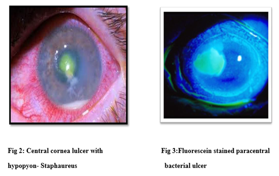
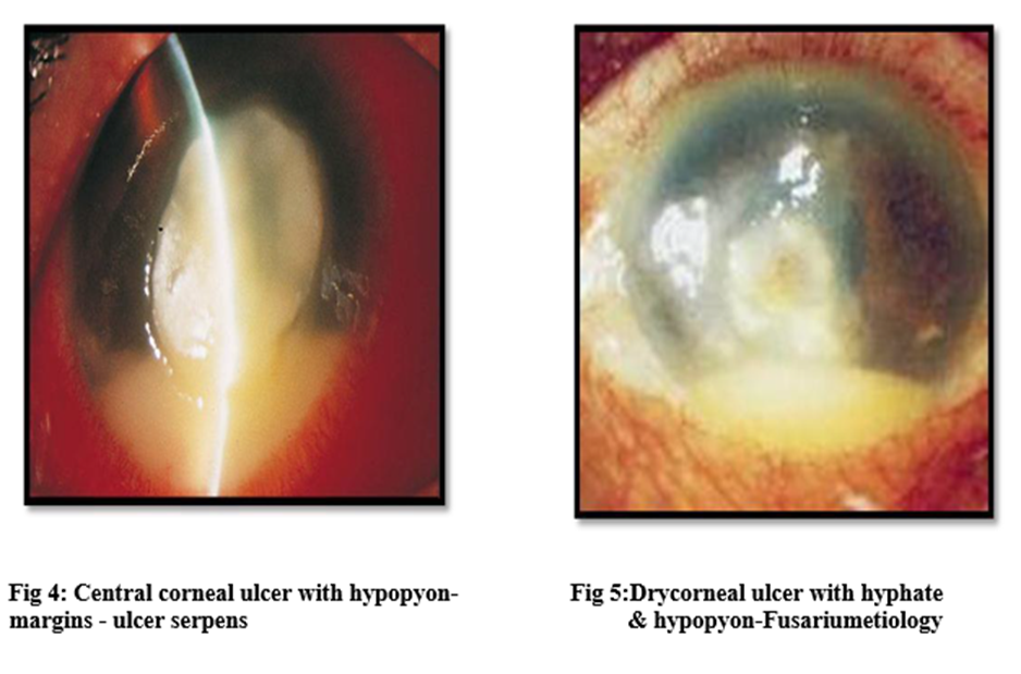
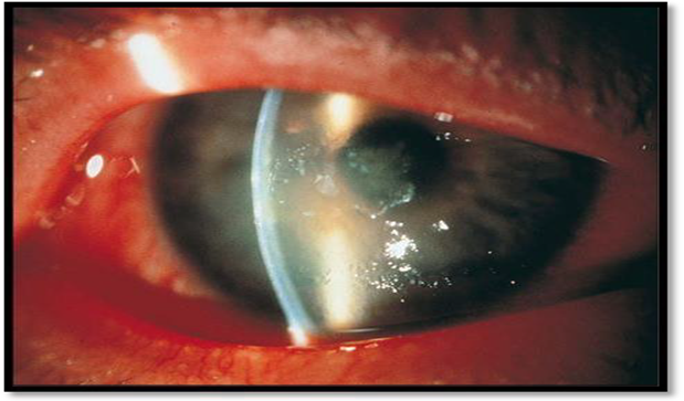
Fig 6:Feathery stromal infiltrates typical of Fungal keratitis
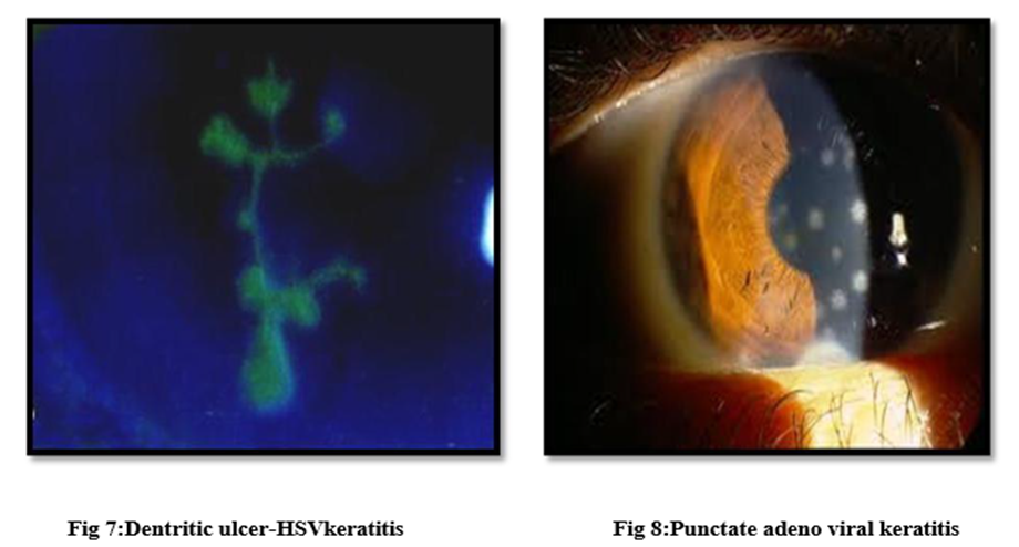
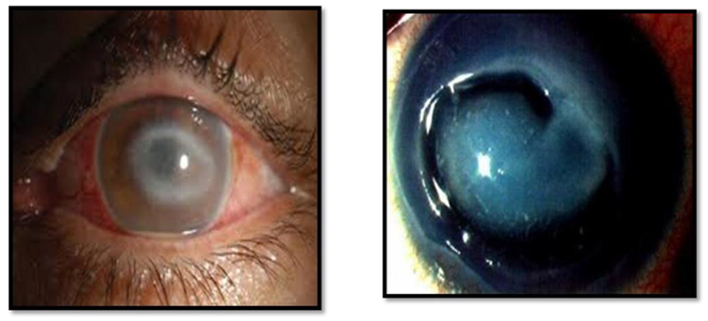
Fig 9: Disciform keratitis-HZVkeratitis Fig 10:Acanthamoebic keratitis- Ring Ulcer
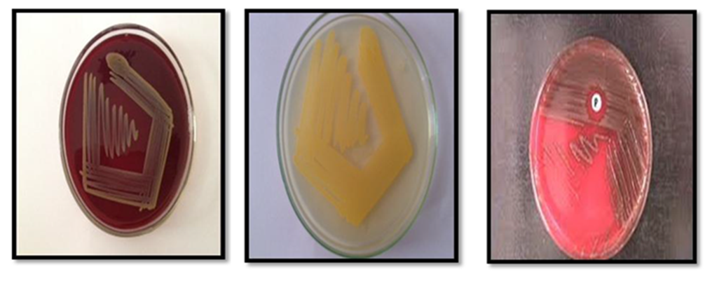
Fig11: Staphaureus colonies on blood & nutrient agar, Streptococcus colonies on blood agar
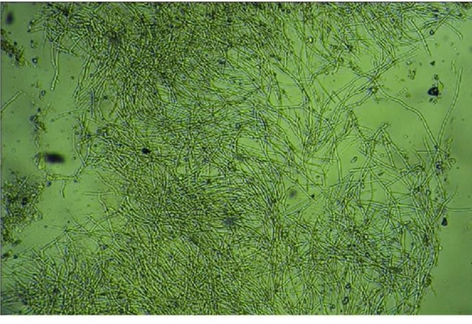
Fig 12:KOH wet mount with fungal hyphae.
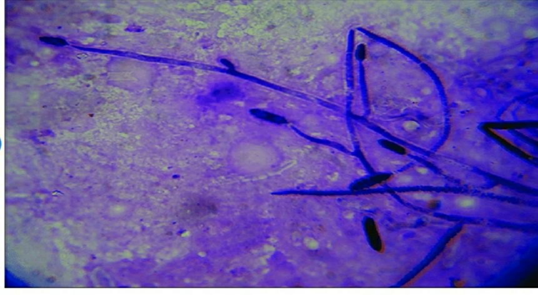
Fig 13: Gramstain with fungal hyphae
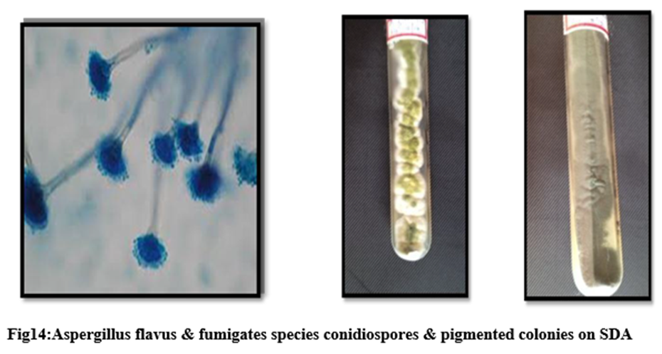
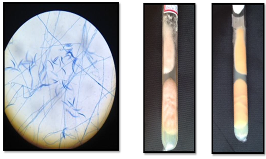
Fig15:Grampositive fungus Fusarium with macroconidiaon microscopy & buff colored colonies on SDA.
DISCUSSION:
In this study, infectious keratitis was more common in males (80%) than females. The age distribution showed a bimodal pattern, with peaks in the 21–30 years (25%) and >60 years (20%) age groups. This bimodal pattern is consistent with earlier epidemiological reports from India, which show that corneal ulcers affect both young adults and elderly individuals.1
Sixty percent of patients belonged to low socioeconomic status and 40% to middle class. This reflects the rural and semi urban background of our population, where agricultural and manual labor are common and access to protective eye care may be limited. In previous national surveys, corneal ulceration was more common in economically disadvantaged groups, which is in line with our findings.1
As reported in earlier South Indian epidemiological studies, both sexes tend to develop corneal ulcers in the middle decades of life, when they are more physically active and at higher risk of ocular trauma.4,5 Our cohort showed a high risk pattern in this age group, although we also noted a second peak in the elderly (>60 years), where age related corneal and systemic disease may contribute to ulcer susceptibility.
Trauma was the most important predisposing factor, present in 31 patients (51.7%). Among trauma related ulcers, injury by stone or insect (42%) was the most common, followed by vegetable matter (35%) and wooden objects (19%). In developing countries, superficial corneal trauma during agricultural work often leads to rapidly progressing corneal ulceration and visual loss.5 In South India, earlier reports emphasized paddy and rice stalk injury as the most common cause of superficial corneal trauma, whereas in our series stone or insect related trauma predominated, reflecting local occupational and environmental exposure.4
Duration of symptoms <10 days was seen in 75% of patients, showing that most individuals seek care early. This is important because short duration ulcers with mild severity are more likely to respond to medical therapy. In contrast, delays beyond 10–21 days are associated with deeper stromal involvement, hypopyon, and poor visual outcomes.3,6
Paracentral ulcers were the most common (46.7%), followed by central (25.0%) and peripheral (18.3%) locations. Paracentral and central ulcers are more likely to threaten the visual axis and cause significant visual impairment.6 Hypopyon was present in 28.3% of cases and was significantly more common in bacterial and fungal keratitis (p=0.04), which is in agreement with earlier South Indian series, where hypopyon was more frequently observed in bacterial keratitis than in fungal keratitis.6
As described in clinical reports on fungal keratitis, our fungal cases commonly presented with long duration of symptoms, dry raised necrotic slough, and satellite lesions, while bacterial keratitis usually showed a short history, greyish white infiltrate, and purulent slough. 3,6 Viral keratitis was clinically suspected in the presence of vesicular lesions on the face and lids (herpes zoster), punctate epithelial keratitis (herpes simplex), and viral type keratoconjunctivitis (adenovirus), in line with established descriptions.3,6
Gram stain examination identified organisms in 8/30 (26.7%) of corneal scrapings and showed good agreement with culture results. This is higher than the 63% concordance reported in earlier work on contact lens related corneal ulcers and rural suppurative keratitis, suggesting that in our setting Gram stain is reasonably useful for guiding early empiric therapy.7,8 In contrast, KOH wet mount was positive in 6/30 (20.0%) cases, and all KOH positive scrapings grew fungus on culture. This high concordance supports the use of KOH wet mount as a rapid bedside method for diagnosing mycotic keratitis, as emphasized by laboratory based studies from India.9
Foster reported that the use of multiple media, including blood and chocolate agar, gives the highest yield of positive cultures in keratomycosis.10 In our study, blood and chocolate agar were positive in 8/30 (26.7%) cultures, with organisms isolated from 50% of all cultured cases (15/30). This yield is comparable to other series from developing countries, where culture positivity ranges from around 40–60%.11 In a study from Ghana, culture positive findings were reported in 57.8% of suppurative keratitis cases, which is similar to our 50% yield.11
Among culture positive cases, 8 (13.3% of all 60 patients) were pure bacterial growth, 6 (10.0%) were pure fungal growth, and 1 (1.7%) was Acanthamoeba. Staphylococcus aureus and Streptococcus pneumoniae each accounted for 5.0% of all cases, underlining the importance of Gram positive cocci in infectious keratitis. 2 In the developing world, Streptococcus pneumoniae remains one of the most common bacterial causes of corneal ulcer, and it should be considered in the initial empiric regimen until culture results are available. 2,11
Fungal isolates were equally divided between Fusarium and Aspergillus (3 each, 5.0% of total 60 each). This dual predominance of Fusarium and Aspergillus spp. is similar to that reported from South Florida and from Ghana, where filamentous fungi are the main cause of fungal keratitis. 11,12 In South India, similar patterns of Fusarium dominated or mixed filamentous fungal keratitis have been reported, which is consistent with our findings.12
Clinically, 15 cases (25%) were suggestive of viral keratitis, including 3 (20%) herpes zoster, 5 (33%) herpes simplex, and 7 (47%) adenoviral keratoconjunctivitis, consistent with the spectrum of viral keratitis described in earlier clinical and textbook sources. 3,6
CONCLUSION:
The present study identified trauma (52%) as the primary risk factor for infectious keratitis. Bacterial etiology was the most common clinically (42%), followed by fungal (32%) and viral keratitis (25%, clinical diagnosis). Staphylococcus aureus and Streptococcus pneumoniae were the main bacterial pathogens, while Fusarium and Aspergillus spp. dominated among fungal isolates. Most ulcers were mild (65%) and 75% of patients presented within 10 days, indicating a window for early medical management. However, delayed presentation still carries a risk of poor visual outcome. Public health strategies targeting rural trauma prevention, farmer and laborer education, and early initiation of empiric anti bacterial and anti fungal therapy are essential for reducing blindness from corneal ulcers in Telangana.
REFERENCES:
1. Govt. of India, National Survey on Blindness: 1999–2001, Report 2 knexch.
2. Bharathi M. J., Ramakrishnan R. et al. Epidemiology of bacterial keratitis in a referral centre in South India. Indian J Med Microbiol 2003;21(4):239–245.
3. Madhukar K. Reddy. Current perspectives in infectious keratitis. Indian J Ophthalmol 1994;42(4):171–192.
4. Erie J. C., Nevitt M. P., Hodge D. O., Ballard D. J. Incidence of ulcerative keratitis in a defined population from 1950 through 1988. Arch Ophthalmol 1993;111:1665–1671.
5. Tylefors B. Epidemiological patterns of ocular trauma. Aust N Z J Ophthalmol 1992;20:95–98.
6. Thomas A. P., Leck K. A., Myatt M. Characteristic clinical features as an aid to the diagnosis of suppurative keratitis caused by filamentous fungi. Br J Ophthalmol 2005;89:1554–1558.
7. Galentine G. Corneal ulcers associated with contact lens wear. Arch Ophthalmol 1984;102(8):891–894.
8. Williams G., Mc Clellan K., Billson F. Suppurative keratitis in rural Bangladesh; the value of gram stain in planning management. Int Ophthalmol 1991;15(2):131–135.
9. Vajpayee R. B., Angra S. K., Sandramouli S., Honavar S. G., Chhabra V. K. Laboratory diagnosis of keratomycosis: comparative evaluation of direct microscopy and culture results. Br J Ophthalmol 2003;87(7):834–838.
10. Forster R. K., Reblla G. The diagnosis and management of keratomycoses. I. Cause and diagnosis. Arch Ophthalmol 1975;93(11):975–978.
11. Hagan M., Wright E., Newman M., Dolin P., Johnson G. Causes of suppurative keratitis in Ghana. Br J Ophthalmol 1995;79:1024–1028.
12. Leisegang T. J. Fungal keratitis. In: Kaufman H. E., Barron B. A., McDonald M. B., eds. The Cornea, 2nd ed. Boston/Toronto/London: Butterworth‑Heinemann; 1998. p. 219–240.