Beyond the Mediastinum: En bloc Resection of Lung, Pericardium, and SVC for Invasive Thymoma, a Case Report and Review.
- ASHUTOSH GUPTA , PROFESSOR SURGICAL ONCOLOGY, Pt. Jawaharlal Nehru Memorial Medical College, Raipur, Chhattisgarh.
- K K SAHU , ASSOCIATE PROFESSOR CVTS, Pt. Jawaharlal Nehru Memorial Medical College, Raipur, Chhattisgarh.
- KISHAN SONI , ASSOCIATE PROFESSOR SURGICAL ONCOLOGY, Pt. Jawaharlal Nehru Memorial Medical College, Raipur, Chhattisgarh.
- ADITI DIWAN , ASSISTANT PROFESSOR, SURGICAL ONCOLOGY, Pt. Jawaharlal Nehru Memorial Medical College, Raipur, Chhattisgarh.
- GUNJAN AGRAWAL , ASSISTANT PROFESSOR, MAXILLOFACIAL SURGERY, Pt. Jawaharlal Nehru Memorial Medical College, Raipur, Chhattisgarh.
- VIVEK CHOUDHARY , DEAN, RADIOTHERAPY, Pt. Jawaharlal Nehru Memorial Medical College, Raipur, Chhattisgarh.
- SONAM , Pt. Jawaharlal Nehru Memorial Medical College, Raipur, Chhattisgarh.
- SAMRIDHA , Pt. Jawaharlal Nehru Memorial Medical College, Raipur, Chhattisgarh
- LAVANYA , Pt. Jawaharlal Nehru Memorial Medical College, Raipur, Chhattisgarh.
Article Information:
Abstract:
Background: Thymic epithelial tumors (TETs) are rare malignancies arising in the anterior mediastinum, with thymoma being the most common subtype. Although many thymomas are encapsulated, locally advanced tumors may invade adjacent mediastinal structures, making surgical management technically challenging. Complete surgical resection remains the most important prognostic factor and the cornerstone of treatment. We report a case of invasive thymoma successfully managed with multivisceral en bloc resection. CasePresentation: A 40-year-old man presented with chest pain, breathlessness, fatigue, and diplopia. Contrast-enhanced computed tomography (CT) of the chest revealed a 77 × 72 mm mass in the pretracheal superior mediastinum with loss of fat planes with the ascending aorta and superior vena cava (SVC). CT-guided biopsy and immunohistochemistry were suggestive of invasive thymoma, most likely WHO type B3. The patient was also found to have acetylcholine receptor antibodies consistent with myasthenia gravis and was started on medical management. Following multidisciplinary team discussion, surgical resection was planned. Through a median sternotomy, en bloc resection of the mediastinal mass was performed along with non-segmental resection of the right lung, resection of the involved pericardium, and tangential resection of the SVC with primary repair. The plane with the ascending aorta was preserved. Histopathological examination confirmed invasive thymoma (WHO type B3) with lung parenchymal invasion. An R0 resection was achieved, and the patient remains disease-free at 6 months follow-up. Conclusion: This case highlights that aggressive multivisceral resection for locally advanced thymoma can achieve complete tumor clearance and favorable early outcomes when performed with careful surgical planning and a multidisciplinary approach.
Keywords:
Article :
INTRODUCTION:
Thymomas and thymic carcinomas (thymic epithelial tumors, TETs) are among the rarest neoplasms with an incidence of 0.15 cases per 100,000 [1,2]. Formally, TETs consist of thymomas, thymic carcinomas, and thymic neuroendocrine tumors (NET). Thymoma is the most common primary anterior mediastinal tumor in adults[2]. It is well accepted that the main effective treatment for a thymic epithelial tumours (TETs) is complete surgical resection [3,4]. These tumours usually develop in the anterior mediastinum in proximity to several important structures and organs, such as the pericardium, great vessels, lungs, and heart. In cases with advanced Masaoka Koga stage, or a large-sized tumour and aggressive histological types, such as type B2/B3 or thymic cancer, invasion of adjacent organs can easily occur [5]. In such circumstances, achieving complete resection can be technically challenging, and incomplete resection is sometimes unavoidable. Here, we report a case of invasive thymoma which was successfully managed with surgical resection.
CASE REPORT :
A 40-year-old man presented to the Surgical Oncology outpatient department with complaints of chest pain, breathlessness, fatigue, and diplopia. Contrast-enhanced computed tomography (CT) of the chest revealed a 77 × 72 mm lesion in the pretracheal superior mediastinum, with loss of fat planes with the ascending aorta and the superior vena cava(SVC) [Image 1] . Cardiopulmonary examination and the remainder of the physical examination were unremarkable. We performed a CT-guided biopsy, which suggested a poorly differentiated neoplasm, with sparse immature T cells and occasional B cells. Immunohistoschemistry was positive for cytokeratin (CK) and p63 and negative for CD5 and CD117, suggesting the diagnosis of invasive thymoma, most likely WHO Type B3. Further blood investigations revealed positive AChR antibodies for myasthenia gravis(MG). Medical management of MG was initiated with pyridostigmine and prednisolone.
Following multidisciplinary team (MDT) discussion, surgical management was planned in collaboration with the cardiothoracic surgical team. After adequate preoperative optimisation, the patient was taken up for surgery. The tumour was resected via median sternotomy, and an en bloc excision of the mediastinal mass was performed along with non-segmental resection of the right lung, resection of the involved pericardium, and tangential resection of the SVC with primary repair [Image 2 and 3] . The plane with the ascending aorta could be delineated and was preserved.
The postoperative course was uneventful. Gross examination of the resected specimen revealed a soft-to-firm, irregular mass measuring 11 × 11 × 5 cm. Final histopathological evaluation confirmed invasive thymoma, Type B3, with microscopic invasion of the capsule and extension into the adjacent lung parenchyma. An R0 resection was achieved, and the patient was kept under observation. He has remained asymptomatic and disease-free at 6 months of follow-up.
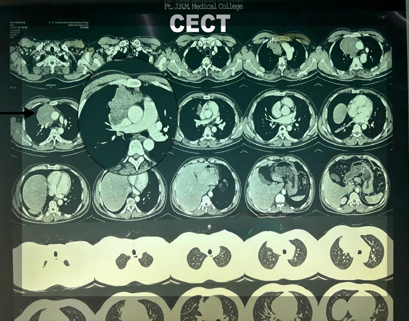
Figure 1. Image 1 : Contrast-enhanced computed tomography (CT) of the chest revealed a 77 × 72 mm lesion in the pretracheal superior mediastinum, with loss of fat planes with the ascending aorta and the superior vena cava(SVC).
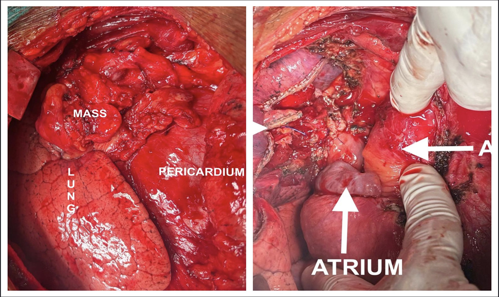
Figure 2. (a): Thymic mass adherent to the lung and pericardium (b) tumor bed after en bloc resection, horizontal arrow depicting the ascending aorta.
DISCUSSION:
Thymic neoplasms are rare tumors, accounting for less than 1% of all adult malignancies. Peak incidence occurs in the fourth and sixth decades of life, with no sexual predilection [6]. Most thymomas are solid neoplasms that are encapsulated and localized to the thymus, but approximately one-third of these invade the tumor capsule and the surrounding structures [7].
About a third to half of all persons are asymptomatic, and the mass is often identified as an incidental finding on imaging performed for an unrelated problem. Other ways it can manifest include symptoms of compression of surrounding organs from the expansile mass, in the form of superior vena cava syndrome, dysphagia, or chest pain. It is often associated with systemic and autoimmune paraneoplastic disorders such as myasthenia gravis, which occurs in 30–40% of cases at diagnosis, the most common being myasthenia gravis [8,9]. The tumor does tend to recur locally but is unlikely to metastasize hematogenously or to regional lymphatics.
Imaging is an essential part of the workup of thymomas to aid in diagnosis and proper staging. It is important to distinguish thymic hyperplasia (thymus symmetrically enlarged with smooth borders and preservation of normal thymic shape) from a thymoma (a well-defined round or oval mass located anterior to the great vessels and heart), as this will direct the mode of treatment. CT also differentiates invasive from noninvasive thymoma. The presence of a lobulated or irregular contour, areas of low attenuation, and multifocal calcification is suggestive of invasive thymoma [10].
The recognition of thymomas and thymic carcinomas as separate entities was introduced in the second edition of the WHO Classification of Tumors in 1999, and the nomenclature of the major thymoma types has been maintained ever since [11]. Based on the morphology and architecture of the epithelial cells and their proportion to lymphocytes, thymomas are divided into types A, AB, B1, B2, B3, and other rare types. Thymic carcinomas are pathologically similar to extrathymic carcinomas and typically show squamous differentiation [12]. Since 2015, all thymomas and thymic carcinomas have been recognized as malignant tumors [13].
Various staging systems of thymomas have been defined based on the degree of invasiveness, but the most common one is the Masaoka-Koga system, which has been widely used because it is a prognostic indicator for thymic malignancy and a predictor of tumor recurrence [14]. Seventy to eighty percent of tumors are classified as stage I (completely encapsulated), while stages II through IV demonstrate the invasive form of the disease. In 2017, for the first time, a structured TNM classification for thymic malignancies was proposed by the International Association for the Study of Lung Cancer (IASLC) and the International Thymic Malignancies Interest Group (ITMIG), on commission by the Union for International Cancer Control (UICC) and the American Joint Committee on Cancer (AJCC), and is now widely used [15].
Owing to a lack of prospective studies in this rare disease, data from retrospective studies still advocate for surgical resection of the entire thymus gland and surrounding mediastinal fat. Various approaches include median sternotomy, lateral thoracotomy, and minimally invasive approaches such as VATS (video-assisted thoracic surgery) or RATS (robotic-assisted thoracic surgery) [16]. Minimally invasive approaches are preferred for localized thymic tumors [17]. When a combined resection including invaded organs is necessary, a median sternotomy is generally the chosen surgical approach.
About 40% of thymic tumors are invasive, and management of these advanced thymomas with Masaoka stages III–IV is still challenging. Unresectable disease can be defined as an extensive tumor involving middle mediastinal organs (trachea, great arteries, and/or heart). The exact role of induction chemotherapy in locally advanced thymoma patients remains controversial. Given the rarity of the tumor, there have been no randomized controlled trials conducted to date. Anthracyclines and cisplatin form the basis of most protocols. Various studies have used the cisplatin, doxorubicin, vincristine, and cyclophosphamide (ADOC) regimen, and the cisplatin, doxorubicin, and cyclophosphamide (PAC) regimen [18]. In a meta-analysis comprising 12 studies and a total of 286 patients, there was a pooled response rate to induction therapy of 59%, a complete resection rate of 73%, a 5-year survival of 87%, and a 10-year survival of 76% [19].
If complete resection is not achievable after 2–4 cycles, or if the patient is not deemed a suitable surgical candidate, definitive radiotherapy is recommended as part of a sequential chemotherapy strategy. A total dose of 60–66 Gy in 30–33 fractions may be considered with cisplatin and etoposide chemotherapy. However, for advanced, non-resectable, non-irradiable, or metastatic (stage IVB) tumors, chemotherapy should be offered as a single agent. The aim is to decrease tumor-related symptoms [20].
PD-L1 positivity is seen in around 67% of thymic malignancies [21]. Recent phase II clinical trials on the role of pembrolizumab in thymic malignancies have shown an overall response rate ranging between 19% and 28% in the recurrent setting [22,23]. However, as previously described, patients with autoimmune conditions may have reduced immune tolerance. Immune-related adverse events (irAEs) occurred in 15% to 62% of patients treated with immune checkpoint inhibitors (ICIs) in various trials [22,23].
The prognosis of thymomas is variable, ranging from an indolent, non-invasive course to an aggressive nature with infiltration of surrounding mediastinal tissues and metastasis. When present at an early stage, the tumor is usually well encapsulated; advanced disease is a less common presentation and is a well-recognized strong risk factor for both local recurrence and generalized metastasis. Due to its indolent nature, even after complete resection, thymomas can recur and should be managed as if they were newly diagnosed tumors. To detect early recurrence, lengthy follow-up is recommended. The time to recurrence has been reported to range from 6 to 234 months [24]. The International Thymic Malignancy Interest Group (ITMIG) recommends annual chest CT for 5 years after resection, followed by alternating chest CT and chest X-ray until year 11, and then annual chest radiography thereafter, as late recurrences are not uncommon [25].
CONCLUSION:
Surgery is the initial treatment of choice for all patients with thymoma. Invasive thymomas with extensive mediastinal involvement pose significant surgical challenges. However, complete en bloc resection remains the cornerstone of treatment and offers the best chance for long-term disease control. This case highlights that even in the presence of invasion into adjacent structures such as the lung, pericardium, and superior vena cava, radical multivisceral resection can be safely performed in carefully selected patients. With meticulous surgical planning and multidisciplinary management, favorable oncologic and functional outcomes can be achieved.
Disclosure statement
No potential conflict of interest was reported by the authors.
REFERENCES:
1. Riedel, R. F. (2006). Thymoma: Benign appearance, malignant potential. The Oncologist, 11(8), 887–894. https://doi.org/10.1634/theoncologist.11-8-887
2. Hsu, C. H., Chan, J. K., Yin, C. H., Lee, C. C., Chern, C. U., & Liao, C. I. (2019). Trends in the incidence of thymoma, thymic carcinoma, and thymic neuroendocrine tumor in the United States. PLoS ONE, 14(12), e0227197. https://doi.org/10.1371/journal.pone.0227197
3. Maggi, G., Giaccone, G., Donadio, M., Ciuffreda, L., Dalesio, O., Leria, G., Trifiletti, G., Casadio, C., Palestro, G., Mancuso, M., & Calciati, A. (1986). Thymoma: A review of 169 cases, with particular reference to results of surgical treatment. Cancer, 58(3), 765–776.
4. Curran, W. J., Kornstein, M. J., Brooks, J. J., & Turrisi, A. T. (1988). Invasive thymoma: The role of mediastinal irradiation following complete or incomplete surgical resection. Journal of Clinical Oncology, 6(11), 1722–1727.
5. Funaki, S., Shintani, Y., Fukui, E., et al. (2020). Surgical treatment strategies for invasive thymoma. Journal of Thoracic Disease, 12(12), 7619–7625. https://doi.org/10.21037/jtd-19-3045
6. Kumawat, M. K., Sharma, N., Lal Jakhar, S., Kumar, H. S., & Kumar, R. (2024). A review of literature on thymoma: An indolent tumor of anterior mediastinum. Asian Pacific Journal of Cancer Care, 9(2), 353–356. https://doi.org/10.31557/apjcc.2024.9.2.353-356
7. Acharya, P. T., Ali, S., Stanescu, A. L., Phillips, G. S., & Lee, E. Y. (2019). Pediatric mediastinal masses: Role of MR imaging as a problem-solving tool. Magnetic Resonance Imaging Clinics of North America, 27(2), 227–242. https://doi.org/10.1016/j.mric.2019.01.003
8. Romi, F. (2011). Thymoma in myasthenia gravis: From diagnosis to treatment. Autoimmune Diseases, 2011, 474512. https://doi.org/10.4061/2011/474512
9. Bernard, C., Frih, H., Pasquet, F., et al. (2016). Thymoma associated with autoimmune diseases: 85 cases and literature review. Autoimmunity Reviews, 15(1), 82–92. https://doi.org/10.1016/j.autrev.2015.09.005
10. Tomiyama, N., Müller, N. L., Ellis, S. J., Cleverley, J. R., Okumura, M., Miyoshi, S., Kusumoto, M., Johkoh, T., Yoshida, S., Mihara, N., Honda, O., Kozuka, T., Hamada, S., & Nakamura, H. (2001). Invasive and noninvasive thymoma: Distinctive CT features. Journal of Computer Assisted Tomography, 25(3), 388–393.
11. Marx, A., Chan, J. K. C., Chalabreysse, L., Dacic, S., Detterbeck, F., French, C. A., et al. (2022). The 2021 WHO classification of tumors of the thymus and mediastinum: What is new in thymic epithelial, germ cell, and mesenchymal tumors? Journal of Thoracic Oncology, 17(2), 200–213. https://doi.org/10.1016/j.jtho.2021.10.010
12. Oramas, D. M., & Moran, C. A. (2022). Thymoma: Histologically a heterogeneous group of tumors. Seminars in Diagnostic Pathology, 39(2), 99–104. https://doi.org/10.1053/j.semdp.2021.06.002
13. Marx, A., Chan, J. K., Coindre, J. M., et al. (2015). The 2015 World Health Organization classification of tumors of the thymus: Continuity and changes. Journal of Thoracic Oncology, 10(10), 1383–1395. https://doi.org/10.1097/JTO.0000000000000654
14. Detterbeck, F. C., et al. (2011). The Masaoka-Koga stage classification for thymic malignancies: Clarification and definition of terms. Journal of Thoracic Oncology, 6(7 Suppl 3), S1710–S1716.
15. Detterbeck, F. C., Stratton, K., Giroux, D., Asamura, H., Crowley, J., Falkson, C., Filosso, P. L., Frazier, A. A., Giaccone, G., Huang, J., et al. (2014). The IASLC/ITMIG thymic epithelial tumors staging project: Proposal for an evidence-based stage classification system. Journal of Thoracic Oncology, 9(Suppl 2), S65–S72. https://doi.org/10.1097/JTO.0000000000000290
16. Liou, D., Berry, M., Brown, L., et al. (2024). The Society of Thoracic Surgeons expert consensus document on the surgical management of thymomas. The Annals of Thoracic Surgery, 118, 975–1004.
17. Zhang, X., Gu, Z., & Fang, W. (2018). Minimally invasive surgery in thymic malignancies: The new standard of care. Journal of Thoracic Disease, 10(Suppl 14), S1666–S1670. https://doi.org/10.21037/jtd.2018.05.168
18. Berruti, A., Borasio, P., Gerbino, A., Gorzegno, G., Moschini, T., Tampellini, M., et al. (1999). Primary chemotherapy with adriamycin, cisplatin, vincristine and cyclophosphamide in locally advanced thymomas: A single institution experience. British Journal of Cancer, 81(5), 841–845. https://doi.org/10.1038/sj.bjc.6690773
19. Hamaji, M., Ali, S. O., & Burt, B. M. (2015). A meta-analysis of induction therapy for advanced thymic epithelial tumors. The Annals of Thoracic Surgery, 99(5), 1848–1856. https://doi.org/10.1016/j.athoracsur.2014.12.048
20. Girard, N., Ruffini, E., Marx, A., et al. (2015). Thymic epithelial tumours: ESMO clinical practice guidelines for diagnosis, treatment and follow-up. Annals of Oncology, 26(Suppl 5), v40–v55.
21. Katsuya, Y., Horinouchi, H., Asao, T., Kitahara, S., Goto, Y., Kanda, S., Fujiwara, Y., Nokihara, H., Yamamoto, N., Watanabe, S., Tsuta, K., & Ohe, Y. (2016). Expression of programmed death 1 (PD-1) and its ligand (PD-L1) in thymic epithelial tumors: Impact on treatment efficacy and alteration in expression after chemotherapy. Lung Cancer, 99, 4–10. https://doi.org/10.1016/j.lungcan.2016.05.007
22. Giaccone, G., Kim, C., Thompson, J., et al. (2018). Pembrolizumab in patients with thymic carcinoma: A single-arm, single-centre, phase 2 study. The Lancet Oncology, 19(3), 347–355. https://doi.org/10.1016/S1470-2045(18)30062-7
23. Cho, J., Kim, H. S., Ku, B. M., et al. (2019). Pembrolizumab for patients with refractory or relapsed thymic epithelial tumor: An open-label phase II trial. Journal of Clinical Oncology, 37(24), 2162–2170. https://doi.org/10.1200/JCO.2017.77.3184
24. Bae, M. K., Lee, C. Y., Lee, J. G., et al. (2013). Predictors of recurrence after thymoma resection. Yonsei Medical Journal, 54(4), 875–882. https://doi.org/10.3349/ymj.2013.54.4.875
25. Fang, W., Detterbeck, F., Ackman, J. B., Marom, E. M., Roden, A. C., Mendogni, P., Marino, M., Lococo, F., Molina, T. J., Rimner, A., Girard, N., Ventura, L., Ricciardi, S., Cangir, A. K., & Szolkowska, M. (2026). International Thymic Malignancy Interest Group standard definitions, policies, and reporting measures for thymic epithelial tumors. Journal of Thoracic Oncology, 21(1), 102–111. https://doi.org/10.1016/j.jtho.2025.09.1757.