OUTCOME OF DISTAL TIBIA FRACTURE TREATED WITH PRIMARY ILIZAROV EXTERNAL FIXATOR.
- Deependra Sonkar , Associate Professor, Department of Orthopaedics, Gandhi Medical College and Hamidia Hospital, Bhopal.
- Manish Singh Rajpoot , Assistant Professor, Department of Orthopaedics, Gandhi Medical College and Hamidia Hospital, Bhopal.
- Shubham Rawat , Junior resident, Department of Orthopaedics, Gandhi Medical College and Hamidia Hospital, Bhopal.
Article Information:
Abstract:
Background: Distal third tibial fractures are challenging injuries due to poor soft tissue coverage, subcutaneous bone location, and compromised vascularity. Achieving stable fixation while preserving biology is difficult, particularly in open fractures. Ilizarov external fixation offers a minimally invasive method that enables early weight bearing and promotes fracture healing [6-9]. Materials and Methods: A prospective observational study of 30 patients with distal third tibial fractures treated with primary Ilizarov external fixation was conducted between April 2024 and October 2025. The study population predominantly consisted of males (96.7%), with the most common age group being 51–60 years (26.7%). The most common mechanism of injury was fall from height (40%), followed by road traffic accidents (23.3%). Fractures were classified using AO/OTA and Gustilo–Anderson classification [5,6], with Grade IIIA fractures being most common (50%), followed by Grade II (26.7%) and Grade IIIB (23.3%). Functional outcomes were assessed using ASAMI criteria. Results: All fractures achieved union (100%). The majority of patients required fixation for 120–129 days (36.7%), with a mean union time of 18.4 weeks. According to ASAMI criteria, excellent outcomes were observed in 73.3%, good in 20%, and fair in 6.7%, resulting in good to excellent outcomes in 93.3% of patients. Superficial pin tract infection occurred in 6.7% and was managed conservatively. No cases of deep infection, non-union, or implant failure were observed. Conclusion: Ilizarov external fixation is a reliable and effective modality for distal third tibial fractures, providing excellent union rates and good functional outcomes with minimal complications, even in high-grade open fractures.
Keywords:
Article :
INTRODUCTION:
Distal third tibial fractures account for a significant proportion of lower limb injuries and are frequently associated with high-energy trauma such as road traffic accidents and falls from height. Owing to the subcutaneous location of the distal tibia and its relatively poor vascular supply, these fractures are prone to complications including delayed union, non-union, infection, and soft tissue compromise (1,2).
Management of distal tibial fractures remains challenging. Conventional methods such as intramedullary nailing and plating, although widely used, may not always be ideal in distal fractures due to difficulties in achieving stable fixation in the metaphyseal region and the risk of further soft tissue injury (3,4). Open reduction and plating in particular have been associated with wound complications and infection, especially in open fractures (5).
The Ilizarov external fixation system provides a minimally invasive alternative that allows stable fixation while preserving the biological environment of the fracture. By utilizing circular ring constructs and tensioned wires, it enables controlled micromotion at the fracture site, promotes osteogenesis, and allows early weight bearing (6,7). This makes it particularly advantageous in fractures with compromised soft tissue conditions.
Despite its established role, there remains variability in reported outcomes (12–14), particularly with respect to union time, functional recovery, and complication rates. Therefore, the present study was undertaken to evaluate the clinical and functional outcomes of distal third tibial fractures treated with primary Ilizarov external fixation, with emphasis on union rates, functional scoring, and associated complications.
MATERIALS AND METHODS:
A prospective observational study was conducted at a tertiary care center between April 2024 and October 2025, including 30 patients with open distal third tibial fractures up to Gustilo–Anderson type II, IIIA and IIIB in patients with age ≥18 years while excluding pathological fractures and patients with severe polytrauma or neurovascular compromise.
Ethical approval was obtained from the institutional ethics committee, and informed consent was obtained from all patients.
Fractures were classified using AO/OTA classification and Gustilo–Anderson classification [5,6]. The majority of fractures were AO type 42 (80%), followed by AO type 43 (20%).
All patients were treated using Ilizarov ring external fixation under fluoroscopic guidance. Closed reduction was preferred. Tensioned wires and olive wires were used to achieve stable fixation.Postoperatively, early mobilization and weight bearing were encouraged. Patients were followed at regular intervals with clinical and radiological assessment until fracture union.
Outcome Measures:
Radiological union
Duration of external fixation application
Functional outcome using ASAMI criteria
Complications
RESULTS:
A total of 30 patients were included in the study. The majority were males (96.7%), with the most common age group being 51–60 years (26.7%) as shown in Table 1
Table 1: Patient Demographics and Injury Characteristics
|
Parameter |
Number (%) |
|
Total patients |
30 |
|
Male |
29 (96.7%) |
|
Female |
1 (3.3%) |
|
Most common age group |
51–60 years (26.7%) |
|
Mode of injury |
|
|
• Fall from height |
12 (40%) |
|
• Road traffic accident |
7 (23.3%) |
|
Gustilo–Anderson Classification |
|
|
• Grade II |
8 (26.7%) |
|
• Grade IIIA |
15 (50%) |
|
• Grade IIIB |
7 (23.3%) |
The most common mechanism of injury was fall from height (40%), followed by road traffic accidents (23.3%).
Functional Outcome (ASAMI):
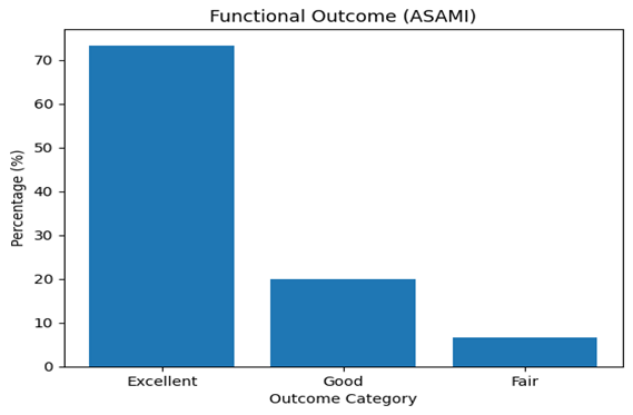
Figure 1: Bar graph showing distribution of functional outcomes based on ASAMI criteria.
Thus, good to excellent outcomes were achieved in 93.3% of patients. All fractures achieved union (100%). The majority required frame fixation for 120–129 days (36.7%), with an average union time of 18.4 weeks.
Complications:
Superficial pin tract infection occurred in 6.7% of patients and was managed conservatively. No cases of deep infection, non-union, or implant failure were observed and summarised in Table 2.
Table 2: Clinical and Functional Outcomes
|
Parameter |
Value |
|
Union rate |
30 (100%) |
|
Mean union time |
18.4 weeks |
|
Most common frame duration |
120–129 days (36.7%) |
|
ASAMI Outcome |
|
|
• Excellent |
22 (73.3%) |
|
• Good |
6 (20%) |
|
• Fair |
2 (6.7%) |
|
Complications |
|
|
• Pin tract infection |
2 (6.7%) |
|
• Deep infection |
0 |
|
• Non-union |
0 |
|
• Implant failure |
0 |
ILLUSTRATIVE CASES
Case 1:
A 48-year-old male presented with a distal third tibial fracture following a road traffic accident. On clinical examination, there was swelling, deformity, and tenderness over the distal leg with intact distal neurovascular status. Radiographs revealed a comminuted distal third tibial fracture with associated fibular fracture (Figure 1A).
The patient was managed with primary Ilizarov external fixation. Closed reduction was achieved under fluoroscopic guidance, and a circular external fixator was applied using tensioned wires and rings to obtain stable fixation. Post-operative radiographs confirmed satisfactory alignment and fixation (Figure 1B).
Early mobilization and weight bearing were initiated. The patient was followed up at regular intervals. At final follow-up, radiographs demonstrated satisfactory fracture union with good alignment (Figure 1C). Functional outcome assessed using ASAMI criteria was excellent.
This case highlights the effectiveness of Ilizarov external fixation in achieving stable fixation and reliable union in distal third tibial fractures.
Case 2:
A 20-year-old male presented following assault on 17 August 2025. The patient sustained an open injury to the left leg with a wound over the distal third of the tibia.
On examination, there was a compound fracture with exposed bone and significant soft tissue injury. Distal neurovascular status was intact. Radiographs revealed a comminuted fracture of the distal third of the tibial shaft, classified as AO/OTA 42-C, associated with a posterior malleolus fracture (Fig 2A-B). The injury was classified as Gustilo–Anderson Grade IIIA.
The patient was managed with primary Ilizarov ring external fixation following thorough wound debridement. Reduction was achieved using ligamentotaxis, and stable fixation was obtained with tensioned wires and appropriate frame configuration (Fig 2C).
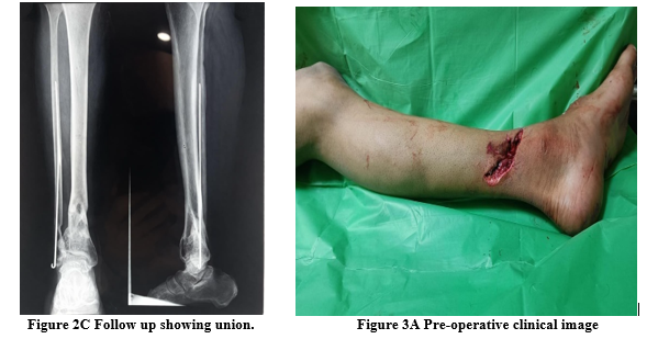
Early mobilization and weight bearing were initiated. Serial radiographs demonstrated progressive fracture healing, and complete union was achieved without major complications (Fig 2D). Functional outcome was assessed as excellent according to ASAMI criteria.
Case 3:
A 40-year-old female presented with a history of fall resulting in injury to the left leg. On examination, there was swelling and tenderness over the distal third of the tibia without significant soft tissue compromise. Distal neurovascular status was intact.
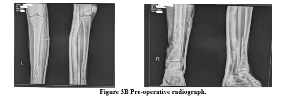
Radiographs revealed a distal third tibial shaft fracture, classified as AO/OTA 42-A (Fig 3A). The injury was classified as Gustilo–Anderson Grade I.
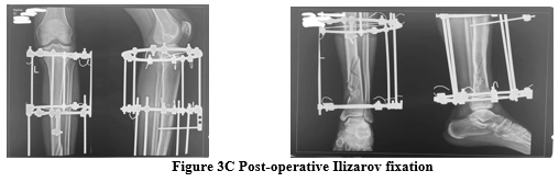
The patient was managed with primary Ilizarov ring external fixation. Reduction was achieved using ligamentotaxis, and stable fixation was obtained with tensioned wires and appropriate frame configuration (Fig 3B).
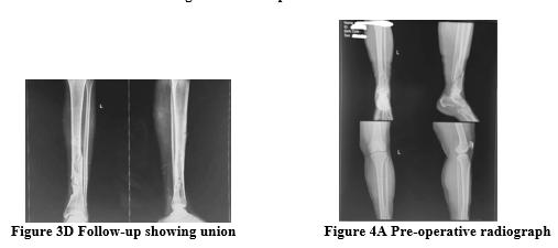
Early mobilization and weight bearing were initiated. Serial radiographs demonstrated progressive fracture healing (Fig 3C), and complete union was achieved within the expected time frame. Functional outcome was assessed as excellent according to ASAMI criteria.
DISCUSSION:
The present study demonstrates a high union rate of 100% with a mean union time of 18.4 weeks, indicating the effectiveness of Ilizarov external fixation in the management of distal third tibial fractures. These findings are comparable to those reported by Wani et al. (17–20 weeks) and Tanna et al., who also observed high union rates with the use of circular external fixation (8,9).
The favorable outcomes observed in this study can be attributed to the biological and mechanical advantages of the Ilizarov system. Preservation of periosteal blood supply, minimal disruption of soft tissues, and stable fixation collectively create an optimal environment for fracture healing. In addition, controlled axial micromotion at the fracture site promotes callus formation, which may explain the consistent union observed across all cases.
Functional outcomes were also encouraging, with 93.3% of patients achieving good to excellent results based on ASAMI criteria. Similar findings have been reported by Ahmad et al., who highlighted the role of early weight bearing and stable fixation in improving functional recovery (10). Early mobilization in the present study likely contributed to reduced joint stiffness and better rehabilitation outcomes.
When compared to alternative fixation methods, Ilizarov external fixation offers distinct advantages in distal tibial fractures. Studies by Vallier et al. and Boraiah et al. have reported higher rates of wound complications and soft tissue problems associated with plating techniques (11,12). Intramedullary nailing, while less invasive, may be associated with malalignment and inadequate distal fixation in metaphyseal fractures, as described by Guo et al. (13).
The complication rate in the present study was low, with only superficial pin tract infections observed in 6.7% of cases, all of which were managed conservatively. The absence of deep infection, non-union, or implant failure further supports the safety profile of this technique, particularly in open fractures and cases with compromised soft tissue.
Overall, the findings of this study reinforce the role of Ilizarov external fixation as a reliable and biologically favorable method for managing distal third tibial fractures, particularly in complex and high-grade injuries(12–14).
CONCLUSION:
Primary Ilizarov external fixation in an open wound is a reliable and better treatment modality for distal third tibial fractures. It provides stable fixation, promotes biological healing, and results in excellent functional outcomes with minimal complications.
CLINICAL MESSAGE
Ilizarov external fixation enables reliable fracture union and good functional outcomes even in high-grade distal tibial fractures and should be considered a primary treatment option in cases with compromised soft tissue.
FIGURE LEGENDS
Figure 2: Open distal tibial fracture treated with Ilizarov fixation showing progression from injury to union
Figure 3: Distal tibial fracture managed with Ilizarov fixation demonstrating sequential healing
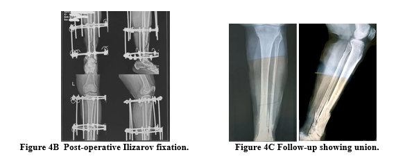
Figure 4: Comminuted distal third tibial fracture treated with Ilizarov external fixation showing progressive healing and restoration of alignment.
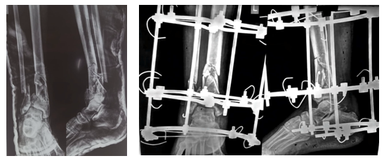 Figure 2A Pre-operative radiograph Figure 2B Post-op Ilizarov fixation
REFERENCES:
1. Court-Brown CM, McBirnie J. The epidemiology of tibial fractures. J Bone Joint Surg Br. 1995;77(3):417–21.
2. Patil S, Montgomery RJ. Management of distal tibial fractures. Injury. 2006;37(2):118–26.
3. Vallier HA, Le TT, Bedi A. Radiographic and clinical comparisons of distal tibia fractures. J Orthop Trauma. 2008;22(5):307–11.
4. Boraiah S, Kemp TJ, Erwteman AS, et al. Outcome following open reduction and internal fixation of distal tibia fractures. J Bone Joint Surg Am. 2010;92(1):146–54.
5. Guo JJ, Tang N, Yang HL, Tang TS. A prospective randomized trial comparing intramedullary nailing and plating for distal tibia fractures. J Bone Joint Surg Br. 2010;92(7):984–8.
6. Ilizarov GA. The tension-stress effect on the genesis and growth of tissues. Part I. Clin Orthop Relat Res. 1989;238:249–81.
7. Ilizarov GA. The tension-stress effect on the genesis and growth of tissues. Part II. Clin Orthop Relat Res. 1989;239:263–85.
8. Paley D. Current techniques of limb lengthening. J Pediatr Orthop. 1988;8(1):73–92.
9. Paley D, Catagni MA, Argnani F, et al. Ilizarov treatment of tibial nonunions with bone loss. Clin Orthop Relat Res. 1989;241:146–65.
10. Marsh JL, Slongo TF, Agel J, et al. Fracture and dislocation classification compendium—2007. J Orthop Trauma. 2007;21(10 Suppl):S1–133.
11. Gustilo RB, Anderson JT. Prevention of infection in open fractures. J Bone Joint Surg Am. 1976;58(4):453–8.
12. Wani NB, Syed B, Qadri AQ, et al. Role of Ilizarov fixator in distal tibial fractures. Strategies Trauma Limb Reconstr. 2011;6(2):63–7.
13. Tanna DD. Ilizarov method for tibial fractures: Indian experience. Indian J Orthop. 2010;44(3):243–9.
Ahmad MA, Sivaraman A, Zia A, Rai A. Functional outcome of distal tibia fractures treated with Ilizarov. J Clin Orthop Trauma. 2019;10(Suppl 1):S123–8.