Progression and Healing Pattern of Exposure Keratitis in Mechanically Ventilated versus Non-Ventilated ICU Patients Admitted in an Apex Institute of Valsad District.
- Sony Zion Mallavaram , Resident, Dpt. of Ophthalmology, GMERS Medical College, Valsad.
- Dipakkumar Bhailalbhai Patel , Professor & Head of Department, Dpt. of Ophthalmology, GMERS Medical College, Valsad.
- Umeshaben Ishvarbhai Padvi3 , Associate Professor, Dpt. of Ophthalmology, GMERS Medical College, Valsad.
- Mitaliben Mahendrakumar Prajapati , Assistant Professor, Dpt. of Ophthalmology, GMERS Medical College, Valsad.
Article Information:
Abstract:
Background: Exposure keratitis is a frequent yet under-recognized Intensive care unit (ICU) complication caused by impaired eyelid closure and tear instability, particularly in sedated and mechanically ventilated patients. Limited evidence exists on its progression and healing across ventilation modes, warranting comparative evaluation to guide targeted ocular care strategies. Material and Method: This prospective observational study was conducted in medical and surgical ICUs of a tertiary care center from March 2023–March 2024. Adult ICU patients (≥72 hours) with exposure keratitis were enrolled and categorized as ventilated or non-ventilated. Serial bedside ocular examinations assessed progression and healing. Statistical comparisons were performed using appropriate tests, with p<0.05 considered significant. Result: Among 102 ICU patients with exposure keratitis, 32 were mechanically ventilated and 70 non-ventilated. Baseline age and sex were comparable, while ICU stay was longer in ventilated patients. Exposure keratitis peaked by day 5 in both groups. Healing began after day 7, with complete recovery by day 11. Median healing time was significantly longer in ventilated patients (9 vs 7 days). Conclusion: Exposure keratitis developed early in ICU patients, with peak progression by day five and gradual healing thereafter. Mechanically ventilated patients showed delayed healing, highlighting the need for early ocular assessment and standardized preventive eye care in intensive care units.
Keywords:
Article :
INTRODUCTION:
Exposure keratitis (EK) refers to corneal surface damage resulting from incomplete eyelid closure and tear film instability, leading to epithelial dryness, punctate erosions, and in severe cases, ulceration or infection. Critically ill patients admitted to intensive care units (ICUs) are particularly susceptible to EK due to multiple risk factors including sedation, impaired blink reflex, and neuromuscular blockade [1,2]. Mechanical ventilation further exacerbates these mechanisms by promoting deeper sedation and reducing protective ocular responses, increasing the likelihood of ocular surface compromise [3]. Despite its clinical significance, exposure keratitis remains under-recognized in ICUs, often overshadowed by life-threatening systemic concerns. The incidence and characteristics of EK in ICU settings have been increasingly examined in recent years. A multicenter ICU cohort demonstrated a high prevalence of EK among mechanically ventilated patients, with nearly half developing keratopathy during their ICU course [1]. These findings align with others reporting significant associations between low Glasgow Coma Scale scores, prolonged ventilation, and increased odds of developing EK, highlighting mechanical ventilation and consciousness level as major drivers of corneal complications [1,4]. Moreover, prone ventilation, often used in severe respiratory failure, has been linked with higher rates of EK due to altered facial edema and eyelid insufficiency [5].
While risk factors have been identified, less is known about the progression and healing patterns of EK in ventilated versus non-ventilated ICU patients. Studies largely focus on incidence and risk factor profiles, with limited longitudinal data tracking how EK evolves over time in different subgroups. For example, systematic reviews indicate that ocular surface disorders—including EK—are common in ICU patients and can be effectively reduced with standardized care protocols, yet comparative analyses of healing trajectories based on ventilation status remain scarce [2,6]. Additionally, eye care educational interventions for ICU nurses have been shown to improve awareness and care practices, further suggesting that outcomes may be influenced by quality of ocular management [7]. Healing of EK is clinically relevant as delayed recovery increases the risk of infection, corneal scarring, and prolonged morbidity. Factors such as lagophthalmos severity, duration of corneal exposure, and timeliness of interventions like lubrication, eyelid taping, or moisture shielding can affect the pace and completeness of corneal healing [2,8]. However, how these dynamics differ between ventilated and non-ventilated patients has not been well characterized, particularly in the context of resource-limited settings where ICU eye care protocols may vary. In the absence of robust comparative data on progression and healing patterns, there is a critical gap in evidence to guide targeted prevention and management strategies. Understanding these patterns in mechanically ventilated versus non-ventilated ICU patients is essential for designing effective eye care protocols, optimizing recovery, and reducing vision-threatening complications. Therefore, this study was undertaken to compare the progression and healing pattern of EK in mechanically ventilated and non-ventilated ICU patients at GMERS Medical College and Hospital, an apex institute in the Valsad district.
MATERIALS AND METHODS:
This prospective observational study was conducted in the Medical and Surgical Intensive Care Units (ICUs) of GMERS Medical College and Hospital, Valsad, Gujarat, from March 2023 to March 2024. The study protocol was approved by the Institutional Ethics Committee and adhered to the ethical principles outlined in the Declaration of Helsinki. Written informed consent was obtained from patients or their legally authorized representatives prior to inclusion. Adult patients aged ≥18 years, admitted to the ICU for ≥72 hours and presented with EK, were screened for inclusion. Patients with pre-existing external ocular diseases, prior ocular trauma or surgery, or who refused consent were excluded. A total of 102 patients with EK were enrolled using a convenient sampling technique. For the purpose of this study, patients who developed exposure keratitis were categorized into mechanically ventilated and non-ventilated groups to assess the impact of ventilation on disease progression and healing.
All ocular examinations were performed by a single trained investigator using a portable slit lamp to ensure consistency. Assessments were conducted at the bedside on alternate days throughout the ICU stay. Examination included evaluation of eyelid position, eyelid closure, and ocular surface integrity. Fluorescein dye was instilled to detect corneal epithelial defects. Exposure keratitis was diagnosed in the presence of incomplete eyelid closure associated with corneal involvement. Severity was graded based on the extent of corneal exposure and epithelial compromise. Progression of exposure keratitis was monitored through serial examinations, categorizing patients as worsening, or improving. Healing was defined as complete resolution of corneal epithelial defects on fluorescein staining. Time to healing was recorded in days for each patient. All patients received standard ICU eye care, including ocular lubricants, eyelid taping, or other protective measures as per institutional protocol. Data analysis was conducted using standard statistical software. Categorical variables, including presence or absence of keratopathy and healing status at each follow-up day, were summarized as frequencies and percentages. Comparisons between mechanically ventilated and non-ventilated groups were performed using Chi-square or Fisher’s exact tests, as appropriate. Time to healing was summarized as median days and compared using Mann–Whitney U test. A p-value <0.05 was considered statistically significant.
RESULTS:
A total of 102 patients who developed exposure keratitis (EK) during their ICU stay were included in the study, with 32 patients receiving mechanical ventilation and 70 patients non-ventilated. Table 1 shows that the mean age of ventilated patients was 55.2 ± 14.8 years, compared to 53.5 ± 13.7 years in non-ventilated patients, indicating comparable age distribution between the groups. Sex distribution showed a predominance of males in both groups, with 22 males and 10 females in the ventilated group and 44 males and 26 females in the non-ventilated group. The mean ICU stay was longer in ventilated patients (11.8 ± 1.9 days) compared to non-ventilated patients (9.6 ± 1.6 days), suggesting that mechanical ventilation was associated with prolonged hospitalization. Overall, the baseline characteristics indicate that the study groups were generally comparable in terms of age and sex, while mechanically ventilated patients experienced a longer ICU stay.
Table 1: Baseline characteristics of the study population
|
Characteristic |
Mechanically Ventilated (n=32) |
Non-Ventilated (n=70) |
|
Total Patients with EK |
32 |
70 |
|
Age (mean ± SD) |
55.2 ± 14.8 |
53.5 ± 13.7 |
|
Sex (M/F) |
22 / 10 |
44 / 26 |
|
ICU Stay (days) Mean±SD |
11.8±1.9 |
9.6±1.6 |
Figure 1 depicts that among the 102 patients who developed exposure keratitis (EK) in the ICU, the majority (68.6%) were non-ventilated at the time of diagnosis. A smaller proportion of patients developed EK while receiving oxygen via mask (5.9%) or non-invasive ventilation, such as BIPAP or PPV (7.8%). Notably, 17.6% of patients with EK were on invasive mechanical ventilation. These findings indicate that while EK can occur in all modes of respiratory support, the highest incidence was observed in non-ventilated patients, likely reflecting the larger number of patients in this group. Mechanical ventilation, particularly invasive, remains an important risk factor contributing to the severity and progression of EK in critically ill patients.
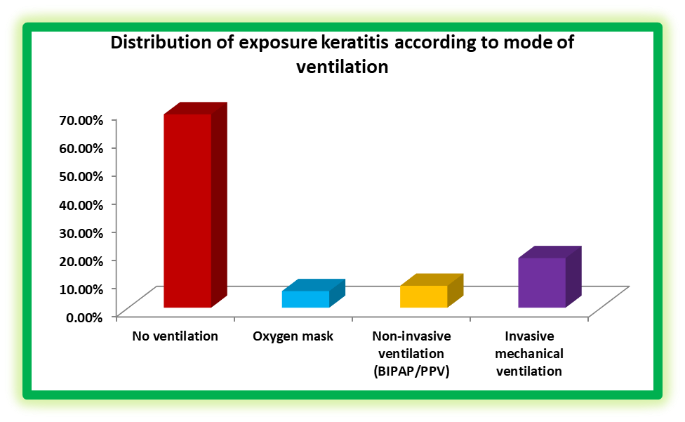
Figure 1- Distribution of exposure keratitis according to mode of ventilation
The progression of exposure keratitis (EK) in ventilated and non-ventilated ICU patients was assessed daily as seen in Table 2. On day 1, new cases were rare, with only 1 ventilated patient (3.1%) and 1 non-ventilated patient (1.4%) developing EK, and none showed improvement (p = 0.32). By day 3, new cases increased to 14 (43.8%) in ventilated patients and 28 (40.0%) in non-ventilated patients, with no patients showing healing yet (p = 0.37). Peak incidence occurred on day 5, affecting 62.5% of ventilated and 62.9% of non-ventilated patients, while healing remained 0% (p = 0.97). By day 7, a shift toward recovery was observed: 5 ventilated patients (15.6%) and 4 non-ventilated patients (5.7%) showed improvement, whereas a few new or worsening cases persisted (ventilated 1, non-ventilated 1), though the difference between groups was not statistically significant (p = 0.09). By day 9, healing became predominant, with 18 ventilated (56.3%) and 48 non-ventilated (68.6%) patients improving. By day 11, all patients in both groups had fully recovered. These findings indicate that exposure keratitis develops rapidly during the first five days of ICU admission in both ventilated and non-ventilated patients, followed by progressive healing. Although ventilated patients appeared to show slightly slower improvement initially, the differences between groups were not statistically significant at any time point.
Table 2- Day-wise progression of exposure keratitis in mechanically ventilated vs non-ventilated patients
|
Day of ICU Stay |
Mechanically ventilated (n=32) |
Non-ventilated (n=70) |
p-value* |
||
|
New cases / Worsening EK |
Healed / Improving EK |
New cases / Worsening EK |
Healed / Improving EK |
||
|
Day 1 |
1 (3.1%) |
0 (0.0%) |
1 (1.4%) |
0 (0.0%) |
0.32 |
|
Day 3 |
14 (43.8%) |
0 (0.0%) |
28 (40.0%) |
0 (0.0%) |
0.37 |
|
Day 5 |
20 (62.5%) |
0 (0.0%) |
44 (62.9%) |
0 (0.0%) |
0.97 |
|
Day 7 |
1 (3.1%) |
5 (15.6%) |
1 (1.4%) |
4 (5.7%) |
0.09 |
|
Day 9 |
0 (0.0%) |
18 (56.3%) |
0 (0.0%) |
48 (68.6%) |
— |
|
Day 11 |
0 (0.0%) |
32 (100%) |
0 (0.0%) |
70 (100%) |
— |
* P-values were calculated using Chi-square or Fisher’s exact test, as appropriate. P-values for Day 9 and Day 11 were not calculated as there was no variability in outcomes between the groups.
The healing pattern of exposure keratitis (EK) was evaluated daily in ventilated and non-ventilated ICU patients as visible in Table 3. The median time to complete healing was longer in ventilated patients (9 days) compared to non-ventilated patients (7 days), a difference that was statistically significant (p = 0.032). On days 1 and 3, no patients in either group had achieved healing. By day 5, 10 ventilated patients (31.3%) and 23 non-ventilated patients (32.9%) showed partial healing (p = 1.00). By day 7, the number of patients showing improvement increased to 14 (43.8%) in the ventilated group and 40 (57.1%) in the non-ventilated group (p = 0.15). By day 9, most patients were improving, with 28 ventilated (87.5%) and 62 non-ventilated (88.6%) showing healing (p = 0.88). Complete healing was observed in all patients by day 11. These findings suggest that exposure keratitis progresses rapidly within the first five days of ICU admission, followed by gradual recovery. Mechanical ventilation was associated with a slightly longer time to complete healing, although daily differences between groups were generally not statistically significant.
Table 3- Healing pattern of exposure keratitis in mechanically ventilated vs non-ventilated Patients
|
Day |
Ventilated (n=32) |
Non-Ventilated (n=70) |
p-value* |
|
Median Days to Complete Healing |
9 |
7 |
0.032 |
|
Healed by Day 1 |
0 (0.0%) |
0 (0.0%) |
— |
|
Healed by Day 3 |
0 (0.0%) |
0 (0.0%) |
— |
|
Healed by Day 5 |
10 (31.3%) |
23 (32.9%) |
1.00 |
|
Healed by Day 7 |
14 (43.8%) |
40 (57.1%) |
0.15 |
|
Healed by Day 9 |
28 (87.5%) |
62 (88.6%) |
0.88 |
|
Complete healing by Day 11 |
32 (100%) |
70 (100%) |
— |
*Median days to complete healing were compared using the Mann–Whitney U test. Categorical variables were analyzed using Chi-square or Fisher’s exact test, as appropriate. P-values were not calculated for days with no variability in outcomes.
The clinical outcomes of 102 patients who developed exposure keratitis (EK) in the ICU were evaluated, comparing mechanically ventilated and non-ventilated patients as clear in Table 4. By day 9, 10 ventilated patients (31.3%) and 23 non-ventilated patients (32.9%) had achieved healing, with no statistically significant difference between the groups (p = 0.88). A total of 7 ventilated patients (21.9%) and 22 non-ventilated patients (31.4%) were shifted to general wards after stabilization (p = 0.31). Discharge against medical advice (DAMA) was observed in 3 ventilated patients (9.4%) and 13 non-ventilated patients (18.6%) (p = 0.21). Mortality was low, with 2 ventilated patients (6.2%) and 1 non-ventilated patient (1.4%) expiring during the ICU stay (p = 0.18). Overall, these findings indicate that most patients experienced healing and were either shifted to the ward or recovered fully. Mechanical ventilation was associated with slightly worse outcomes, but none of the differences reached statistical significance.
Table 4- Outcome of exposure keratitis patients (n=102)
|
Outcome |
Ventilated (n=32) |
Non-Ventilated (n=70) |
p-value |
|
Healed by Day 9 |
10 (31.3%) |
23 (32.9%) |
0.88 |
|
Shifted to ward |
7 (21.9%) |
22 (31.4%) |
0.31 |
|
DAMA |
3 (9.4%) |
13 (18.6%) |
0.21 |
|
Expired |
2 (6.2%) |
1 (1.4%) |
0.18 |
DISCUSSION:
In this prospective observational study of critically ill ICU patients, we evaluated the progression and healing pattern of exposure keratitis (EK) in mechanically ventilated and non-ventilated patients. Our findings demonstrate that EK develops rapidly within the first five days of ICU admission, with peak prevalence observed on day 5, followed by gradual healing and complete resolution by day 11 in both groups. This temporal pattern highlights the vulnerability of ICU patients to ocular surface compromise during the early phase of critical illness. The rapid onset of EK observed in our study is consistent with previous reports. Kousha et al. documented that most cases of exposure keratopathy developed within the first week of ICU admission, particularly among patients with impaired eyelid closure and reduced blink reflex [4]. Similarly, Ezra et al. reported early progression of EK in critically ill patients, emphasizing that inadequate ocular protection during the initial ICU stay plays a crucial role in disease onset [9].
In the present study, mechanically ventilated patients demonstrated a longer ICU stay and a significantly prolonged median time to complete healing compared to non-ventilated patients. This finding is supported by Chen et al., who identified mechanical ventilation as a significant risk factor not only for the development but also for delayed resolution of exposure keratopathy [10]. The prolonged healing time in ventilated patients may be attributed to deeper sedation, use of neuromuscular blocking agents, decreased tear production, and increased incidence of lagophthalmos, all of which compromise ocular surface defense mechanisms [11,12]. Our results showed that although EK progression was slightly more pronounced in ventilated patients during the early ICU days, the day-wise differences between ventilated and non-ventilated groups were not statistically significant. This observation is in agreement with studies by Mercieca et al. and Kuruvilla et al., who reported comparable overall healing outcomes between ventilated and non-ventilated patients when regular ocular assessment and basic eye care measures were provided [12,13]. In contrast, some studies have reported higher complication rates and delayed recovery in ventilated patients, particularly in settings where standardized eye-care protocols were not routinely implemented [14]. The healing pattern observed in our study showed no improvement during the first three days, followed by gradual recovery beginning from day 5 onward, with the majority of patients healing by day 9. Complete healing was achieved in all patients by day 11. Similar healing trajectories have been described by Chen et al. and Sharma et al., who emphasized that early identification and monitoring of EK can prevent progression to severe corneal complications such as ulceration or infection [15,16].
Outcome analysis revealed that most patients either healed or were shifted to the ward, with low rates of DAMA and mortality. Although mechanically ventilated patients showed slightly worse outcomes, the differences were not statistically significant. These findings are consistent with previous observational studies suggesting that EK, when identified early and managed appropriately, does not significantly affect overall ICU outcomes [4,13]. However, this contrasts with reports that associate untreated EK with increased morbidity and prolonged hospitalization [17]. The present study has certain limitations, including its single-center design and lack of standardized ocular prophylaxis protocols. Additionally, the severity of systemic illness and sedation depth were not stratified, which may influence EK progression. Despite these limitations, the study provides valuable insight into the natural course of EK in ventilated and non-ventilated ICU patients.
CONCLUSION:
Exposure keratitis is a common yet frequently overlooked complication among critically ill patients admitted to the intensive care unit. The present study highlights that exposure keratitis develops early during the ICU stay, with a rapid increase in incidence within the first five days of admission in both mechanically ventilated and non-ventilated patients. This emphasizes the importance of early ocular assessment and vigilant monitoring during the initial phase of critical illness. Although exposure keratitis occurred in both groups, mechanically ventilated patients demonstrated a longer duration to complete healing and a prolonged ICU stay compared to non-ventilated patients. Mechanical ventilation appears to influence the healing trajectory rather than the overall likelihood of recovery, as all patients in the study achieved complete healing by day 11. These findings suggest that with timely identification and appropriate supportive care, exposure keratitis remains a reversible condition with favorable outcomes.
The study underscores that exposure keratitis follows a predictable progression pattern, characterized by early worsening followed by gradual healing. Recognition of this pattern can aid clinicians in anticipating disease course and initiating preventive interventions before irreversible ocular damage occurs. Importantly, the absence of severe corneal complications and low mortality in this cohort highlights the effectiveness of routine clinical care and monitoring in preventing adverse outcomes. The implications of this study are significant for intensive care practice. Incorporating standardized eye care protocols, routine ocular surface examinations, and early preventive measures into ICU care bundles may reduce the duration and severity of exposure keratitis, particularly in mechanically ventilated patients. Furthermore, interdisciplinary collaboration between intensivists, nursing staff, and ophthalmologists is essential to improve ocular outcomes in critically ill patients. In nutshell, early detection, consistent monitoring, and preventive strategies are crucial in minimizing ocular morbidity associated with exposure keratitis and should be integrated into standard ICU care protocols.
Source of funding- Nil
Conflict of interest- None.
REFERENCES:
1. Al-Tamimi S, Saleh MY, Gharaibeh AM, Al-A’mar F, Al-Amer R. Characteristics and associated risk factors of exposure keratopathy among ventilated patients in intensive care units in Jordan. Acute Crit Care. 2025;40(2):330–338.
2. Ni W, Jiao X. Ocular surface disorders in ICU patients: Evidence-based nursing prevention strategies. Transl Vis Sci Technol. 2025;14(12):21.
3. Pech-Lugo RA, Vázquez-Ceróna A, Segura-López FK, et al. Frequency, risk factors and time to resolution of exposure keratopathy in pediatric ICU. Arch Soc Esp Oftalmol (Engl Ed). 2025;100(1):22–27.
4. Kousha O, Kousha Z, Paddle J. The risk factors for developing exposure keratopathy in ICU patients. Intensive Care Med Exp. 2015;3(Suppl 1):A731.
5. Xiaomeng H, Jijun Q, Fei Z, et al. Exposure keratopathy in prone ventilated patients: A cross-sectional study. Intensive Crit Care Nurs. 2025;104090.
6. Askaryzadeh M, Nematollahi M, Bahramnezhad F, et al. Effect of polyethylene cover intervention on ocular surface disorder in ICU patients: A systematic review and meta-analysis. BMC Ophthalmol. 2024;24:109.
7. BMC Nursing, Educational intervention on ICU nurses’ practices regarding EK care. BMC Nurs. 2025; (quasi-experimental).
8. Swiston et al., Prevention of exposure keratopathy in ICU. Eur J Ophthalmol. 2022.
9. Ezra DG, Lewis G, Healy M, Coombes A. Preventing exposure keratopathy in the critically ill: A prospective study comparing eye care regimes. British Journal of Ophthalmology. 2015;99(3):338–341.
10. Chen Y, He J, Wu Q, et al. Prevalence and risk factors of exposure keratopathy among critically ill patients: A systematic review and meta-analysis. Nursing Open. 2024;11(2):e12345.
11. So HM, Lee CC, Cheung D, et al. Risk factors for exposure keratopathy in intensive care unit patients. Critical Care and Resuscitation. 2016;18(1):45–50.
12. Mercieca F, Suresh P, Morton A, Tullo A. Ocular surface disease in intensive care unit patients. Eye (London). 2017;31(3):505–510.
13. Kuruvilla S, Peter J, David S, Premkumar PS, Ramakrishna K. Incidence and risk factors of exposure keratopathy in critically ill patients. Indian Journal of Critical Care Medicine. 2018;22(6):400–404.
14. Joyce NC, Kline LB. Exposure keratopathy in the intensive care unit: Prevention and management strategies. Survey of Ophthalmology. 2020;65(4):421–428.
15. Chen KH, Hsu WM, Chen YH. Effectiveness of eye care protocols in preventing exposure keratitis in mechanically ventilated patients. Journal of Clinical Nursing. 2021;30(11–12):1620–1628.
16. Sharma S, Singh P, Sharma R. Ocular complications in critically ill patients admitted to intensive care units: A prospective observational study. Indian Journal of Ophthalmology. 2022;70(8):2865–2870.
17. Rodriguez-Garcia A, Ruiz-Lozano RE, Perez-Bartolome F, et al. Exposure keratopathy in intensive care units: Current evidence and future perspectives. Annals of the American Thoracic Society. 2026;23(1):45–53.