Relationship Between Pulmonary Function Parameters and Thoracic Dimensions Among Medical Students: A Cross-Sectional Observational Study.
- P. Uday Kumar , Assistant Professor, Department of Anatomy, Nova Institute of Medical Sciences and Research Centre, Jaffarguda (V), Abdullapurmet (M), Ranga Reddy Dt., Telangana, India.
- Sudheer Babu. N , Associate Professor, Department of Physiology, Nova Institute of Medical Sciences and Research Centre, Jaffarguda (V), Abdullapurmet (M), Ranga Reddy Dt., Telangana, India.
Article Information:
Abstract:
Background: Pulmonary function is influenced by airway caliber, lung compliance, respiratory muscle performance, and body habitus. Thoracic dimensions represent a simple anthropometric surrogate of chest wall size and mobility, yet evidence relating these measurements to spirometric indices in healthy Indian medical students remains limited. Objectives: To evaluate pulmonary function parameters and thoracic dimensions among medical students and to determine the relationship between thoracic measurements and spirometric indices. Methods: This cross-sectional observational study was conducted among 100 medical students. Age, sex, height, weight, body mass index, spirometric variables, and thoracic dimensions were recorded using standardized procedures. Group comparisons were performed by sex, and correlations between thoracic dimensions and pulmonary function parameters were analyzed. Results: The mean age of participants was 21.1 ± 1.7 years; 52.0% were males. Mean FVC, FEV1, FEV1/FVC, and PEFR were 3.52 ± 0.67 L, 3.07 ± 0.55 L, 87.3 ± 4.8%, and 7.35 ± 1.39 L/s, respectively. Males had significantly higher FVC, FEV1, PEFR, and thoracic dimensions than females. FVC showed positive correlations with inspiratory chest circumference, chest expansion, transverse thoracic diameter, and anteroposterior diameter. Similar associations were observed for FEV1 and PEFR, whereas FEV1/FVC was not significantly related to most thoracic measurements. Most students had normal spirometry. Conclusion: Thoracic dimensions, particularly inspiratory chest circumference and chest expansion, were positively associated with pulmonary function in medical students. These findings support the utility of simple thoracic anthropometry as an adjunct in respiratory assessment among healthy young adults.
Keywords:
Article :
INTRODUCTION:
Pulmonary function testing remains a fundamental component of respiratory assessment because it provides objective information on lung volumes, airflow, and ventilatory mechanics [1,2]. Among the commonly used indices, forced vital capacity [FVC], forced expiratory volume in one second [FEV1], the FEV1/FVC ratio, and peak expiratory flow rate [PEFR] are especially useful for characterizing normal ventilatory status and identifying early functional abnormalities [2]. Contemporary spirometry standards emphasize careful technique, reproducibility, and interpretation against suitable reference values, since age, sex, height, ethnicity, and body build substantially influence measured values [4]. In healthy young adults, these physiological variations are often more reflective of anthropometric and musculoskeletal determinants than of disease processes [4,5]. Thoracic dimensions are simple external anthropometric measures that represent chest wall size, shape, and mobility. Chest circumference during inspiration and expiration, chest expansion, and anteroposterior and transverse thoracic diameters can be measured bedside with minimal equipment. Prior studies have shown that chest wall mobility and thoracic expansion are associated with respiratory muscle strength and lung function, while broader anthropometric attributes such as height, weight, and body mass index also contribute to spirometric variability [6-10]. Evidence from healthy adults and other populations suggests that greater thoracic expansion is generally linked with higher lung volumes, although the strength of the association varies across age groups and clinical settings [6-9].
In India and other South Asian populations, locally relevant data remain important because pulmonary function reference patterns differ across ethnic and regional groups [3-5]. Young adults entering medical education frequently experience changes in physical activity, sedentary behavior, nutrition, and stress, all of which can influence body composition and respiratory performance [5,11]. Medical students therefore form a relevant population for evaluating normal physiological variation in pulmonary function and its relationship with thoracic anthropometry. In addition, simple thoracic measurements could serve as practical adjuncts in screening, teaching, and student health assessment when spirometry is unavailable or used only selectively [6,7,].
However, published evidence specifically examining the relationship between thoracic dimensions and spirometric indices among healthy medical students is limited. A clearer understanding of this relationship can help interpret normal sex-based differences in respiratory performance and identify whether chest wall measurements correspond meaningfully with pulmonary function in young adults [8,9]. Therefore, this study was undertaken to assess pulmonary function parameters and thoracic dimensions among medical students and to determine the relationship between thoracic measurements and spirometric indices in this population. The findings were also intended to describe sex-wise differences in spirometric and thoracic variables within the study cohort.
METHODOLOGY:
Study Design and setting:
This cross-sectional observational study was conducted in the Department of Physiology, Nova Institute of Medical Sciences, Hyderabad, Telangana, India, over the study period from January 2025 to February 2026. The study enrolled undergraduate medical students who were available during the data collection period and consented to participate.
Participants:
Students aged 18 to 25 years were eligible for inclusion. Apparently healthy students of either sex were considered for the study after obtaining written informed consent. Students with a history of chronic respiratory disease, acute respiratory infection at the time of assessment, smoking or vaping, known cardiovascular disease, thoracic cage deformity, neuromuscular disorder, recent thoracic or abdominal surgery, or inability to perform acceptable spirometry were excluded. Demographic details, relevant medical history, and anthropometric measurements were recorded using a predesigned case record form.
Anthropometry and thoracic dimensions:
Height was measured to the nearest 0.1 cm using a stadiometer, and weight was measured to the nearest 0.1 kg using a calibrated weighing scale with participants in light clothing and without footwear. Body mass index [BMI] was calculated as weight in kilograms divided by height in meters squared. Thoracic measurements were obtained in the standing position at the end of quiet expiration and at maximal inspiration using a non-stretchable measuring tape. Chest circumference at expiration and inspiration was recorded in centimeters, and chest expansion was derived as the difference between the two values. Anteroposterior and transverse thoracic diameters were measured at standardized thoracic landmarks using a spreading caliper. Spirometry. Pulmonary function testing was performed using a calibrated spirometer according to ATS/ERS recommendations [1,2]. Participants were instructed regarding the maneuver and allowed practice attempts before formal testing. At least three acceptable forced expiratory maneuvers were obtained, and the best reproducible values were used for analysis. The spirometric parameters recorded were FVC, FEV1, FEV1/FVC ratio, and PEFR. Interpretation of spirometric patterns was guided by standard interpretive strategies and appropriate reference principles [2,3]. A restrictive spirometric pattern was reported only as a pattern on spirometry and not as a definitive diagnosis.
Statistical analysis:
Data were entered into Microsoft Excel and analyzed using SPSS software version 25.0. Continuous variables were summarized as mean ± standard deviation, and categorical variables were expressed as frequency and percentage. Sex-wise comparisons were performed using the independent samples t test for continuous variables and the chi-square test for categorical variables. Pearson correlation analysis was used to assess the relationship between thoracic dimensions and pulmonary function parameters. A p value of less than 0.05 was considered statistically significant.
Ethical considerations:
The study was conducted after approval from the Institutional Ethics Committee of Nova Institute of Medical Sciences. Confidentiality of participant data was maintained throughout the study, and all procedures were performed in accordance with accepted ethical standards for human research.
RESULTS:
A total of 100 medical students were included in the study. The age of the participants ranged from 18 to 25 years, with a mean age of 21.1 ± 1.7 years. Among them, 52 [52.0%] were males and 48 [48.0%] were females. The majority of students belonged to the 21–23 years age group [46.0%], followed by 18–20 years [42.0%] and 24–25 years [12.0%]. The mean height, weight, and body mass index [BMI] of the study population were 166.6 ± 8.0 cm, 61.8 ± 10.2 kg, and 22.2 ± 3.1 kg/m², respectively. The baseline characteristics of the participants are presented in Table 1.
Table 1. Baseline characteristics of study participants [N = 100]
|
Variable |
Category / Value |
n |
% / Mean ± SD |
|
Age group [years] |
18–20 |
42 |
42.0 |
|
|
21–23 |
46 |
46.0 |
|
|
24–25 |
12 |
12.0 |
|
Sex |
Male |
52 |
52.0 |
|
|
Female |
48 |
48.0 |
|
Age [years] |
Overall |
— |
21.1 ± 1.7 |
|
Height [cm] |
Overall |
— |
166.6 ± 8.0 |
|
Weight [kg] |
Overall |
— |
61.8 ± 10.2 |
|
BMI [kg/m²] |
Overall |
— |
22.2 ± 3.1 |
The mean pulmonary function parameters of the study population were as follows: FVC 3.52 ± 0.67 L, FEV1 3.07 ± 0.55 L, FEV1/FVC ratio 87.3 ± 4.8%, and PEFR 7.35 ± 1.39 L/s. Male students demonstrated significantly higher FVC, FEV1, and PEFR compared to female students. However, the mean FEV1/FVC ratio was slightly higher among females. The sex-wise comparison of pulmonary function parameters is shown in Table 2.
Table 2. Comparison of pulmonary function parameters between male and female participants
|
Parameter |
Male [n = 52] |
Female [n = 48] |
Overall |
p value |
|
FVC [L] |
3.92 ± 0.58 |
3.08 ± 0.49 |
3.52 ± 0.67 |
<0.001 |
|
FEV1 [L] |
3.38 ± 0.50 |
2.73 ± 0.42 |
3.07 ± 0.55 |
<0.001 |
|
FEV1/FVC [%] |
86.2 ± 4.8 |
88.4 ± 4.7 |
87.3 ± 4.8 |
0.026 |
|
PEFR [L/s] |
8.14 ± 1.26 |
6.49 ± 1.01 |
7.35 ± 1.39 |
<0.001 |
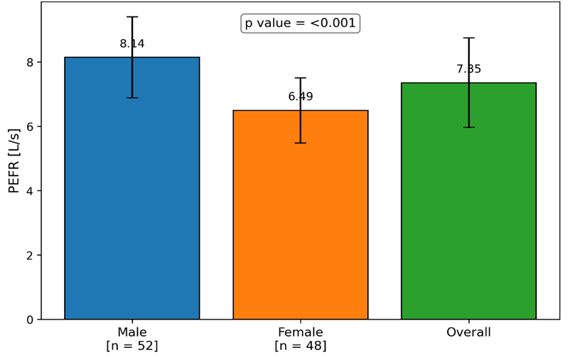
Figure 1 : Comparison of PFER[L/S] between male and female participants
Thoracic dimensions were also significantly greater among males than females. The mean chest circumference at expiration was 82.4 ± 6.1 cm, while the mean chest circumference at full inspiration was 86.9 ± 6.5 cm. The mean chest expansion was 4.5 ± 1.1 cm. The mean anteroposterior thoracic diameter was 19.6 ± 1.6 cm, and the mean transverse thoracic diameter was 27.9 ± 2.3 cm. The detailed thoracic measurements are shown in Table 3.
Table 3. Comparison of thoracic dimensions between male and female participants
|
Parameter |
Male [n = 52] |
Female [n = 48] |
Overall |
p value |
|
Chest circumference at expiration [cm] |
84.9 ± 5.8 |
79.7 ± 5.4 |
82.4 ± 6.1 |
<0.001 |
|
Chest circumference at inspiration [cm] |
89.8 ± 6.0 |
83.7 ± 5.5 |
86.9 ± 6.5 |
<0.001 |
|
Chest expansion [cm] |
4.9 ± 1.0 |
4.0 ± 0.9 |
4.5 ± 1.1 |
<0.001 |
|
Anteroposterior thoracic diameter [cm] |
20.4 ± 1.4 |
18.7 ± 1.3 |
19.6 ± 1.6 |
<0.001 |
|
Transverse thoracic diameter [cm] |
28.9 ± 2.1 |
26.8 ± 1.9 |
27.9 ± 2.3 |
<0.001 |
Correlation analysis showed a statistically significant positive relationship between pulmonary function parameters and thoracic dimensions. FVC showed moderate positive correlations with chest circumference at inspiration [r = 0.52, p < 0.001], chest expansion [r = 0.48, p < 0.001], transverse thoracic diameter [r = 0.41, p < 0.001], and anteroposterior diameter [r = 0.36, p < 0.001]. Similar positive correlations were observed for FEV1 and PEFR. In contrast, the FEV1/FVC ratio did not show a significant association with most thoracic measurements. These findings are summarized in Table 4.
Table 4. Correlation between pulmonary function parameters and thoracic dimensions
|
Thoracic parameter |
FVC [r] |
p value |
FEV1 [r] |
p value |
PEFR [r] |
p value |
FEV1/FVC [r] |
p value |
|
Chest circumference at expiration |
0.39 |
<0.001 |
0.35 |
<0.001 |
0.29 |
0.003 |
0.08 |
0.417 |
|
Chest circumference at inspiration |
0.52 |
<0.001 |
0.49 |
<0.001 |
0.35 |
<0.001 |
0.10 |
0.306 |
|
Chest expansion |
0.48 |
<0.001 |
0.44 |
<0.001 |
0.39 |
<0.001 |
0.11 |
0.271 |
|
Anteroposterior diameter |
0.36 |
<0.001 |
0.33 |
0.001 |
0.28 |
0.005 |
-0.06 |
0.541 |
|
Transverse diameter |
0.41 |
<0.001 |
0.38 |
<0.001 |
0.30 |
0.002 |
-0.04 |
0.682 |
On spirometric pattern analysis, 88 students [88.0%] had values within normal limits, 9 [9.0%] showed a mild restrictive spirometric pattern, and 3 [3.0%] demonstrated a borderline obstructive spirometric pattern. The distribution of spirometric patterns is presented in Table 5.
Table 5. Distribution of spirometric patterns among the study participants
|
Spirometric pattern |
n |
% |
|
Normal |
88 |
88.0 |
|
Mild restrictive spirometric pattern |
9 |
9.0 |
|
Borderline obstructive spirometric pattern |
3 |
3.0 |
|
Total |
100 |
100.0 |
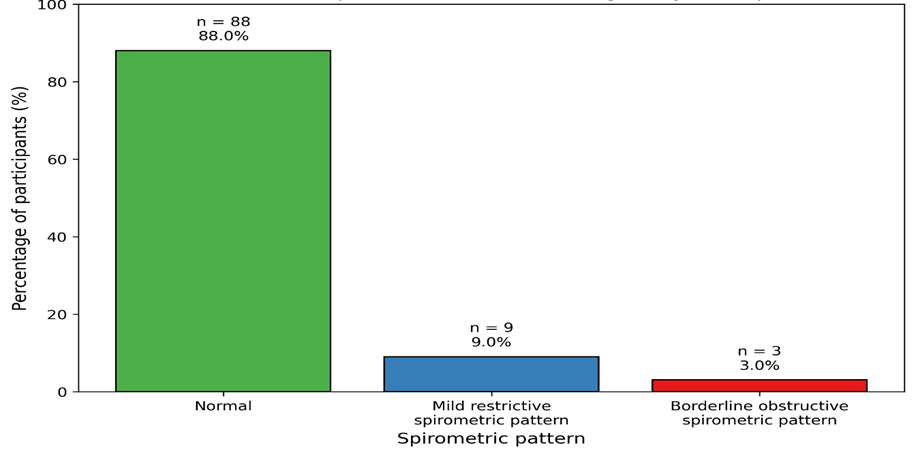
Figure 2: Distribution of spirometric patterns among the study participants
Students with normal spirometry had higher mean chest expansion compared with those showing abnormal spirometric patterns [4.6 ± 1.0 cm vs 3.8 ± 0.9 cm, p = 0.002]. The comparison of chest expansion according to spirometric status is shown in Table 6. Overall, the findings indicate that larger thoracic dimensions, particularly inspiratory chest circumference and chest expansion, were associated with better pulmonary function among medical students. Absolute lung volumes and flow rates were significantly higher in males, whereas the FEV1/FVC ratio remained comparable between sexes with only a marginal female predominance.
Table 6. Comparison of chest expansion according to spirometric status
|
Spirometric status |
n |
Chest expansion [cm] |
p value |
|
Normal spirometry |
88 |
4.6 ± 1.0 |
0.002 |
|
Abnormal spirometric pattern |
12 |
3.8 ± 0.9 |
|
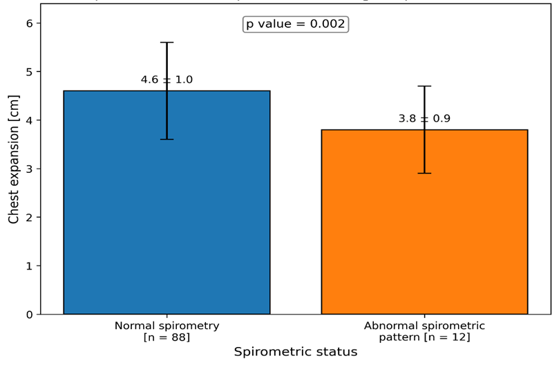
Figure 3: Comparison of chest expansion according to spirometric status
DISCUSSION:
This study evaluated the relationship between pulmonary function parameters and thoracic dimensions among 100 medical students and demonstrated a clear positive association between chest size or mobility and spirometric performance. The overall spirometric values observed in this cohort were within the expected range for healthy young adults, and most participants had normal spirometry. These findings are consistent with the concept that lung function in healthy individuals is strongly shaped by anthropometry, sex, and chest wall mechanics rather than overt disease [12].
Male students showed significantly higher FVC, FEV1, PEFR, and thoracic measurements than female students, whereas the FEV1/FVC ratio was only slightly higher among females. This pattern agrees with earlier work showing that absolute pulmonary volumes and expiratory flow rates are generally higher in males because of larger body size, greater thoracic dimensions, and higher respiratory muscle mass, while the FEV1/FVC ratio tends to remain relatively preserved across sexes [11]. Similar associations between body build and pulmonary function have been reported in healthy university populations and other adult cohorts [10-12].
Among the thoracic variables studied, inspiratory chest circumference and chest expansion showed the strongest correlations with FVC and FEV1. This observation is biologically plausible because inspiratory thoracic excursion reflects chest wall compliance, respiratory muscle action, and the capacity of the rib cage to accommodate inspiratory volume. Previous studies by Derasse et al., Lanza et al., and Reddy et al. also reported meaningful associations between chest expansion or chest wall mobility and lung volumes, supporting the value of simple thoracic measurements as correlates of ventilatory performance [6,7,9]. The present findings further showed that transverse and anteroposterior thoracic diameters were positively related to spirometric values, which aligns with evidence that external chest dimensions contribute to interindividual variability in lung function [8,10,12].
The absence of a significant relationship between most thoracic dimensions and the FEV1/FVC ratio is also noteworthy. Unlike absolute lung volumes, the ratio reflects the relationship between two related spirometric measures and is influenced less by overall body size. Consequently, larger thoracic dimensions are expected to increase both FVC and FEV1 in parallel, producing stronger associations with the individual parameters than with the ratio itself [1-3]. This explains why thoracic anthropometry in this study tracked lung volume and flow more closely than airflow proportion.
A small proportion of students demonstrated abnormal spirometric patterns, predominantly a mild restrictive spirometric pattern. Their lower mean chest expansion compared with students with normal spirometry strengthens the interpretation that reduced chest wall mobility can accompany lower ventilatory capacity even in otherwise healthy young adults [6,7,9]. Taken together, these results indicate that thoracic anthropometry, especially inspiratory circumference and chest expansion, offers useful adjunctive information in student respiratory assessment and reinforces the close physiological link between chest wall dimensions and pulmonary function.
Limitations
This study was conducted in a single medical college and included only apparently healthy medical students, which limits wider generalizability. The cross-sectional design precludes temporal inference. Thoracic measurements were obtained manually and remain subject to observer variability. Additional determinants such as physical activity, nutritional status, hemoglobin level, and diffusion capacity were not assessed. Restrictive patterns identified on spirometry were not confirmed with full lung volume measurements.
CONCLUSION:
In this cross-sectional study of 100 medical students, pulmonary function parameters showed significant positive relationships with thoracic dimensions. Male students had higher absolute spirometric values and larger thoracic measurements than female students, while the FEV1/FVC ratio remained broadly comparable between sexes. Inspiratory chest circumference, chest expansion, and transverse thoracic diameter demonstrated the strongest correlations with FVC, FEV1, and PEFR. Students with abnormal spirometric patterns also showed lower chest expansion than those with normal spirometry. These findings indicate that simple thoracic anthropometric measurements can complement spirometric assessment in healthy young adults and provide physiological insight into sex-based patterns and variations in ventilatory performance within the medical student population and similar settings.
REFERENCES:
1. Graham BL, Steenbruggen I, Miller MR, Barjaktarevic IZ, Cooper BG, Hall GL, et al. Standardization of spirometry 2019 update: an official American Thoracic Society and European Respiratory Society technical statement. Am J Respir Crit Care Med. 2019;200(8):e70-e88.
2. Stanojevic S, Kaminsky DA, Miller MR, Thompson B, Aliverti A, Barjaktarevic I, et al. ERS/ATS technical standard on interpretive strategies for routine lung function tests. Eur Respir J. 2022;60(1):2101499.
3. Quanjer PH, Stanojevic S, Cole TJ, Baur X, Hall GL, Culver BH, et al. Multi-ethnic reference values for spirometry for the 3-95-yr age range: the global lung function 2012 equations. Eur Respir J. 2012;40(6):1324-43.
4. Nayak PK, Satpathy S, Manjareeka M, Samanta P, Mishra J, Pradhan BB. Normal spirometric standards in young adult Indian population. J Basic Clin Physiol Pharmacol. 2015;26(4):321-5.
5. Fulambarker A, Copur AS, Javeri A, Jere S, Cohen ME. Reference values for pulmonary function in Asian Indians living in the United States. Chest. 2004;126(4):1225-33.
6. Derasse M, Lefebvre S, Liistro G, Reychler G. Chest expansion and lung function for healthy subjects and individuals with pulmonary disease. Respir Care. 2021;66(4):661-8.
7. Lanza FC, de Camargo AA, Archija LRF, Selman JPR, Malaguti C, Dal Corso S. Chest wall mobility is related to respiratory muscle strength and lung volumes in healthy subjects. Respir Care. 2013;58(12):2107-12.
8. Debouche S, Pitance L, Robert A, Liistro G, Reychler G. Reliability and reproducibility of chest wall expansion measurement in young healthy adults. J Manipulative Physiol Ther. 2016;39(6):443-9.
9. Reddy RS, Alahmari KA, Silvian PS, Ahmad IA, Kakarparthi VN, Rengaramanujam K. Reliability of chest wall mobility and its correlation with lung functions in healthy nonsmokers, healthy smokers, and patients with COPD. Can Respir J. 2019;2019:5175949.
10. Whittaker AL, Sutton AJ, Beardsmore CS. Are ethnic differences in lung function explained by chest size? Arch Dis Child Fetal Neonatal Ed. 2005;90(5):F423-8.
11. Ebomoyi MI, Iyawe VI. Variations of peak expiratory flow rate with anthropometric determinants in a population of healthy adult Nigerians. Niger J Physiol Sci. 2005;20(1-2):85-9.
12. Shah H, Mali S, Ranga S, Jadhav C, Rukadikar A, Singh AK, et al. Effect of body mass index on cardiorespiratory parameters among medical students: a cross-sectional study. Int J Physiol Pathophysiol Pharmacol. 2022;14(1):4-9.
13. Ogunlana MO, Oyewole OO, Lateef AI, Ayodeji AF. Anthropometric determinants of lung function in apparently healthy individuals. S Afr J Physiother. 2021;77(1):1509.
14. Nowakowski D, Kliś K, Żurawiecka M, Dubrowski A, Wronka I. Influence of socioeconomic and anthropometric factors on respiratory function in female university students. Adv Exp Med Biol. 2017;968:41-48.