Clinico-Mycological Profile of Chronic and Recurrent Dermatophytosis with Associated Risk Factors in a Tertiary Care Centre.
- Manju , Department-Microbiology, World College of Medical Sciences Research And Hospital, Jhjjhar, Haryana.
- Sandeep Kumar , Department-Biochemistry, World College of Medical Sciences Research And Hospital, Jhjjhar, Haryana.
- Priyanka , Department-Microbiology, World College of Medical Sciences Research And Hospital, Jhjjhar, Haryana.
- Sukhveer Singh , Department-Community Medicine, Saradar Patel Medical College, Bikaner.
Article Information:
Abstract:
Background: Dermatophytosis has emerged as a persistent public health problem in tropical countries, with an increasing trend of chronic and recurrent infections. The changing epidemiology, rising antifungal resistance, and widespread misuse of topical steroids contribute significantly to this burden. This study aimed to evaluate the clinical patterns, etiological agents, and associated risk factors in chronic and recurrent dermatophytosis. Methods: A hospital-based cross-sectional observational study was conducted over a period of 6 months in a tertiary care centre. A total of 120 patients with clinically diagnosed chronic (duration >6 months) or recurrent dermatophytosis were included. Detailed history, clinical examination, and risk factor assessment were recorded using a structured proforma. Skin, hair, and nail samples were subjected to direct microscopy using KOH mount and fungal culture on Sabouraud Dextrose Agar. Results: Among 120 patients, 72 (60%) were males and 48 (40%) were females. The most affected age group was 21–40 years (46.7%). Tinea corporis with tinea cruris was the most common clinical presentation (38.3%), followed by isolated tinea cruris (22.5%). KOH positivity was observed in 96 (80%) cases, while culture positivity was seen in 84 (70%) cases. Trichophyton rubrum (52.4%) was the predominant isolate followed by Trichophyton mentagrophytes (32.1%). Major risk factors included topical steroid misuse (48.3%), poor hygiene (41.7%), family history (36.7%), and diabetes mellitus (25%). Conclusions: Chronic and recurrent dermatophytosis is strongly associated with modifiable risk factors such as steroid misuse and poor hygiene. Early diagnosis, appropriate antifungal therapy, and patient education are crucial to reduce disease burden and recurrence
Keywords:
Article :
INTRODUCTION:
Dermatophytosis is a common superficial fungal infection affecting keratinized tissues such as skin, hair, and nails, and is highly prevalent in tropical countries like India due to hot and humid climatic conditions.1,2 Dermatophytes belonging to the genera Trichophyton, Microsporum, and Epidermophyton are responsible for these infections, commonly referred to as tinea or ringworm.3
In recent years, India has witnessed a significant rise in chronic and recurrent dermatophytosis, often described as an emerging epidemic.4 This changing trend has been attributed to widespread misuse of topical corticosteroid combinations, incomplete antifungal therapy, and poor treatment compliance.5
Chronic dermatophytosis is defined as infection persisting for more than six months, while recurrent infection refers to reappearance after clinical resolution.6 Several host and environmental factors, including diabetes mellitus, poor hygiene, occlusive clothing, and close contact within families, contribute to persistence and recurrence of infection.7
Understanding the clinico-mycological pattern along with associated risk factors is essential for effective management and prevention of recurrence. Hence, this study was undertaken to evaluate the clinical patterns, etiological agents, and associated risk factors in chronic and recurrent dermatophytosis.
MATERIALS AND METHODS:
A hospital-based cross-sectional observational study was conducted in the Department of Dermatology in collaboration with the Microbiology Laboratory of a tertiary care centre over a period of six months after obtaining Institutional Ethics Committee approval. A total of 120 patients with clinically diagnosed chronic and recurrent dermatophytosis were included after obtaining informed consent. Patients on antifungal therapy within the last four weeks and those with non-dermatophyte infections were excluded.
Relevant clinical details including age, sex, duration, site of involvement, recurrence, treatment history, and associated risk factors such as diabetes, steroid use, hygiene, and occlusive clothing were recorded. Samples were collected from affected sites after cleaning with 70% alcohol, including skin scrapings, nail clippings, and hair samples under aseptic precautions.
All specimens were examined by direct microscopy using 10% KOH mount (20% for nails) and cultured on Sabouraud Dextrose Agar with and without antibiotics. Identification of dermatophytes was based on colony characteristics and microscopic features using Lactophenol Cotton Blue mount. Data were analyzed using descriptive statistics and expressed as percentages.
RESULTS:
In the present study, a total of 120 patients with chronic and recurrent dermatophytosis were included. Among them, 72 (60%) were males and 48 (40%) were females, showing a male predominance. The most commonly affected age group was 21–40 years (46.7%), followed by 41–60 years (26.7%). Majority of the patients belonged to lower socioeconomic status and had a positive family history in 44 (36.7%) cases (Table 1).
Table 1: Sociodemographic details
|
Variables |
Number |
% |
|
Age (years) |
|
|
|
0–20 |
18 |
15.0 |
|
21–40 |
56 |
46.7 |
|
41–60 |
32 |
26.7 |
|
>60 |
14 |
11.6 |
|
Gender |
|
|
|
Male |
72 |
60.0 |
|
Female |
48 |
40.0 |
|
Family history |
|
|
|
Present |
44 |
36.7 |
|
Absent |
76 |
63.3 |
The duration of disease was more than 12 months in 66 (55%) patients, while 54 (45%) had duration between 6–12 months. The most common clinical presentation observed was combined tinea corporis and tinea cruris (38.3%), followed by tinea cruris (22.5%) and tinea corporis (17.5%) (Table 2).
Table 2: Clinical features of dermatophytosis
|
Variables |
Number |
% |
|
Duration of disease |
|
|
|
6–12 months |
54 |
45.0 |
|
>12 months |
66 |
55.0 |
|
Type of dermatophytosis |
|
|
|
Tinea corporis + cruris |
46 |
38.3 |
|
Tinea cruris |
27 |
22.5 |
|
Tinea corporis |
21 |
17.5 |
|
Tinea faciei |
10 |
8.3 |
|
Tinea pedis |
8 |
6.7 |
|
Tinea unguium |
5 |
4.2 |
|
Mixed infections |
3 |
2.5 |
|
Variables |
Number |
% |
Out of 120 cases, 96 (80%) were positive on direct microscopy by KOH mount, while 84 (70%) were culture positive. Among the culture-positive cases, Trichophyton rubrum was the most common isolate (52.4%), followed by Trichophyton mentagrophytes (32.1%) (Table 3).
Table 3: Distribution of dermatophyte species (n=84)
|
Species |
Number |
% |
|
T. rubrum |
44 |
52.4 |
|
T. mentagrophytes |
27 |
32.1 |
|
Microsporum spp. |
9 |
10.7 |
|
E. floccosum |
4 |
4.8 |
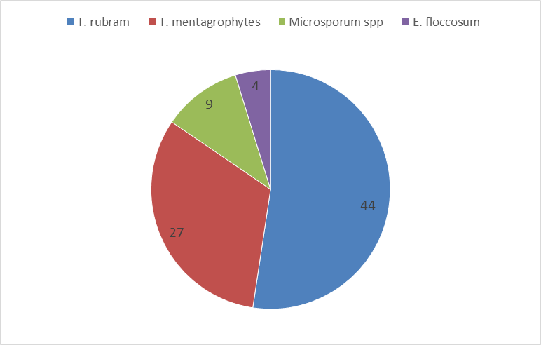
Figure : 1Distribution of dermatophyte species
The various risk factors associated with chronic and recurrent dermatophytosis were analyzed. The most common risk factor observed was topical steroid misuse in 58 (48.3%) patients, followed by poor hygiene in 50 (41.7%) and family history in 44 (36.7%) patients. Diabetes mellitus was present in 30 (25%) cases. Multiple risk factors were observed in several patients (Table 4).
Table 4: Risk factors associated with dermatophytosis (n=120)
|
Risk factors |
Number |
% |
|
Topical steroid use |
58 |
48.3 |
|
Poor hygiene |
50 |
41.7 |
|
Family history |
44 |
36.7 |
|
Diabetes mellitus |
30 |
25.0 |
|
Occlusive clothing |
28 |
23.3 |
|
Animal exposure |
10 |
8.3 |
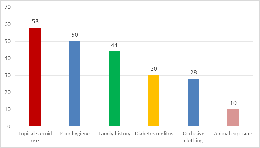
Figure: 2 Risk factors associated with dermatophytosis
DISCUSSION:
Dermatophytosis continues to be a common public health problem in tropical countries like India due to favorable climatic conditions such as heat and humidity, along with poor hygiene and overcrowding, which facilitate fungal growth and transmission. The increasing trend of chronic and recurrent dermatophytosis has further emphasized the need for regional epidemiological studies.
In the present study, males constituted 60% of cases, showing male predominance. Similar findings were reported by Hosthota et al., where males accounted for 61.3% of cases, and Surendran et al., who also observed male predominance.8,9 Similarly a study conducted by Jain et al noted tinea infections were common among men (67.5%) The higher prevalence among males in the present study may be attributed to increased physical activity, sweating, and occupational exposure.10
Dermatophytosis was most commonly seen in the age group of 21–40 years (46.7%) in the present study. Hosthota et al. reported maximum incidence in 21–30 years (37.3%), while Vineetha et al. observed peak incidence in the 20–40 year group. suggesting that young adults are more susceptible due to increased exposure and sweating.8,7
The most common clinical presentation in the present study was combined tinea corporis and tinea cruris (38.3%), followed by isolated tinea cruris (22.5%) and tinea corporis (17.5%). In contrast, Hosthota et al. reported tinea cruris as the most common type (50%), followed by tinea corporis (18.4%) and tinea unguium (11.9%). Similarly, Similar findings were reported by Majeed N et al., Kumar S et al.11,12 The higher frequency of mixed infections in the present study may reflect the increasing trend of extensive and chronic dermatophytosis.
In the present study, Trichophyton rubrum was the predominant isolate (52.4%), followed by Trichophyton mentagrophytes (32.1%). Comparable findings were reported by Chithra et al., where T. rubrum accounted for 58.1% and T. mentagrophytes for 31.8% of isolates. Hosthota et al. also reported T. rubrum as the commonest isolate (33%) followed by T. mentagrophytes (20%).13,7 These findings are similar to a study conducted by Mohanty et al.23 Other researchers who reported T. rubrum as predominant isolate in their studies, were Bindu V et al in 2002 - 66.2%, Sumana et al in 2004 - 60%, Peerapur et al in 2004-43.7%.14-17
Direct microscopy (KOH mount) was positive in 80% of cases in the present study, while culture positivity was 70%. Chithra et al. reported higher positivity rates with KOH (95.17%) compared to culture (88.96%).13 Similarly, Agarwal et al. reported KOH positivity of around 84–90% and culture positivity ranging from 60–80%. The higher sensitivity of KOH mount makes it a useful rapid screening tool, while culture remains essential for species identification.18
The present study identified topical steroid misuse as the most common risk factor (48.3%), followed by poor hygiene (41.7%), family history (36.7%), and diabetes mellitus (25%). Hosthota et al. reported poor hygiene (32.1%), steroid use (23.9%), and diabetes (20.1%) as major risk factors. Chithra et al. reported diabetes mellitus in 10.3% of cases and significant association with occupational exposure.8,13
The presence of multiple risk factors in many patients in the present study suggests that chronic and recurrent dermatophytosis is multifactorial in origin. Continued exposure to infection, inappropriate treatment, and persistence of underlying conditions contribute to the increasing burden of disease.
Overall, the findings of the present study are consistent with previous literature, while also highlighting the emerging trend of mixed infections and the significant role of modifiable risk factors such as steroid misuse in the persistence and recurrence of dermatophytosis.
CONCLUSION:
Chronic and recurrent dermatophytosis is increasingly common, particularly among young adults. Mixed infections were frequent, with Trichophyton rubrum as the predominant isolate. KOH mount proved to be a useful rapid diagnostic tool. Topical steroid misuse, poor hygiene, and diabetes were major contributing factors. Early diagnosis, appropriate treatment, and patient education are essential to prevent recurrence.
REFERENCES:
1. Havlickova B, Czaika VA, Friedrich M. Epidemiological trends in skin mycoses worldwide. Mycoses. 2008;51(Suppl 4):2–15.
2. Verma S, Madhu R. The great Indian epidemic of superficial dermatophytosis: An appraisal. Indian J Dermatol. 2017;62(3):227–36.
3. Singh S, Beena PM. Profile of dermatophyte infections in Baroda. Indian J Dermatol Venereol Leprol. 2003;69(4):281–3.
4. Nenoff P, Verma SB, Vasani R, Burmester A, Hipler UC, Wittig F, et al. The current Indian epidemic of superficial dermatophytosis due to Trichophyton mentagrophytes—A molecular study. Mycoses. 2019;62(4):336–56.
5. Dogra S, Uprety S. The menace of chronic and recurrent dermatophytosis in India: Current scenario. Indian Dermatol Online J. 2016;7(5):343–6.
6. Bishnoi A, Vinay K, Dogra S. Emergence of chronic dermatophytosis in India: A challenge to dermatologists. Indian Dermatol Online J. 2019;10(3):284–6.
7. Vineetha M, Sheeja S, Celine MI, et al. Profile of dermatophytosis in a tertiary care center in Kerala, India. Indian J Dermatol. 2019;64(4):266–71.
8. Hosthota A, Gowda T, Manikonda R. Clinical profile and risk factors of dermatophytoses: a hospital based study. Int J Res Dermatol 2018;4:508-13.
9. Surendran KA, Bhat RM, Boloor R, et al. A clinical and mycological study of dermatophytic infections. Indian J Dermatol 2014;59(3):262-7
10. Jain N, Sharma M, Saxena VN. Clinico-mycological profile of dermatophytosis in Jaipur, Rajasthan. Indian J Dermatol Venereol Leprol. 2008;74:274-5.
11. Majeed N, Narayanankutty S, Rajan R, et al. Clinicomycological study of dermatophytosis in a tertiary care centre. J Acad Clin Microbiol 2016;18(2):110-3.
12. Kumar S, Mallya PS, Kumari P. Clinico-mycological study of dermatophytosis in a tertiary care hospital. Int J Sci Stud 2014;1(6):27-32.
13. Chithra T et al. Clinico-Mycological Profile of Dermatophytosis in a Tertiary Care Hospital. Int. J Med. Pharm. Res., 7 (2): 388‐395, 2026
14. Mohanty JC, Mohanty SK, Sahoo RC, Sahoo A, Praharaj N. Incidence of dermatophytosis in Orissa. Indian J Med Microbiol. 1998;16:78-80.
15. Bindu V, Pavithran K. Clinico - mycological study of dermatophytosis in Calicut. Indian J DermatolVenereol Leprol. 2002;68:259-61.
16. Sumana V, Singaracharya MA. Dermatophytosis in Khammam. Indian J Microbiol. 2004;47:287-9
17. Peerapur BV, Inamdar AC, Puspha PV, Shrikant B.Clinico mycological study of dermatophytosis in Bijapur. Indian J Med Microbiol. 2004;22:273-4
18. Agarwal US, Saran J, Agarwal P. Clinicomycological study of dermatophytes in a tertiary care centre in northwest India. Indian J Dermatol Venereol Leprol. 2014;80:194.