Anesthetic Management of EUA/ Biopsy for Menorrhagia in Von Recklinghausen’s Disease - A Case Report.
- Dinesh K. , Postgraduate in Anesthesia, Department of Anaesthesia, Dhanalakshmi Srinivasan Medical College and Hospital, Siruvachur, Perambalur, Tamil Nadu, India.
- Sirpikha V. , Postgraduate in Anesthesia, Department of Anaesthesia, Dhanalakshmi Srinivasan Medical College and Hospital, Siruvachur, Perambalur, Tamil Nadu, India.
- Geetha J. , Professor, Department of Anaesthesia, Dhanalakshmi Srinivasan Medical College and Hospital, Siruvachur, Perambalur, Tamil Nadu, India.
Article Information:
Abstract:
Background: Aim: To describe the clinical presentation and multisystem involvement in a 45-year-old female patient diagnosed with von Recklinghausen’s syndrome (Neurofibromatosis type 1), highlighting its neurocutaneous and systemic manifestations. Materials and Methods: This is a case report of a 45-year-old female patient presenting with multiple system involvement. Clinical evaluation included detailed history taking, physical examination, and assessment of neurocutaneous features such as neurofibromas and skin pigmentation. Additional investigations were carried out to evaluate skeletal, pulmonary, hematological, and gynecological involvement. Results: The patient exhibited characteristic neurocutaneous manifestations, including multiple neurofibromas and abnormal pigmentation. Associated systemic involvement was observed in the skeletal, pulmonary, hematological, and gynecological systems. Based on the clinical findings and multisystem involvement, a diagnosis of von Recklinghausen’s syndrome (Neurofibromatosis type 1) was established. Conclusion: von Recklinghausen’s syndrome is a multisystem neurocutaneous disorder with variable clinical presentation. Early recognition of characteristic skin lesions along with evaluation for systemic involvement is essential for timely diagnosis and comprehensive management of the condition. Abbreviations : EUA: Examination Under Anesthesia, NF: Neurofibromatosis.
Keywords:
Article :
HISTORY:
45 years old female patient with severe anemia and menorrhagia for 3 months presented for evaluation. She has had three full term normal deliveries and has good exercise tolerance. Due to her restricted activity, she did not have any exertional dyspnea.
CLINICAL FEATURES:
The patient was thin, pale, short statured (height 134cm and weight-24kg BMI- 21.43Kg/m2) with a large bony lump on the left posterior thorax, gross lower thoracic scoliosis of 75 degrees with convexity to the right, multiple neurofibromata all over the body along with café-au-lait spots (coffee with milk colored) macules more so well seen in the lower limbs.
She had profuse bleeding per vagina during her menstrual periods in the last 3 months which lasted 8-10 days per month. She also passed few clots. She had three full term normal deliveries and all are fine. None of her daughters had neurofibromata.
Respiratory System
Chest deformed due to scoliosis, respiratory rate 20/ min. Breath holding was 10-15 seconds.
SpO2 was 98% on room air, she was unable to blow a balloon. Pulmonary function tests showed moderate obstruction with reduced vital capacity. Maximum expiratory flow was 33% force vital capacity was 1.72 Litres. FEV1/FVC had predicted percentage at 84%.
Cardiovascular System
Her pulse rate was 88 per minute and blood pressure 90/60 mmHg in the right upper limb in sitting position. Her hemoglobin was 8.5g% and was planned for blood transfusion before the major procedure.
Abdomen
Patient was unable to lie flat in supine position due to spine deformity. Firm swelling just felt in the suprapubic region and bimanual palpation confirmed a 12 weeks fibroid uterus as a firm to hard mass. Cervix was normal. No other organomegaly.
Central Nervous System
Patient was conscious, oriented, higher functions appeared normal. She was comfortable at rest. She had an upper motor neuron right facial nerve palsy and brisk deep tendon jerks. There was no clonus, no history of seizures and no clinically detectable sensory deficits.
Preparation and Optimization
• One unit of packed cells was kept ready to be transfused on the table if needed.
• Preparation of respiratory system in view of restrictive lung disease, probable interstitial lung disease, or pulmonary fibrosis as a component of Von Recklinghausen was by incentive spirometry and budecort nebulization 8th hourly for desensitization and humidification of the airway.
Conduct of Anesthesia
The nature of the uterine mass raised a doubt of the neurofibroma or leiomyoma due to the clinical presentation. A peri menopause lady with previous 3 normal vaginal deliveries and leiomyoma would be expected to present a larger mass with multiple fibroids. Hence the rare cause of uterine neurofibroma in this case of extensive neurofibromatosis was thought of and patient was posted for examination under anesthesia and diagnostic dilatation and curettage.
She was shifted to OT in a lateral propped up position, proper padding and pillows were supportive to make her supine as to prevent pulling on the pedunculated neurofibromata around lumbosacral region.
Standard monitors were connected and baseline parameters were noted. She was oxygenated with a poly mask and lithotomy was allowed after a slow intravenous administration of pentazocine 30mg and promethazine 12.5mg with 30 mg propofol. She was well under and tolerated the procedure. Being a perimenopausal woman with previous normal deliveries the os was patulous and there was no need for cervical dilatation. She remained hemodynamically stable and shifted to post-operative ward. Chest physiotherapy was continued for 48 hours in the postoperative period. She was discharged with an advice to return for the biopsy report and major surgery- namely hysterectomy if needed. Profound bleeding per vagina settled with one shot of tranexamic acid 1 gram intravenous and curettage of endometrium.
DISCUSSION:
Neurodegenerative disorders present and enormous challenge because of the complexity of the nervous system, the broad clinical and genetic heterogenicity characteristic of these diseases and the progressive and generally irreversible nature of their neuropathology.[1]
Neurofibromatosis type 1(optic nerve glioma, sarcoma and pheochromocytoma) Neurofibromatosis type 2 (acoustic neuroma and other CNS tumors) constitute the list of rare inherited cancer syndromes often due to sporadic mutation or due to inheritance of germ-line mutations in a cancer predisposition gene.[2]
Neurofibromatosis is a group of autosomal dominant neurocutaneous disorders marked by a predisposition to tumor formation particularly involving nerve sheaths. Three types of neurofibromatosis as distinct clinical entities with differing gene mutations and manifestations have been described namely NF1, NF2 and Schwanomatosis.[3]
NF1 is characterized by the presence of multiple neurofibromas, with multiple body organ involvement, café au lait macules (CALM), axillary or inguinal freckling, hamartomatous Lisch nodules of iris and disease specific bony dysplasia. This is von Recklinghausen disease in which patients have greater risks of musculoskeletal cardiovascular and nervous system abnormalities. NF1 is caused by a loss of function mutation in 1 allele of NF1 gene on 12q chromosome resulting in 50% loss of function of neurofibromin, a tumor suppressor protein ubiquitously expressed.[4]
NF2 has an autosomal dominant inheritance pattern and is characterized by development of bilateral vestibular schwannomas and meningiomas. Neurofibromas do not occur in this syndrome.[5]
The third type of neurofibromatosis is Schwanomatosis is the rarest of the 3 characterized by the presence of multiple non-intradermal peripheral and spinal schwannomas. Localized or diffuse chronic pain or asymptomatic masses are common presentations. The absence of vestibular schwannoma differentiates it from NF2.[6] SMARCB1 and LZTR1 are the common gene mutations either spontaneous or inherited with reduced penetrance.
Our patient in this case report belongs to NF1 and only NF1 shall be elaborated in this discussion. Approximately half of all NF1 cases is inherited while other half results from denovo mutations. This gene is located on Chromosome 17q 11.2 encodes the protein neurofibromin. Neurofibromin is a tumor suppressor protein in the RAS/MAPK and mTOR pathways. RAS is a GDPase that activates the downstream mitogen-activated protein kinase (MAPK) and the mammalian target of rapamycin (mTOR) cell proliferation pathway.
Inadequate neurofibroma activity result in a higher risk of tumors including malignant peripheral nerve sheath tumors (MPNST) optic pathway gliomas and pheochromocytomas. Mosaicism can occur resulting in generalized segmental or gonadal expression of NF1 gene. The segmental NF1 gene has pigment changes, tumors or both and is limited to one or more body segments. Our patient had a uterine fibroid turned out to be sarcoma and the café au lait macules were restricted to both lower extremities. Gonadal NF1 gene mutations occur when mutations affect only ova or sperm. The NF1 gene has complete penetrance with highly variable expressivity.[7]
Diagnostic Criteria for Neurofibromatosis Type 1
An international consensus group revised the diagnostic criteria for NF1 in 2021, with the presence of two of the following be required for the diagnosis.
1. Presence of more than or equal to six CALMS greater than 5mm in prepubertal individuals and more than 15 mm in post pubertal individuals.
2. Presence of more than or equal to 2 neurofibromas or more than one plexiform neurofibroma.
3. Axillary or inguinal freckling.
4. Optic pathway glioma.
5. Presence of more than 2 iris Lisch nodules on choroidal abnormalities.
6. Distinct bony lesions, including sphenoid dysplasia, anterolateral bowing of tibia, or pseudoarthrosis of ling bone.
7. First degree relative with NF1 or heterozygous pathognomic NF1, variant allele fraction of 50% in apparently normal tissues (e.g.) white blood cells.[8]
Management
CALMS and painless neurofibromas do not need treatment and excision of the neurofibromata often recurs. Plexiform neurofibromas are disfiguring infiltrative and difficult to remove and may involve the airway and have an 8 to 13 % malignant potential.
Imatinib, a tyrosine kinase inhibitor has been shown to reduce the size of plexiform neurofibroma. MEK inhibitor targeting downstream RAS, Selumatinib has also shown partial response in reducing the size of plexiform neurofibromas.[9]
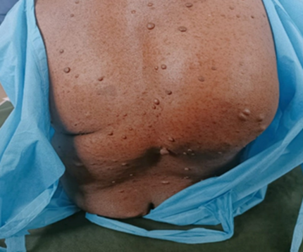
Figure 1: Cutaneous neurofibromata with spine deformity
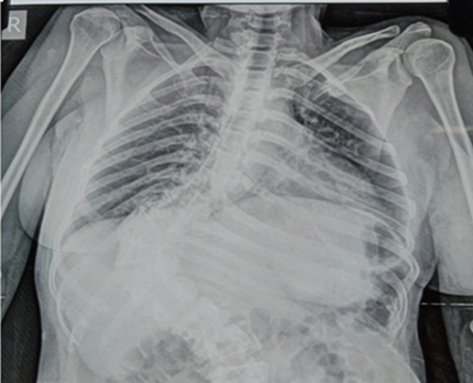
Figure 2: X-ray chest standing radiograph showing crowding of ribs and gross spine curvature
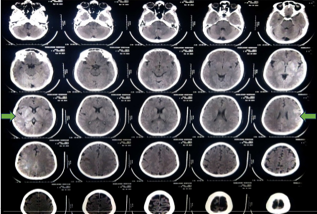
Figure 3: CT brain showing demyelination changes in basal ganglia
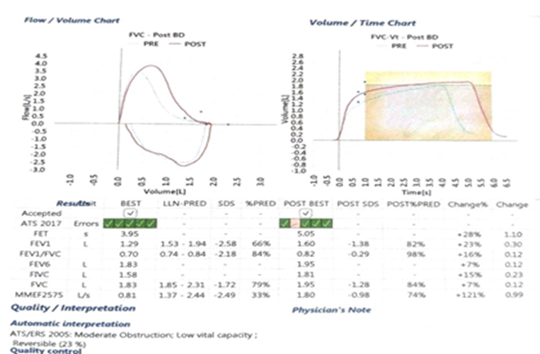
Figure 4: Spirometry showing low vital capacity and moderate obstruction
Figure 5: Clinical parameters
|
Hb |
8.5 |
|
BMI |
24.7 |
|
PR |
88/min |
|
BP |
90/60mmHg |
|
sPO2 |
98%RA |
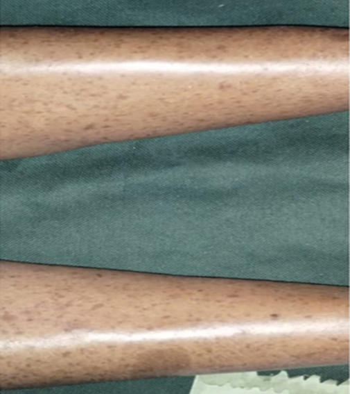
Figure 6: Café-au-lait macules (CALM) in lower limbs
Most patients with NF1 do not require treatment. Subcutaneous neurofibromas may be painful or disfiguring and can be excised surgically through recurrence is common. Genetic counselling is important and must be provided to all patients and families in whom NF1 is present.[10]
Scoliosis
In this case report the gross thoracic scoliosis is the prominent deformity which may conceal a spinal cord neurofibroma and is the cause of the poor respiratory reserve in this patient.
The scoliosis research society has defined scoliosis as a lateral curvature of the spine greater than 10 degrees as measured using Cobb method on a standing radiograph. It is a friplanar deformity of lordosis, rotation, and lateral wedging of vertebrae. Scoliosis is a combination of angular displacement and lateral displacement of spines.
Apart from congenital and traumatic causes of scoliosis bony dysplasia of neurofibromatosis forms an important cause of scoliosis.
The scoliosis in the case report is a double major curve deformity where two balancing curves of equal structural change and magnitude would manifest. Thoracic curve is major and lumbar curve is structural.
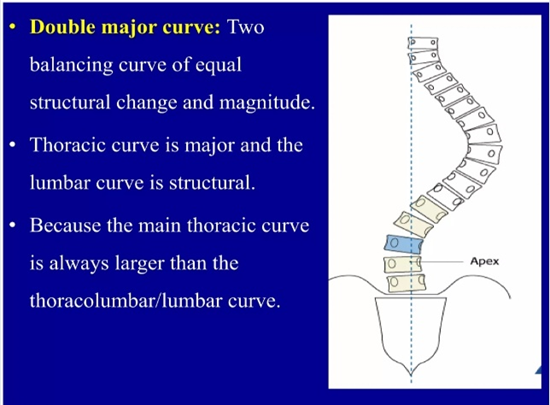
Figure 7: Double Major Curve
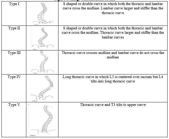
Our patient belongs to King classification type II thoracic dominant S shaped curve. If the deformity is progressive and painful patient may be advised back exercises and bracing.
Figure 8: Kings Classification of AIS
Genetic Counselling for Autosomal Dominant Disorders
Genetic counselling for autosomal dominant disorders focuses on 50% risk of transmission per pregnancy if one parent is affected, as only one mutated gene is required to cause the condition.
Counselling provides essential information to the affected individual and family members in an understandable and compassionate manner about family risks, testing options and reproductive choices including pre-implantation genetic testing or prenatal diagnosis.
The core components of counselling include:
1. Risk Evaluation
There is 50% chance that the mutation would pass on to each child.
2. Variable Expressivity and Penetrance
Family members may not show symptoms and severity of the disorder may vary.
3. De Novo Mutations
Sometimes none of the parents has the gene but the child may present due to new mutation.
4. Family Testing
Counsellors guide in identifying relatives who may possess the risk of developing the disorder.
5. Reproductive Options
Prenatal diagnosis by amniocentesis or chorionic villus biopsy or preimplantation genetic testing during IVF may be suggested for early diagnosis and appropriate management.
CONCLUSION:
This case report is a classic case of Neurofibromatosis Type 1 (Von Recklinghausen disease) with neurofibromata, spine deformity, café-au-lait macules in lower limbs, right facial palsy, and demyelination changes in basal ganglia with rare involvement of uterine fibroid which turned out to be sarcoma. Though this patient presented only for treatment or diagnosis etiology of menorrhagia, a complete holistic approach to her system involvement made anesthesia and procedure phase safe and pleasant. Every patient attending a clinic should receive a thorough evaluation, identification, and advice regarding physiotherapy for scoliosis, and immediate attention if progressive neurological deficit occurs and offered proper counselling to the family members regarding genetics and plan for the next child.
An interdisciplinary approach goes a long way in benefitting patients with inherited genetic disorders with organ system involvement. The patient may present for a different symptom but it is the duty of every physician to identify the syndrome and direct patients towards treatment, rehabilitation, and genetic counselling
REFERENCES:
[1] Kolodny EH, Sathe S. Inherited neurodegenerative diseases. Chap- 24.17. In: Ogg GS, Warrell DA, Cox TM, et al, eds. Oxford textbook of Medicine. 5th edn. Oxford University Press 2010.
[2] Rosalind A Eeles RA. Genetics of inherited cancers. Chap- 6.3. In: Ogg GS, Warrell DA, Cox TM, et al, eds. Oxford Textbook of Medicine. 5th edn. Oxford University Press 2010.
[3] Le C, Thomas A, Liew F. Neurofibromatosis. StatPearls [Internet]. Treasure Island (FL): StatPearls Publishing 2026.
[4] Poplausky D, Young JN, Tai H, et al. Dermatologic Manifestations of Neurofibromatosis Type 1 and Emerging Treatments. Cancers (Basel) 2023;15(10):2770.
[5] Raj P, Bhatia S, Soin N, et al. Classification of Schwannoma. Genetic updates and international consensus recommendation. Neurology Journal 2025;38(5):533-42.
[6] Moskova V, Esparza S, Smith MJ, et al. Clinical features of Schwanomatosis. Oncologist 2012;17(10):1317-22.
[7] Wilson BN, John AM, Handler MZ, et al. Neurofibromatosis Type 1: developments in genetics and treatment. J Am Acad Dermatol 2021;84(6):1667-76.
[8] Legius E, Messiaen L, Wolkenstein P, et al. Revised diagnostic criteria for Neurofibromatosis. Genet Med 2021;23(8):1506-13.
[9] Gross AM, Glassberg B, Wolters PL, et al. Selumetinib in children with NF1 and asymptomatic inoperable plexiform neurofibroma. Neuro-Oncology 2022;24(11) 1978-88.
[10] James A, Baskovich J, Ruben I. Congenital and developmental neurocutaneous disorders. Chap- 444. In: Goldman L, Cooney KA, eds. Goldman-Cecil Medicine. 23rd edn. Vol. 2. Elsevier/ South Asian Ed. First printed 2008: p. 2789, 2792, 2793.
[11] Moore DW. Spone-Ortho bullets. In: King Classification of spine. https://share.google/42Cy1Fzi8uMQCrjqW
[12] Genetic counselling for autosomal dominant diseases. NIH https://pubmed.ncbi.nlm.nih.gov