MORPHOLOGICAL AND MORPHOMETRIC STUDY OF MENTAL FORAMEN AND ITS CLINICAL SIGNIFICANCE.
- Jatinder Kour , Senior Resident, Department of Anatomy AIIMS Jammu, India.
- Yusra Magrey , Senior Resident, Department of Anatomy GMC Anantnag, India.
- Sangeeta Gupta , Professor and Head, Department of Anatomy GMC Jammu, India.
Article Information:
Abstract:
Background: The mental foramen is an oval/circular opening on the anterior surface of the mandible, located below or between the premolars, approximately midway between the alveolar crest and lower border of the mandible. Variations of the mental foramen are often encountered ranging from difference in shape and position to the presence of accessory foramen or even complete absence in some cases. It is very much important to locate the mental foramen and to determine its accurate position in case of implantation in interforaminal area to prevent the complication. Aims and Objectives: Thus, the present study was undertaken to study the size, shape and location of mental foramen and to determine the clinical significance of mental foramen in oral and maxillofacial surgeries and local anesthetic procedures. Methodology: This observational study was conducted in the Post Graduate Department of Anatomy, Government Medical College, Jammu for a period of 18 months on 60 samples of mandible. The various parameters for mental foramen were measured such as shape/ size, distances from mandibular landmarks, position of the mental foramen in relation to the mandibular teeth on the right and left sides of the mandible. Results: It was found in our study that in most of the cases the foramen was oval in shape. The location of the mental foramina was symmetrical in all the bones. The mental foramen located below the root of first molar was found in none of the bones. Conclusion: The study came to the conclusion that mental foramen is a significant anatomical landmark. Its morphological and morphometric parameters provide valuable information to dental surgeons for effective localization of the neurovascular bundle passing through mental foramen thus avoiding complications from local anesthetic, surgical and other invasive procedures.
Keywords:
Article :
INTRODUCTION:
The mental foramen is an oval/circular opening on the anterior surface of the mandible, located below or between the premolars, approximately midway between the alveolar crest and lower border of the mandible1. The mental foramen gives path to mental nerve and vessels. The mental nerve emerges at the mental foramen and divided into four branches angular, medial and lateral inferior labial and mental branch2. It supplies the lower lip, gum and also the buccal vestibule anterior to the first mandibular molar3.
The mental foramen is located slightly below among toothless individuals, it is observed that it is in line with longitudinal axis of the 2nd premolar in 63% individuals and generally lies at the level of vestibular fornix and about a finger's breadth above the inferior border of the mandible4. Variations of the mental foramen are often encountered ranging from difference in shape and position to the presence of accessory foramen or even complete absence in some cases5. Both the mental and accessory mental foramen show ethnic variations. So, the knowledge of mental foramen morphometry can help the dental surgeons to apply for nerve blocks in surgeries related to the lower jaw. Accessory mental foramen morphometry will help in preventing accessory nerve injury during periapical surgery. If it is not blocked, the paraesthesia will be less2.
Also it is very much important to locate the mental foramen and to determine its accurate position in case of implantation in interforaminal area to prevent the complication. It was reported that the minimum distance between mental foramen and an implant should be up to 6 mm as any invasive procedure done in this region can lead to damage of the neurovascular bundles and can also result in paresthesia 6.
Thus, the present study was undertaken to study the size, shape and location of mental foramen and to determine the clinical significance of mental foramen in oral and maxillofacial surgeries and local anesthetic procedures.
AIMS AND OBJECTIVES:
· To study the shape, location and size of mental foramen.
· To study the clinical significance of mental foramen in oral and maxillofacial surgeries and local anaesthetic procedures.
METHODOLOGY :
This observational study was conducted in the Post Graduate Department of Anatomy, Government Medical College, Jammu for a period of 18 months from February 2021 up to August 2022. The study consists of 60 samples of mandible with the following inclusion and exclusion criteria:
INCLUSION CRITERIA: Mandible should be:
I. Normal
II. Complete
III. Healthy
IV. Dry mandible.
EXCLUSION CRITERIA: Mandible should not be:
I. Mutilated
II. Deformed
III. Destroyed Samples.
The various parameters for mental foramen were measured on the right and left sides of the mandible such as shape of the mental foramen i.e. oval or round. The distances from the most anterior portion of the anterior border of the mental foramen was measured from various mandibular landmarks. The position of the mental foramen in relation to the mandibular teeth was measured and expressed as a percentage for each of the six positions described by Tebo and Telford. Size of the mental foramen was measured with the help of a vernier caliper in terms of Transverse / Vertical diameters of mental foramen passing through the center of the foramen. The Ethical clearance was obtained from the Institutional Ethics Committee before the commencement of the study.
Statistical analysis: The results of afore mentioned study were measured, tabulated and analyzed. Data was entered in a Microsoft Excel Spreadsheet. Continuous variables were summarized as mean and standard deviation. Categorical variables were summarized as percentages. All the data was analysed using statistical package for social science (SPSS) software and appropriate statistical analytical tests. Comparison of study parameters was done using Independent t-test. The level of significance for the present study was fixed at a P-value of less than 0.05.
RESULTS:
Table 1: Shape of the mental foramen
|
Shape |
Right Side |
Left Side |
||
|
No. |
% |
No. |
% |
|
|
Round |
19 |
31.7 |
20 |
33.3 |
|
Oval |
41 |
68.3 |
40 |
66.7 |
Table 1 depicts the distribution of the shape of mental foramen of the bones under study. The results of present study inferred that on the right-side oval shape was found in 41 bones (68.3%) and round shape in 19 bones (31.7%) whereas on the left side oval shape was found in 40 bones (66.7%) and round shape was found in 20 (33.3%) mental foramen of the bones.
Table 2: Distance of mental foramen from mandibular landmarks
|
Landmarks |
Mean Distance on Right Side |
Mean Distance on Left Side |
P value |
|
Symphysis Menti |
23.44±1.62 |
23.55±1.67 |
0.355 |
|
Posterior Border of Ramus |
58.79±3.78 |
58.81±3.60 |
0.912 |
|
Alveolar Crest |
11.86±1.48 |
12.15±1.42 |
0.016* |
|
Base of Mandible |
12.49±1.58 |
12.60±1.51 |
0.367 |
*Statistically significant (P<0.05, Paired t-test)
Table 2 depicts the distance of mental foramen from mandibular landmarks. The results of present study revealed that the mean distance from symphysis menti on the right side was 23.44±1.62mm and on left side was 23.55±1.67mm, from posterior border of ramus on the right 58.79±3.78mm and on left side was 58.81±3.60mm, from base of mandible on right side was 12.49±1.58mm and on left side was 12.60±1.51mm and mean distance from alveolar crest on right side was 11.86±1.48mm and on left side was 12.15±1.42mm. Statistical analysis showed that there was a statistically significant difference between right and left sides in relation to mean distance from alveolar crest (P=0.016). The mean distance of mental foramen from alveolar crest was significantly higher on left side compared to that on right side.
Table 3: Showing distribution of the mean size of the mental foramen
|
|
Right Side |
Left Side |
P value |
|
Transverse Diameter |
2.20±0.62 |
2.17±0.56 |
0.243 |
|
Vertical Diameter |
1.91±0.56 |
1.92±0.52 |
0.637 |
Table 3 depicts the size of mental foramen in terms of transverse/vertical diameter. In afore mentioned study mean right side transverse diameter was 2.20±0.62mm and mean vertical diameter was 1.91±0.56mm whereas left side mean transverse diameter was 2.17±0.56mm and mean vertical diameter was 1.92±0.52mm. Statistical analysis showed that there was no statistically significant difference between right and left sides in relation to mean transverse diameter (P=0.243) and mean vertical diameter (P=0.637).
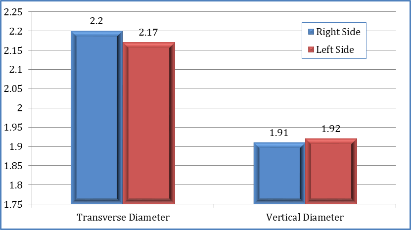
Fig. 1: Bar graph showing size of the mental foramen according to Transverse and vertical diameter.
Table 4: Showing position of mental foramen with relation to mandibular teeth
|
Position of mental foramen |
Right Side |
Left Side |
P value |
|
Anterior to first premolar |
5 (8.3%) |
5 (8.3%) |
1.00 |
|
Apex of first premolar |
8 (13.3%) |
8 (13.3%) |
1.00 |
|
Between Apex of two premolars |
21 (35%) |
21 (35%) |
1.00 |
|
Apex of second premolar |
22 (36.7%) |
22 (36.7%) |
1.00 |
|
Posterior to secondpremolar |
4 (6.7%) |
4 (6.7%) |
1.00 |
|
Belowtherootof first molar |
0 (0%) |
0 (0%) |
1.00 |
Table 4 depicts the position of mental foramen with relation to mandibular teeth. In afore mentioned study, it was concluded that the position of mental foramen was anterior to first premolar was found in 5 (8.3%) bones on right and left side, at apex of first premolar was found in 8 (13.3%) bones on left and right side, between the apex of two premolars was found in 21 (35%) bones on left and right side, at apex of second premolar was found in 22 (36.7%) bones on left and right side and posterior to second premolar was found in 4 (6.7%) bones on left and right side. However, mental foramen present below the root of first molar was neither found on right side nor on the left side. Statistical analysis showed that there was no statistically significant difference between right and left sides in any of the parameter (P=1.000).
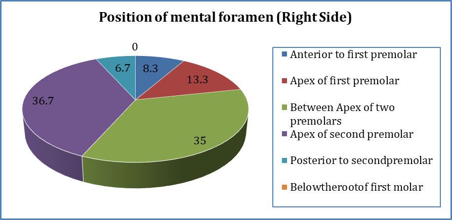
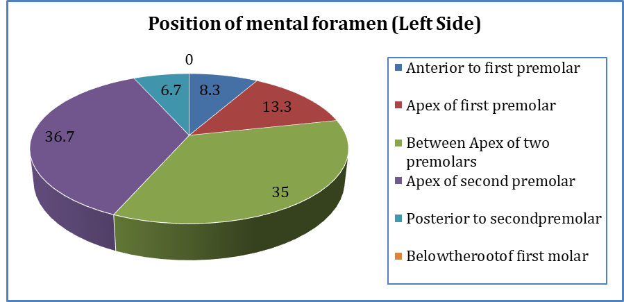
Fig. 2: Pie chart showing position of mental foramen in relation with mandibular teeth (Right and Left)
DISCUSSION:
In present study, oval shaped mental foramina were observed in 41 (68.3%) bones on right side and 40 (66.7%) on left side whereas while taking into consideration the round shaped mental foramina, the statistics fell at 19 (31.7%) bones on right side and 20 (33.3%) on left side. The findings are positively supported by the evidence derived from the results of the study conducted by Singh D et al., (2019) who concluded that 71.87% bones mental foramina were oval in shape on right side and 60.93% on left side, whereas 28.12% bones were round in shape on right side and 39.06% on left side3. In another study conducted by Budhiraja V et al., (2012), it was observed that in most cases 78 (74.3%) bones, the mental foramen was oval in shape on both sides and 27(25.7%) bones were round in shape on both sides, which was again in concordance with the inference of present study5. Similarly, Udhaya K et al., (2013) determined that majority of the mental foramen were in oval shape constituting 75 (83.33%) bones under study and rest 15 (16.67%) bones were having round mental foramina which was again in accordance with the results of afore mentioned study which concluded that oval shaped mental foramina outnumbered the round shaped mental foramina7.
In current study, the mean distance of mental foramen from symphysis menti on the right side was 23.44±1.62 and on left side was 23.55±1.67, from the Posterior Border of Ramus of mandible on the right side was 58.79±3.78 and left side was 58.81±3.60, from the Base of Mandible on right side was 12.49±1.58 and on left side was 12.60±1.51 and mean distance from the Alveolar Crest on right side was 11.86±1.48 and on left side was 12.15±1.42. The results of present study are consistent with the results of the study conducted by Zainab H et al., (2020) who observed that the mean distance between the mental foramen and symphysis menti for right side was 23.21 mm±2.12 and for left side was 23.10 mm±2.418. The mean distance between mental foramen and posterior border of ramus of mandible for right side was 62.58 mm±4.23 and for left side was 63.84 mm±4.72. The mean distance between mental foramen and alveolar crest for right side was 15.23 mm±1.12 and for left side was 16.07 mm±1.01. The mean distance between the mental foramen and base of mandible for right side was 15.61 mm±1.03 and for left side was 15.98 mm±1.06.
In similar study performed by Sankar DK et al., (2011) who observed that the mean horizontal distance between symphysis menti and mental foramen was 27.2 mm on right side and 27.7 mm on the left side, mental foramen and posterior border of ramus was 70.7 mm on both sides, vertical distance between mental foramen and inferior border of mandible was 16.5 mm on right side and 14.3 mm on left side, alveolar crest and mental foramen was 13.7 mm on right side and 16.4 on left side and these values were far more as compared to the observations of current study thus, showing discordance with the current study10. Another study carried out by Siddiqui AU et al., (2011) analyzed the mean distance between symphysis menti and anterior margin of (mental foramen) MeF was 18.8±12.02mm and 19.6±12.18mm, on the right and left sides respectively, between posterior margin of mental foramen and posterior border of ramus was 48.8±28.6mm on the right side and 47.9 ±28.1mm on the left side and mean distance between alveolar crest and superior margin of mental foramen was 10.2±5.4mm on right side and 10±5.2mm on the left side, mean distance between inferior border of mental foramen and base of mandible was 9.9±5.12mm on right side and 10.1±5.2mm on left side10. These findings documented by Siddiqui AU et al., (2011) were far less thus, again negating the results of the present study10. The results of present study also compared with the study conducted by Kqiku L et al., (2013) where the mean distance from the mental foramen to symphysis menti was 24.76±4.42mm on right side and 24.92±3.65 on left side, mean distance of the mental foramen to the posterior border of the mandibular ramus was 67.02±6.23mm on right side and 67.98±5.40 on left side, mean distance of the mental foramen to the base of mandible was 14.58±2.09mm on right side and 14.77±1.97mm on left side and mean distance of the mental foramen to the alveolar crest was 20.59±1.70mm on right side and 20.17±1.33 on left side, as the statistical correlation can be made out in two parameters whereas it cannot be established in others11. This difference in the observations can be attributed to the ethnicity, build, sex, environmental factors and nutritional status of the population under study.
In present study, mean transverse diameter of mental foramen was 2.20±0.62mm and mean vertical diameter was 1.91±0.56mm on right side whereas on left side mean transverse diameter was 2.17±0.56mm and mean vertical diameter was 1.92±0.52mm. In similar study conducted by Rai R et al., (2014) observed that the mean vertical diameters were 2.3±0.64mm on right and 2.29±0.6mm on left sides whereas the mean horizontal diameters were 2.63±0.85mm and 2.61±0.85mm on right and left sides respectively12. In another morphometrical study conducted by Singh D et al., (2019), it was observed that the mean transverse diameter of mental foramen was 3.45±0.70mm and 2.90±0.87mm on right and left side respectively whereas the mean vertical diameter was 2.33± 0.56mm and 2.23±0.94mm on right and left side respectively3. Both the study groups documented the values of diameter of mental foramina slightly more than those observed in current study.
The present study concluded that the mental foramen present anterior to first premolar was found in 5 (8.3%) bones on right and left side, at the apex of first premolar was found in 8 (13.3%) bones on left and right side, between the apex of two premolars was found in 21 (35%) bones on left and right side, at the apex of second premolar was found in 22 (36.7%) bones on left and right side and posterior to second premolar was found in 4 (6.7%) bones on left and right side. However, mental foramen located below the root of first molar was neither found on right side nor on the left side. The findings are correlated with the study conducted by Siddiqui AU et al., (2011) who documented that the commonest position of the mental foramen was at the apex of second premolar 41 (44.08%) on right side and 43 (46.23%) on left side followed by position between apex of two premolars 39 (41.93%) on right side and 33 (35.48%) on left side, 6 (6.45%) on right side and 8 (8.60%) on left side both at the position of apex of first premolar and posterior to second premolar, 1 (1.07%) are anterior to first premolar on both sides and none at the level of first molar10.
In another study conducted by Singh D et al., (2019) it was reported that in 56 (67.46%) bones mental foramen was located below the apex of second premolar on right side and 54 (65.06%) bones on left side, 18 (21.68%) bones between the apex of two premolars on right side and 17 (20.48%) bones on left side, 7 (8.43%) bones at posterior to second premolar on right side and 9 (10.84%) bones on left side, 1 (1.20%) bones at apex of first molar on right side and 2 (2.40%) on left side, 1 (1.20%) bones below the root of first molar on both sides and none at the level of anterior to the first premolar3.
The observations of current study inferred that the position of mental foramen was symmetrical bilaterally in all the bones under study. This fact was in discordance with the results of above studies.
CONCLUSION:
The present observational study examined the morphological and morphometric parameters of the mental foramen, which are important in nerve block and various surgical procedures such as sagittal split osteotomy, orthognathic procedures, dental implant surgeries, and other maxillofacial procedures. These parameters help surgeons and anesthetists avoid injuring the neurovascular bundle. Thus, the study came to the conclusion that mental foramen is a significant anatomical landmark.
REFERENCES:
1. Standring S. Gray’s Anatomy. The Anatomical Basis of Clinical practice, 39th Ed. Elesevier Ltd., UK. 2005; 27:481-484.
2. Raman RK, Choudhary G. Study on Morphology and Morphometric of Mental Foramen in Adult Human Mandibles of Kathmandu Valley Population. Int J Sci Res 2015; 4(8):1947-1949.
3. Singh D, Patnaik P, Gupta N. Morphology and Morphometric Analysis of the Mental Foramen in Dry Adult Human Mandibles from North India. Int J Sci Healthcare Res 2019;4(1): 220-226.
4. Soikkonen K, Wolf J, Ainamo A, Xie
a. Q. Changes in the position of the mental foramen as a result of alveolar atrophy. J Oral Rehabil 1995; 22 (11):
b. 831–833.
5. Budhiraja V, Rastogi R, Lalwani R, Goel P, Bose SC. Study of Position, Shape, and Size of Mental Foramen Utilizing Various Parameters in Dry Adult Human Mandibles from North India. Int. Sch Res Notices 2012; 961429.
6. Zmyslowska-Polakowska E, Radwanski M, Ledzion S, Leski M, Zmyslowska A, Lukomska-Szymanska M. Evaluation of Size and Location of a Mental Foramen in the Polish Population Using Cone-Beam Computed Tomography. Biomed Res Int 2019;1659476.
7. Udhaya K, Saraladevi KV, Sridhar J. The Morphometric Analysis of the Mental Foramen in Adult Dry Human Mandibles: A study on the South Indian Population. J Clin Diagn Res 2013;7(8):1547-1551.
8. Zainab H, Sylvia S, Ahmed MK. Study of anatomical variations of mental foramen in dry adult human mandibles and its clinical importance in Karnataka population. Indian J Clin Anat Physiol 2020;7(3):327
9. Sankar DK, Bhanu SP, Susan PJ. Morphometrical and morphological study of mental foramen in dry dentulous mandibles of South Andhra population of India. Indian J Dent Res 2011; 22(4): 542-546.
10. Siddiqui AU, Daimi SR, Mishra PP, Doshi SS, Date JY, Khurana G. Morphological and morphometric analysis of mental foramen utilizing various assessment parameters in dry human mandibles. Int J Stud Res 2011;1(1):19-22.
11. Kqiku L, Weiglein A, Kamberi B, Hoxha V, Meqa K, Städtler P. Position of the mental foramen in Kosovarian population. Coll Antropol 2013;37(2):545-549.
12. Rai R, Shrestha S, Jha S. Mental foramen: a morphological and morphometrical study. Int J Healthcare Biomed Res 2014;2(4):144-150.