The Measurement of Pancreatic Volume in Adults by using CT scan and its Correlation with various Demographic Markers.
- Yusra Magrey , Senior Resident, Department of Anatomy GMC Anantnag, India
- Jatinder Kour , Senior Resident, Department of Anatomy AIIMS Jammu, India.
- Nusrat Jabeen , Professor and Head, Department of Anatomy GMC Jammu, India.
Article Information:
Abstract:
Background: The pancreas is an essential organ which produces enzymes to digest the food and hormones to lower down the blood sugar level. It was reported that the several pancreatic disorders alter the anatomy of pancreas. The pancreatic volume is an important clinical marker of disease progression and altered volume of pancreas is associated with infective, infiltrative, immunological, infestative and malignant conditions. Aims and Objectives: To measure the normal pancreatic volumes in adults by using CT scan and correlation of pancreatic volume measurements with various demographic markers. Methodology: This cross-sectional observational type of study was done for assessment of normal pancreatic volumes by using Computed Tomography, in adults attending a tertiary care hospital. The study was conducted in the Post Graduate Department of Anatomy in collaboration with the department of Radio diagnosis and Imaging, Government Medical College, Jammu for a period of 18 months from February 2021 up to August 2022. The patients presenting for CT abdomen were selected as per the inclusion and exclusion criteria. Results and observations: In our study there were 61% males and 39% were females. The mean pancreatic volume in females was 114.90±18.21 cm3 and in males was 122.60±16.47 cm3. The overall mean pancreatic volume of the study population was 119.59±17.49 cm3. Conclusion: The present observational study concluded that mean pancreatic volume was higher in males than in females in our study population as there was significant difference in pancreatic volume among male and female patients.
Keywords:
Article :
INTRODUCTION:
Pancreas is one of the largest digestive glands. The pancreas is an essential organ which produces enzymes to digest the food and hormones to lower down the blood sugar level. The length of the pancreas in adults, measures 12- 15cm. In adults, it has an average volume of 70-80cm3, although this varies considerably between individuals (with a range of 40-170 cm3) and tends to be greater in males. Pancreatic volume increases with age, to reach a peak in the fourth decade 1.
It was reported that the several pancreatic disorders alter the anatomy of pancreas. The pancreatic volume is an important clinical marker of disease progression 2 and altered volume of pancreas is associated with infective, infiltrative, immunological, infestative and malignant conditions.
The ailments of pancreas can be determined by various investigations (laboratory and radiographic) which helps in the treatment and prognosis of the diseases / ailments. The pancreatic volume can be measured by using radiographic studies (CT scan and MRI) of abdominal organs to make an appropriate diagnosis. 3 The various studies suggest that as the alteration in organ volume and its factors can be accurately measured with CT 2.
Thus, the present study was undertaken to estimate the pancreatic volume in adults by using the computed tomography.
Aims and Objectives
1. To measure the normal pancreatic volume in adults by using CT scan.
2. To correlate the pancreatic volume measurements with various demographic markers.
METHODOLOGY:
This cross-sectional observational type of study was done for assessment of normal pancreatic volumes by using Computed Tomography, in adults attending a tertiary care hospital. The study was conducted in the Post Graduate Department of Anatomy in collaboration with the department of Radio diagnosis and Imaging, Government Medical College, Jammu for a period of 18 months from February 2021 up to August 2022.
The patients presenting for CT abdomen were selected as per the following inclusion and exclusion criteria:
INCLUSION CRITERIA
A. The patients of either sex, in the age group of 20-50 years, who after the routine clinical evaluation, will undergo CT examination in the Department of Radio-diagnosis, due to various indications for the abdominal CT.
B. Only those CT images, which will be declared as ‘normal’ by the radiologists will be included in this study.
EXCLUSION CRITERIA
A. Presence of either clinical or CT signs of any pancreatic or peri- pancreatic pathology.
B. Patients with previous pancreatic surgery.
C. Patients with difficult identification of pancreatic margins from the adjacent structures.
D. Patients with various pathological conditions affecting the adjacent organs (Stomach, peritoneum, or retroperitoneum) such as large tumours, fluid collections, massive lymph node enlargement with consecutive compression, infiltration or displacement of the pancreas.
E. Pregnant women
F. Patients with history of Diabetes Mellitus, Malaria.
Imaging Protocol
Contrast enhanced CT (CECT) examination of abdomen was done using Somatom definition flash 256 slice multidetector CT scan at Government Medical College, Jammu. The following measurement (in centimeters) were taken, after identification of normal pancreas on CT scans:
· Cranial-caudal (CC) dimensions of the entire pancreas (CCpancreas), body and tail (CC body/tail), body (CCbody) and head (CChead) will be taken.
· Anterior-posterior (AP) diameters of the tail (APtail) and body (APbody) from the splenic vein to the anterior contour of the pancreatic body, and head (APhead) at the level on which the pair of the superior mesenteric vein and artery is demonstrated.
· Length of body and tail (LLbody/tail) - It will be measured as a maximal linear distance from the pancreatic neck to the tip of the pancreatic tail.
The pancreatic volume was computed by employing the following standard formula:
PV= (AP tail + AP body)/ 2 X Lengths body & tail X CC body + (AP head / 2) 2 X 3.14 X CC head
The data was collected in a pre-defined proforma and was entered in a Microsoft Excel Spreadsheet. All the data was analyzed using statistical package for social science (SPSS) software and appropriate statistical analytical tests.
RESULTS:
In our study the subjects were categorized in 3 categories according to age, i.e. 20-30 years, 31-40 years and 41-50 years, among the majority the female subjects were in the age group of 31-40 years and male subjects were in the age group of 20-30 years. The majority of the subjects were males.
Table 1. Cranio-caudal pancreatic head length (cm)
|
Age Group |
Females N |
Mean |
SD |
P |
Males N |
Mean |
SD |
|
20–30 |
10 |
3.8000 |
0.50332 |
23 |
4.2391 |
0.65626 |
|
|
31–40 |
18 |
4.1056 |
0.62447 |
22 |
3.9364 |
0.58923 |
|
|
41–50 |
11 |
4.0727 |
0.49415 |
16 |
3.7625 |
0.50448 |
|
|
Total |
39 |
4.0179 |
0.56189 |
61 |
4.0049 |
0.61764 |
Table 1 depicts that the mean cranio-caudal pancreatic head length in females was 4.01±0.56 cm and in males was 4.00±0.61 cm. The overall mean cranio-caudal pancreatic head length of the study population was 4.01±0.59 cm. The mean Cranio-caudal pancreatic head length among different age groups is not statistically significant.\
Table 2. Cranio-caudal pancreatic body length
|
Age Group |
Females N |
Mean |
SD |
Males N |
Mean |
SD |
|
20–30 |
10 |
3.5500 |
0.69322 |
23 |
3.0174 |
0.45890 |
|
31–40 |
18 |
3.2944 |
0.51958 |
22 |
3.4091 |
0.50229 |
|
41–50 |
11 |
3.0364 |
0.36952 |
16 |
3.4813 |
0.41828 |
|
Total |
39 |
3.2872 |
0.55402 |
61 |
3.2803 |
0.50260 |
Table 2. depicts Cranio-caudal pancreatic body length among various age groups. The mean cranio-caudal pancreatic body length in females was 3.28±0.55 cm and in males was 3.28±0.50 cm. The overall mean cranio- caudal pancreatic body length of the study population was 3.28±0.52 cm. The mean Cranio-caudal pancreatic body length among different age groups is not statistically significant.
Table 3. Antero-posterior pancreatic head length
|
Age Group |
Females N |
Mean |
SD |
Males N |
Mean |
SD |
|
20–30 |
10 |
3.2300 |
0.3772 |
23 |
3.7470 |
0.4669 |
|
31–40 |
18 |
3.3160 |
0.6428 |
22 |
3.6540 |
0.6559 |
|
41–50 |
11 |
3.6630 |
0.6682 |
16 |
3.6250 |
0.6308 |
|
Total |
39 |
3.3928 |
0.6058 |
61 |
3.6820 |
0.5769 |
Table 3. depicts antero-posterior pancreatic head length among various age groups. In our study the mean antero-posterior pancreatic head length in females was 3.39±0.60 cm and in males was 3.68±0.57 cm. The overall mean antero- posterior pancreatic head length of the study population was 3.56±0.60 cm. The mean antero-posterior pancreatic head length among different age groups is not statistically significant.
In our study, the mean antero-posterior pancreatic body length in females was 2.39±0.41 cm and in males was 2.38±0.44 cm. The overall mean antero- posterior pancreatic body length of the study population was 2.38±0.43 cm. The mean antero-posterior pancreatic body length among different age groups is not statistically significant.
Table 4. Antero-posterior pancreatic tail length
|
Age Group |
Females (N) |
Females (Mean) |
Females (SD) |
Males (N) |
Males (Mean) |
Males (SD) |
|
20-30 |
10 |
2.3800 |
0.37653 |
23 |
2.3130 |
0.43307 |
|
31-40 |
18 |
2.2167 |
0.35521 |
22 |
2.4227 |
0.32940 |
|
41-50 |
11 |
2.1455 |
0.42039 |
16 |
2.3375 |
0.36492 |
|
Total |
39 |
2.2385 |
0.38018 |
61 |
2.3590 |
0.37744 |
|
P values |
0.359 |
0.608 |
Table 4. depicts antero-posterior pancreatic tail length among various age groups. The mean antero-posterior pancreatic tail length in females was 2.23±0.38 cm and in males was 2.35±0.37 cm. The overall mean antero- posterior pancreatic tail length of the study population was 2.31±0.38 cm. The mean antero-posterior pancreatic tail length among different age groups is not statistically significant.
In our study, the mean body/tail pancreatic length in females was 10.39±0.73 cm and in males was 10.36±0.76 cm. The overall mean body/tail pancreatic length of the study population was 10.38±0.75 cm. The mean Body/ Tail pancreatic length among different age groups is not statistically significant.
In our study the mean pancreatic volume in females was 114.90±18.21 cm3 and in males was 122.60±16.47 cm3. The overall mean pancreatic volume of the study population was 119.59±17.49 cm3.
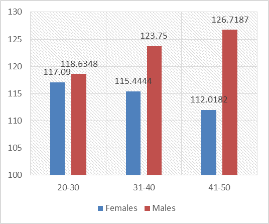
Figure 1. Pancreatic Volume
In our study Correlation analysis between pancreatic dimensions and pancreatic volume showed that the pancreatic volume had a statistically significant positive correlation with cranio-caudal body length (r = 0.502, P<0.001), antero-posterior head length (r = 0.494, P<0.001), antero-posterior body length (r = 0.284, P=0.004), antero-posterior tail length (r = 0.203, P=0.043) and length body/tail (r = 0.213, P=0.033).
DISCUSSION:
The determination of the pancreatic volume (PV) has significant potential for analyzing the various clinical diagnosis. A significant change in the pancreatic volume signifies the pathological conditions of the pancreatic endocrine or exocrine function.
In our study the subjects were categorized in 3 categories according to age, i.e. 20-30 years, 31-40 years and 41-50 years, among the majority the female subjects were in the age group of 31-40 years and male subjects were in the age group of 20-30 years. The majority of the subjects were males.
The present study found that the mean cranio-caudal pancreatic body length in females was 3.28±0.55 cm and in males was 3.28±0.50 cm. The overall mean cranio-caudal pancreatic body length of the study population was 3.28±0.52 cm. The mean Cranio-caudal pancreatic body length among different age groups is not statistically significant. The results are comparable with the study conducted by Li Lu et al., (2021) who reported that the mean cranio-caudal pancreatic body length in females was 3.48±1.06 cm and in males was 3.03±0.64 cm. The overall mean cranio-caudal pancreatic body length of the study population was 3.26±0.90 cm 4.
In our study the mean antero-posterior pancreatic head length in females was 3.39±0.60 cm and in males was 3.68±0.57 cm. The overall mean antero-posterior pancreatic head length of the study population was 3.56±0.60 cm. The mean antero-posterior pancreatic head length among different age groups is not statistically significant. In similar study conducted by Arua HN et al., (2020) who reported that the mean antero-posterior pancreatic head length in females was 2.67 ± 2.54 cm and in males was 2.68 ± 2.85 cm respectively, which is in agreement with the results of the present study 5.
In our study the mean antero-posterior pancreatic body length in females was 2.39±0.41 cm and in males was 2.38±0.44 cm. The overall mean antero-posterior pancreatic body length of the study population was 2.38±0.43 cm. The mean antero-posterior pancreatic body length among different age groups is not statistically significant. The findings are in concordance with the study conducted by Arua HN et al., (2020) who observed that the mean antero-posterior pancreatic body length in females was 2.09 ± 1.96 cm and in males was 2.15 ± 2.33 cm 5.
The present study found that the mean antero-posterior pancreatic tail length in females was 2.23±0.38 cm and in males was 2.35±0.37 cm. The overall mean antero-posterior pancreatic tail length of the study population was 2.31±0.38 cm. The mean antero-posterior pancreatic tail length among different age groups is not statistically significant. In accordance to these findings Arua HN et al., (2020) and Li Lu et al., (2021) who reported that the mean antero-posterior pancreatic tail length was 1.70 ± 2.21 cm, 1.74 ± 0.47 cm respectively in females and 1.76 ± 1.91 cm, 2.09 ± 0.51 cm respectively in males 4,5.
In our study the mean body/tail pancreatic length in females was 10.39±0.73 cm and in males was 10.36±0.76 cm. The overall mean body/tail pancreatic length of the study population was 10.38±0.75 cm. The mean Body/ Tail pancreatic length among different age groups is not statistically significant. The outcome of the study is in accordance with the study conducted by Li Lu et al., (2021) who found that the mean body/tail pancreatic length in females was 10.40 ± 1.78 cm and in males was 11.54 ± 2.06 cm. The overall mean body/tail pancreatic length of the study population was 10.97 ± 2.01 cm 5.
In our study the observed mean pancreatic volume in females was 114.90±18.21 cm3 and in males was 122.60±16.47 cm3. The overall mean pancreatic volume of the study population was 119.59±17.49 cm3. These findings are correlated with the studies conducted by Yoon J et al., (2020), Li L et al., (2021), Lim SH et al., (2022), Nisa G et al., (2019) and Djuric- Stefanovic A et al., (2012) who reported that the mean pancreatic volume was 62.648±19.094 cm3, 77.44 ± 19.11 cm3, 66.5 cm3, 115.71± 33.60 cm3 and 79.2 ± 24.1 cm3 (2,4,6,7,3).
The correlation analysis between pancreatic dimensions and pancreatic volume showed that the pancreatic volume had a statistically significant positive correlation with cranio-caudal body length (r = 0.502, P<0.001), antero-posterior head length (r = 0.494, P<0.001), antero-posterior body length (r = 0.284, P=0.004), antero-posterior tail length (r = 0.203, P=0.043) and length body/tail (r = 0.213, P=0.033). Similarly, Li Lu et al., (2021) observed that the pancreatic volume had a statistically significant positive correlation with other pancreatic parameters 4.
CONCLUSION:
The present observational study concluded that mean pancreatic volume was higher in males than in females and there was significant difference in pancreatic volume among male and female patients. The overall mean pancreatic volume of the study population was 119.59±17.49 cm3. This data can be used by clinicians to estimate more accurately the degree of atrophy or hypertrophy of organs in certain disorders.
REFERENCES:
1. Standring, S. Pancreas, Gray’s Anatomy. The Anatomical basis of clinical practice, 41st Ed. King’s Col London, UK. 2016;1179.
2. Yoon J, Kim KG, Kim YJ, Lim S, Park YH, Kim D, Kang HT, Lee DH. Distribution and Characteristics of Pancreatic Volume Using Computed Tomography Volumetry. Healthc Inform Res. 2020;26(4):321-327.
3. Djuric-Stefanovic A, Masulovic D, Kostic J, Randjic K, Saranovic D. CT volumetry of normal pancreas: correlation with the pancreatic diameters measurable by the cross-sectional imaging, and relationship with the gender, age, and body constitution. Surg Radiol Anat. 2012;34:811–817.
4. Li L, S Wang, F Wang, GN Huang, D Zhang, GX Wang. Normal pancreatic volume assessment using abdominal computed tomography volumetry. Europe PMC. 2021;100(34): e27096.
5. Arua HN, Idigo FU, Arua CL. Correlation of Ultrasound and Computed Tomography Measurements of the Pancreas in a Normal Adult Nigerian Population. JOP. 2021; 22(1): 21-27.
6. Lim S, Bae JH, Chun EJ, Kim H, Kim SY, Kim KM, et al. Differences in pancreatic volume, fat content, and fat density measured by multidetector-row computed tomography according to the duration of diabetes. Acta Diabetol. 2014;51(5):739-748.
7. Nisa G, Shah BA, Gul S, Bhat SA. Computed tomography based pancreatic volume measurement in Kashmiri population. Int J Anat Res. 2019;7(4.1):6996-7002.