Sinonasal Schwannoma Presenting as a Polypoidal Mass of the Posterior Ethmoid and Sphenoid Sinus: A Rare Case Report with Radiological and Histopathological Correlation
- Dr Rajat Parashar , MS ENT, Assistant Professor, NIIMS, Greater Noida
- Dr Prathamesh Lanjewar , CFN, DNHE, MBBS, Research Associate, NIIMS, Gr. Noida
- Dr Avantika Chaudhary , MBBS, Research Associate, NIIMS, Gr. Noida
Article Information:
Abstract:
Background: Sinonasal schwannomas are rare benign tumors arising from peripheral nerve sheath cells, accounting for less than 4% of head and neck schwannomas. We report a case of a 49-year-old male presenting with unilateral nasal obstruction and a polypoidal mass arising from the right posterior ethmoid and sphenoid sinus. Imaging revealed a soft tissue attenuating lesion without significant bone destruction. Endoscopic surgical excision was performed. Histopathological examination confirmed schwannoma with characteristic Antoni A and Antoni B areas. This case highlights the importance of considering schwannoma in the differential diagnosis of unilateral sinonasal masses.
Keywords:
Article :
INTRODUCTION:
Schwannomas are benign encapsulated tumors originating from Schwann cells of peripheral nerves. In the head and neck region, they commonly arise from cranial nerves; however, sinonasal schwannomas are rare due to sparse innervation.
They most frequently originate from:
• Branches of the trigeminal nerve (V1, V2)
• Autonomic nerve fibers
Their nonspecific presentation often mimics:
• Nasal polyps
• Inverted papilloma
• Malignancy
Thus, radiological and histopathological confirmation is essential.
Case Presentation:
Patient Details
• Age/Sex: 49-year-old male
• Presenting complaints:
o Progressive unilateral nasal obstruction (right side)
o Occasional nasal discharge
o No epistaxis or visual symptoms
Radiological Findings
CT PNS (Axial & Coronal)
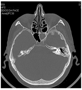
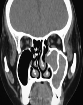
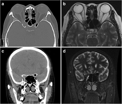
Key Findings:
• Polypoidal soft tissue density mass in:
o Right posterior ethmoid sinus
o Sphenoid sinus
• Mild heterogeneous enhancement
• No significant bone destruction
• No intracranial or orbital extension
👉 Suggestive of benign expansile lesion
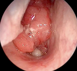
Intraoperative Findings
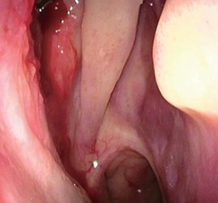
• Polypoidal, lobulated mass
• Moderately vascular
• Origin: posterior ethmoid region extending to sphenoid
• Complete excision done endoscopically
• Minimal intraoperative bleeding
Histopathological Findings
Gross
• Multiple soft tissue fragments
• Largest ~3 × 1.5 × 1 cm
Microscopy
• Biphasic pattern:
o Antoni A areas: hypercellular with nuclear palisading
o Antoni B areas: hypocellular myxoid regions
• Spindle-shaped cells with wavy nuclei
• No atypia or malignancy
Diagnosis
✅ Schwannoma
DISCUSSION:
Sinonasal schwannomas are rare and account for a very small fraction of sinonasal tumors.
Origin
Likely from:
• Posterior ethmoidal nerve (branch of V1)
• Sphenopalatine ganglion fibers
Clinical Features
• Unilateral nasal obstruction (most common)
• Rhinorrhea
• Headache
• Rarely epistaxis
Radiological Features
• Well-defined soft tissue mass
• Mild to moderate enhancement
• Bone remodeling rather than destruction
👉 Helps differentiate from malignancy
Differential Diagnosis
• Antrochoanal polyp
• Inverted papilloma
• Fungal sinusitis
• Esthesioneuroblastoma
• Sinonasal carcinoma
Histological Hallmarks
• Antoni A & B areas
• Verocay bodies (occasionally)
• S-100 positivity (if IHC done)
Treatment
• Endoscopic complete excision (gold standard)
• No role of radiotherapy in benign lesions
Prognosis
• Excellent
• Recurrence rare if completely excised
• Malignant transformation extremely rare
CONCLUSION:
Sinonasal schwannoma should be considered in unilateral sinonasal masses, especially when imaging shows a well-defined lesion without aggressive bone destruction. Endoscopic excision provides excellent outcomes with minimal morbidity.
REFERENCES:
1. Thompson LD. Sinonasal schwannoma. Head Neck Pathol.
2. Shugar JM et al. Schwannoma of nasal cavity. Laryngoscope.
3. Perzin KH, Panyu H. Non-epithelial tumors of nasal cavity.
4. Friedman I et al. Tumors of nasal cavity and sinuses.
5. Barnes L. Surgical pathology of head and neck tumors.