A Histopathological Study of Cutaneous Adnexal Lesions in a Tertiary Care Hospital.
- Sravanthi Gurugubelli , Assistant Professor, Department of Pathology, NRI Institute of Medical Sciences (NRIIMS), Sangivalasa, Visakhapatnam, Andhra Pradesh, India.
- Y. Akhila , Assistant Professor, Department of Pathology, NRI Institute of Medical Sciences (NRIIMS), Sangivalasa, Visakhapatnam, Andhra Pradesh, India.
- Hema Sundar Pydi , Associate Professor, Department of Forensic Medicine & Toxicology, Government of Medical College, Andhra Pradesh, India.
- P.Sandya Rani Guruvelli , Senior Resident, Department of Pathology, NRI Institute of Medical Sciences (NRIIMS), Sangivalasa, Visakhapatnam, Andhra Pradesh, India.
- Vattikuti Satya Veni , Final year Post Graduate, Department of Pathology, NRI Institute of Medical Sciences (NRIIMS), Sangivalasa, Visakhapatnam, Andhra Pradesh, India.
- Palika Sri Keerthika , Final year Post Graduate, Department of Pathology, NRI Institute of Medical Sciences (NRIIMS), Sangivalasa, Visakhapatnam, Andhra Pradesh, India.
- Atla Bhagya Lakshmi , Professor & Head, Department of Pathology, Principal, NRI Institute of Medical Sciences (NRIIMS), Sangivalasa, Visakhapatnam, Andhra Pradesh, India.
Article Information:
Abstract:
Background: The skin is a complex organ composed of multiple adnexal structures, including the pilosebaceous unit, eccrine glands, and apocrine glands. A wide spectrum of lesions may arise from these cutaneous appendages. Although skin adnexal tumors are relatively uncommon, their diagnosis is often challenging because a single lesion may show differentiation toward more than one adnexal line, and the terminology used in dermatopathology is often complex. In addition, distinguishing benign from malignant adnexal neoplasms may be difficult on clinical grounds alone, making histopathological examination essential for definitive diagnosis. Materials and Methods: This observational study was conducted at NRIIMS, Visakhapatnam, over a period of two years. Skin lesion specimens were obtained from patients attending the Department of Dermatology after informed consent. The specimens were received in the Department of Pathology and fixed in 10% neutral buffered formalin. Gross examination was performed, followed by routine tissue processing, paraffin embedding, sectioning, and hematoxylin and eosin staining. Histopathological evaluation was then carried out to establish the diagnosis. Results: Out of 600 skin lesion specimens studied, 23 cases were identified as adnexal lesions, giving an incidence of 3.83%. Among these, benign lesions constituted the majority. Benign neoplastic lesions accounted for 52.19% of cases, non-neoplastic lesions for 21.73%, hamartomas and hyperplasias for 13.04%, and malignant lesions for 13.04%. Thus, benign adnexal lesions were more common than malignant ones in the present study. Conclusion: The present study highlights the diverse histomorphological patterns of skin adnexal lesions. Benign skin adnexal tumors were more frequent than malignant tumors. Careful histopathological evaluation remains the cornerstone for accurate diagnosis and for distinguishing benign from malignant lesions.
Keywords:
Article :
INTRODUCTION:
The skin is a complex and multifunctional organ composed of the epidermis, dermis, and specialized adnexal structures, including the pilosebaceous unit, eccrine glands, and apocrine glands. Because of this structural complexity, a wide variety of lesions may arise in the skin, ranging from inflammatory and developmental conditions to benign and malignant neoplasms [1,2]. Among these, cutaneous adnexal lesions represent a distinctive and heterogeneous group derived from the appendageal components of the skin. These lesions are believed to originate from pluripotent or multipotent stem cells capable of differentiating toward follicular, sebaceous, eccrine, or apocrine structures [8].
Cutaneous adnexal lesions comprise hyperplasias, hamartomas, benign tumors, and malignant tumors, reflecting different degrees of differentiation. The majority of these lesions are benign, while malignant adnexal tumors are relatively uncommon [1]. Despite their low incidence, they are of considerable diagnostic importance because of their broad histomorphological spectrum and the frequent overlap of differentiation patterns within a single lesion. This often makes precise classification difficult, particularly on clinical grounds alone.
Clinically, adnexal lesions usually present as asymptomatic papules, nodules, or plaque-like lesions, with nonspecific features that may mimic other cutaneous conditions [8]. Their diagnosis is further complicated by the large number of entities, rarity of many lesions, and complex terminology used in dermatopathology [7]. Therefore, histopathological examination remains the cornerstone for definitive diagnosis, accurate classification, and distinction between benign and malignant lesions. In selected cases, ancillary techniques such as immunohistochemistry and electron microscopy may aid in understanding histogenesis and confirming the final diagnosis.
Certain adnexal tumors also serve as markers of underlying syndromic associations, such as multiple trichilemmomas in Cowden syndrome and sebaceous adenomas in Muir–Torre syndrome [1]. Hence, recognition of these lesions has both diagnostic and clinical significance. The present study was undertaken to identify and classify various cutaneous adnexal tumor-like lesions and tumors, determine their line of differentiation, and analyze their frequency according to age, sex, and anatomical site.
Objectives
The present study was undertaken to evaluate the histomorphological spectrum of cutaneous adnexal tumor-like lesions and tumors, to determine their adnexal line of differentiation, and to assess their frequency in relation to age, sex, and anatomical site.
MATERIALS AND METHODS:
Study Design
The present study was a hospital-based observational descriptive study conducted to evaluate the histopathological spectrum of cutaneous adnexal lesions. The study was designed to identify, classify, and analyze lesions of adnexal origin based on their morphological features and clinicopathological distribution.
Study Setting
The study was carried out in the Department of Pathology at NRI Institute of Medical Sciences [NRIIMS], Visakhapatnam. All skin biopsy specimens received from the Department of Dermatology and other concerned clinical departments during the study period were subjected to histopathological examination in the pathology laboratory.
Study Period
The study was conducted over a period of two years.
Study Sample
A total of 600 skin biopsy specimens were received in the Department of Pathology during the study period. Among these, 23 cases were identified as cutaneous adnexal lesions on histopathological examination and were included in the present study. These cases formed the basis for detailed morphological analysis.
Inclusion Criteria
All skin biopsy specimens showing histopathological evidence of adnexal origin were included in the study. This included adnexal tumor-like lesions as well as true adnexal tumors demonstrating follicular, sebaceous, eccrine, or apocrine differentiation.
Exclusion Criteria
Skin lesions not showing adnexal differentiation on histopathological examination were excluded from the study. Non-adnexal lesions of the epidermis, epidermal tumors, and other cutaneous lesions unrelated to skin appendages were not considered for analysis.
Specimen Collection and Fixation
Skin biopsy specimens were obtained from patients presenting with cutaneous lesions after obtaining informed consent. The specimens were received in the Department of Pathology in 10% neutral buffered formalin. Proper fixation was ensured before further gross and microscopic examination.
Gross Examination
Each specimen was examined grossly for size, shape, color, consistency, and external surface features. Relevant gross findings were noted, and representative sections were selected for histopathological processing.
Histopathological Processing
After gross examination, the specimens were subjected to routine tissue processing. The tissues were dehydrated in graded alcohol, cleared in xylene, and embedded in paraffin wax. Paraffin blocks were prepared, and thin sections were cut using a microtome.
Staining Technique
The tissue sections were stained with hematoxylin and eosin [H&E] stain according to standard laboratory protocol. The stained slides were then examined under light microscopy for histomorphological evaluation.
Histopathological Evaluation
Microscopic examination was performed in all cases to establish the diagnosis and determine the type of adnexal lesion. The lesions were categorized based on their histological architecture, cytological features, and line of differentiation. Wherever applicable, the lesions were classified into non-neoplastic lesions, hamartomas and hyperplasias, benign tumors, and malignant tumors.
Study Variables
The lesions were analyzed with respect to the following parameters: age of the patient, sex, anatomical site of the lesion, biological behavior, histological type, and line of differentiation. The line of differentiation was assessed as follicular, sebaceous, eccrine, or apocrine based on characteristic histopathological features.
Data Analysis
The collected data were compiled and analyzed descriptively. Frequencies and percentages were calculated for various clinicopathological parameters such as age distribution, sex distribution, site of occurrence, nature of lesion, and histological subtype.The main outcome measures of the study were the frequency of cutaneous adnexal lesions among all skin biopsies, distribution of lesions according to biological behavior, and classification of lesions based on histological type and adnexal differentiation.
Ethical Considerations
The study was conducted on biopsy specimens received for routine diagnostic purposes. Patient confidentiality was maintained throughout the study, and all relevant clinical details were recorded only for academic and diagnostic evaluation.
RESULTS:
The most common age group involved was 51-60 years followed by 31-40 and 10-20 years(Fig 1).
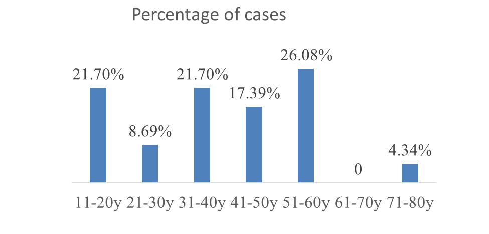
Fig 1: Distribution of cases in relation to the age
• Both sexes show almost equal predilection, 12 cases (52.17%) in male and 11(47.83%) in females (Table1).
Table 1: Frequency of lesions in relation to the sex
|
• Nature of lesion |
• Male • (no. of cases and percentage ) |
• Female • (no. of cases and percentage ) |
|
Non neoplastic |
• 3 (13.04%) |
• 2 (8.69%) |
|
• Hyperplasia and Hamartomas |
• 3 (13.04%) |
• 0 |
|
• Benign |
• 5 (21.73%) |
• 7 (30.4%) |
|
• Malignant |
• 1 (4.34%) |
• 2 (8.69%) |
Most of the lesions are seen on the head and neck site. Out of 23 cases, 11cases (47.82%) were identified on the head and neck site (Table 2).
Table 2: Distribution of lesion in relation to the site
|
Site |
Number of cases |
Percentage (%) |
|
Head &Neck |
11 |
47.82 |
|
Extremities |
8 |
34.78 |
|
Trunk &Abdomen |
4 |
17.40 |
|
Total |
23 |
100 |
Out of 23 cases diagnosed as skin adnexal lesions, 5 were non-neoplastic, 18 were neoplastic. Of the 5 non neoplastic lesions, 2 are cystic lesions and 3 are inflammatory lesions (Table 3). All 3 malignant lesions were seen in the 6th decade. Females however showed more neoplastic lesions.
Table 3: Nature of lesions in relation to percentage of cases
|
Nature of lesion |
No. of cases |
Percentage (%) |
|
Non neoplastic |
5 |
21.73 % |
|
Hyperplasia and hamartoma |
3 |
13.04 % |
|
Benign |
12 |
52.19% |
|
Malignant |
3 |
13.04 % |
|
Total |
23 |
100% |
In our study most of the lesion are from follicular differentiation. Out of 23cases, 9 cases (48%) are follicular differentiation. Least are apocrine differentiation lesions, 1 case (4%) [ Fig 2 and Table 4].
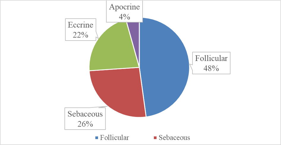
Fig 2: Frequency of lesions according to their differentiation
Table 4: Distribution of lesions in relation to line of differentiation
|
Line of differentiation |
Non neoplastic |
Hyperplasias and Hamartomas |
Benign |
Malignant |
|
Follicular(11) |
4(36.36%) |
1(9.09%) |
5(45.45%) |
1(9.09%) |
|
Sebaceous(6) |
1(16.66%) |
2(33.34%) |
1(16.66%) |
2(33.34) |
|
Eccrine(5) |
- |
- |
5(100%) |
- |
|
Apocrine (1) |
- |
- |
1(100%) |
- |
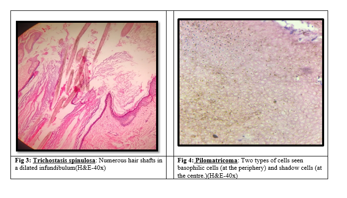
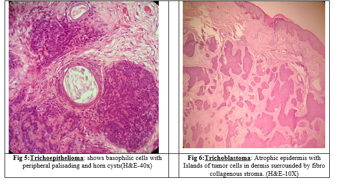
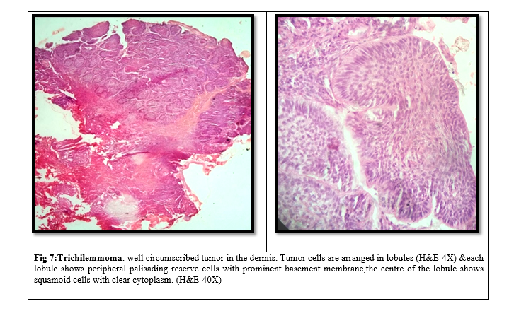
In our study the lesions of follicular differentiation are 11 in number and the conditions are Trichilemmal cyst, keratosis pilaris, Hidradenitis suppurativa, Trichostasis spinulosa (Fig3), Dilated pore of winer, Infundibular cyst, Trichoepithelioma(Fig5), Trichoblastoma(Fig6), Trichilemmoma(Fig7), Pilomatricoma(Fig4), Trichilemmal carcinoma.
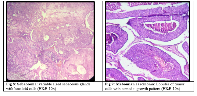
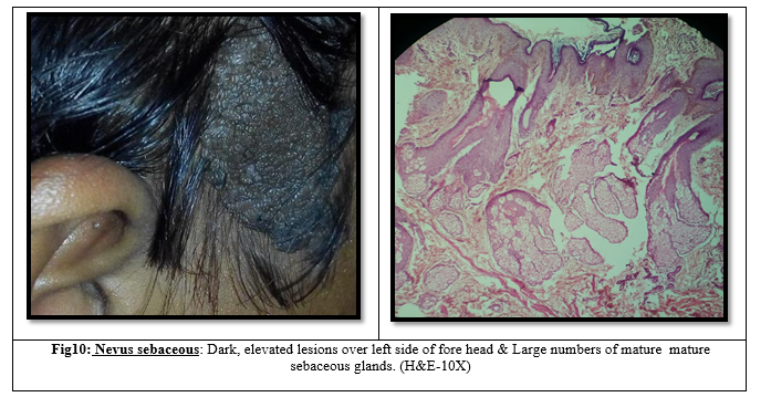
The conditions we got in present study that originated from sebaceous sgland are 6 in number and those are Steatocystoma Multiplex, Nevus Sebaceous of Jadassohn(Fig10), sebaceous hyperplasia, sebaceoma(Fig8), sebaceous carcinoma. A case of Mebomian carcinoma(Fig9), which is also Sebaceous carcinoma that occurs in the sebaceous glands of eye lid, was seen in present study.
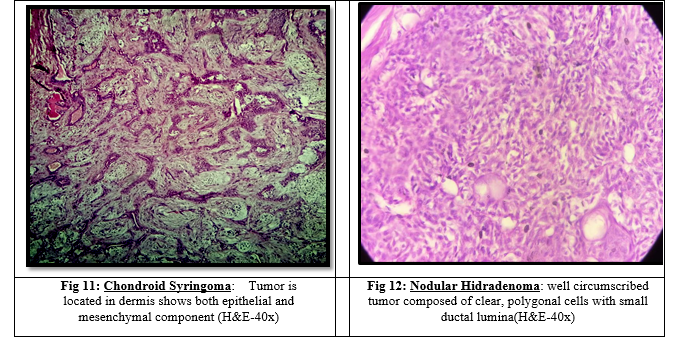
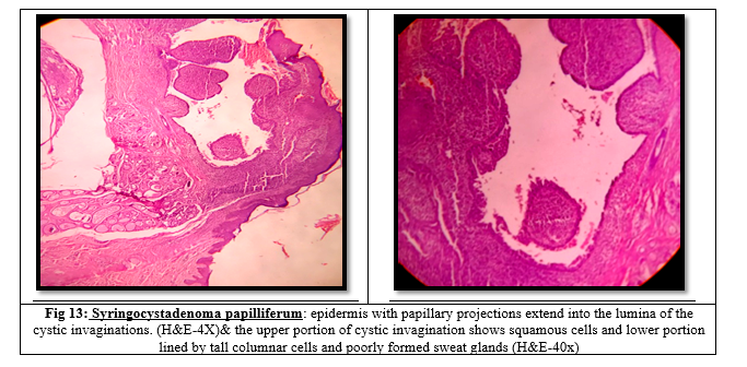
In present study the eccrine and apocrine differentiation conditions are 5 and 1 in number respectively and that lesions are Nodular hidradenoma(Fig12), Papillary eccrineadenoma, Eccrine poroma, chondroid syringoma(Fig11) and Eccrine syringofibradenoma, and Syringocystadenoma papilliferum(Fig13).
DISCUSSION:
The frequency of cutaneous adnexal lesions reported in the literature varies across studies, largely because of differences in study design, sample size, inclusion criteria, and histopathological classification. In the present study, 600 skin biopsy specimens were examined over a period of two years, of which 23 cases were identified as cutaneous adnexal lesions, yielding a prevalence of 3.83%. This finding is higher than that reported by Sejekan et al. [3], who observed a prevalence of 2.6%, and by Subrata Pal et al. [4], who reported a prevalence of 1.32%. The higher prevalence in the present study may be related to differences in case selection, referral pattern, and institutional load.
The mean age at presentation in the present study was 39.17 years, which is comparable to the observations of Sejekan et al. [3] and Subrata Pal et al. [4], who reported mean ages of 38.7 years and 38.4 years, respectively. This suggests that cutaneous adnexal lesions are more frequently encountered in middle-aged individuals. With regard to sex distribution, the present study showed a slight male predominance, with a male-to-female ratio of 1.09:1. This is in close agreement with the studies conducted by Ankith S et al. [10] and Divya et al. [5], who reported male-to-female ratios of 1.07:1 and 1.14:1, respectively.
The most common site of involvement in the present study was the head and neck region, accounting for 47.8% of cases. This observation is consistent with earlier studies by Divya et al. [5] and Sahu et al. [6], who also documented a predominance of lesions in the head and neck region, with frequencies of 73.3% and 60%, respectively. The predilection for this site may be explained by the relatively high concentration of pilosebaceous units and other adnexal structures in this anatomical region.
In the present series, cystic lesions constituted 8.69% of cases, which is comparable to the findings of Solomon et al. [11], who reported 7.6%, and Ankith S et al. [10], who reported 11.7%. Inflammatory lesions accounted for 13.04% of cases in the current study, showing a similar pattern to the observations of Neerja Puri et al. [12] and Moure et al. [14]. Hamartomas and hyperplasias also represented 13.04% of cases in the present study, which is close to the frequencies reported by Radhika et al. [13] at 14.2% and Solomon et al. [11] at 11.8%.
A major finding of the present study was that benign lesions were far more common than malignant lesions. This pattern is in accordance with the observations made by Sejekan et al. [3], Divya et al. [5], and Subrata Pal et al. [4], all of whom reported a predominance of benign adnexal lesions over malignant counterparts. This reinforces the established concept that most cutaneous adnexal lesions encountered in routine histopathological practice are benign in nature, while malignant adnexal tumors remain relatively uncommon.
With respect to line of differentiation, follicular differentiation was the most common in the present study, accounting for 47.8% of cases, followed by sebaceous differentiation. This finding is similar to that reported by Sejekan et al. [3]. In contrast, studies by Subrata Pal et al. [4] and Divya et al. [5] found eccrine differentiation to be the most common pattern. Such differences may reflect regional variation, small sample size, or institutional differences in the spectrum of cases received.
Overall, the present study highlights the diverse histomorphological spectrum of cutaneous adnexal lesions and confirms the importance of histopathological examination in their accurate classification. The frequent overlap in morphological features, together with the rarity of many of these entities, makes histopathology indispensable for precise diagnosis and for distinguishing benign lesions from malignant neoplasms.
CONCLUSION:
The diagnosis of cutaneous adnexal lesions remains challenging because of their wide histomorphological diversity, overlapping patterns of differentiation, and complex nomenclature. The present study demonstrated that benign adnexal lesions are more common than malignant lesions and that follicular differentiation constitutes the predominant line of differentiation in this series. Histopathological examination remains the gold standard for the diagnosis, classification, and assessment of biological behavior of these lesions. Accurate recognition of adnexal tumor-like lesions and tumors is essential not only for correct diagnosis but also for guiding appropriate clinical management and therapeutic intervention. A better understanding of these uncommon lesions can improve diagnostic accuracy and increase awareness of their significance in routine clinical practice.
REFERENCES:
1. David E. Elder et al; Lever’s Histopathology of the skin ;11th Edition; 2015; 1673-1813.
2. James W Peterson et al; Weedon’s Skin Pathology;5th Edition; 2021; 952-1000.
3. Sejekan SV, Biligi DS.Clinicopathological Study of Skin Adnexal Tumors with Special Emphasis on the Line of Differentiation. JMed Sci Health 2022; 8(1):8-13.
4. Subrata Pal, Tanushree Mondal, N S Jamadar, Sajeeb Mondal, Rajashree Pradhan, Clinico-pathological study of skin adnexal tumor – a retrospective review of 242 cases from a tertiary care hospital in India; International Journal of Health and Clinical Research, 2021;4(22):148-150.
5. Divya Tejaswi Gopidesi etal,Skin Adnexal Tumors: A Histopathological analysis of 30 Cases at a Tertiary Care Centre.Saudi J Pathol Microbiol 2020; 5(2):67-71.
6. Sahu, A., Sa, D. K., Naik, S. K., & Agrawal, K. C. (2018). Skin Adnexal Tumors: A histopathological study of 60 cases at a tertiary care centre. Annals of Pathology and Laboratory Medicine, 5(3), A215-220.
7. Samanta M, Mangal N, Bhavani K, Koteeswaran G, Parmar PC. Histopathological study of skin tumours. Trop J Pathol Microbiol. 2018;4(2):195–200.
8. Shukla P, Fatima U, Malaviya AK. Histomorphological and Immunohistochemical Reappraisal of Cutaneous Adnexal Tumours: A Hospital Based Study. Scientifica. 2016;2016:1–9.
9. Vaibhav B, Alka G, Prashant M, Kalpana S. SkinTumors-Histopathological review of 125 cases. Indian Medical Gazette. 2014;148(11):418–445.
10. Sharma A, Paricharak DG, Nigam JS, Rewri S, SoniPB, Omhare A, et al. Histopathological Study of SkinAdnexal Tumours—Institutional Study in South India.Journal of Skin Cancer. 2014:1–4.
11. Solomon R, Yusuf I, Ochicha O. Skin adnexal tumours in Kano, Northern Nigeria. Niger J Basic Clin Sci 2015;12:51-4.
12. Neerja Puri a clinical and histopathological study of cicatricial alopecias Journal of Pakistan Association of Dermatologists 2013;23 (3):272-276.
13. K. Radhika, B. V. Phaneendra, N. Rukmangadha, and M. K. Reddy, “A study of biopsy confirmed skin adnexal tumors: experience at a tertiary care teaching hospital,” Journal of Clinical and Scientific Research, vol. 2, pp. 132–138, 2013.
14. Moure ERD, Romiti R, Machado MCMR, Valente NYS. Primary cicatricial alopecias: a review of histopathologic findingsin 38 patients from a clinical university hospital in São Paulo, Brazil. Clinics. 2008;63:747-52.