Outcome of neonates who presented with Infantile hypertrophic pyloric stenosis (IHPS) in a tertiary care hospital: A Retrospective study.
- Prashant K Zulpi. , Associate Professor, Dept of Pediatric Surgery. SDM College Of Medical Science And Hospital. Sattur , Dharwad. India.
- Gururaj Bangari. , Assistant Professor, Department Of Radiology, SDM College Of Medical Science And Hospital, Sattur, Dharwad India.
- Aishwaraya Manthale. , Senior Resident, Dept of Paediatrics East Point College Of Medical Sciences And Research Centre India..
Article Information:
Abstract:
Background: Infantile hypertrophic pyloric stenosis (IHPS) is one of the most common cause of gastric outlet obstruction in infants with incidence is approximately 1–4 per 1000 live with male to female ratio of 4:1. Most of the time treatment is delayed due to delay in the diagnosis, especially in peripheries. The purpose of this study was to document our experience in management of pyloric stenosis in neonates within our setting. Materials and Methods: This is a retrospective observational study comprising of 24 neonates with IHPS. All neonates were initially evaluated with ultrasound (USG) abdomen to confirm the diagnosis. After resuscitation all neonates were treated surgically with Ramstedt's Pyloromyotomy. All neonates were followed up postsurgery. Results: This study comprised of 24 neonates with IHPS. Out of the 24 Neonates enrolled in this study 16 (66%) were males and 8 (33%) were females with a M: F ratio of 2:1. All neonates presented with nonbilious vomiting with 3 presented as abdominal distension, 2 presented as palpable olive, 2 with visible gastric peristalsis, 1 refusal of feed, 1 decreased urine output, 1 palpable olive and 1 with fever. Most of the neonates had metabolic alkalosis and dyselectrolytemia at presentation. All the patients underwent Ramstedt pyloromyotomy with good outcome and no single mortality. Conclusion: Management of IHPS has very good outcomes in our setting. Earlier referral of patients leads to improved outcomes.
Keywords:
Article :
INTRODUCTION:
Infantile hypertrophic pyloric stenosis (IHPS) is the most common surgical condition of vomiting in infants (1). It is characterized by hypertrophy of the circular muscle of the pylorus, causing pyloric channel narrowing and elongation leading to gastric outlet obstruction.(2) The incidence of HPS is reported to be between 1-4 cases per 1000 live births(3). First born male children are most commonly affected with male to female ratio of 4 to 5:1(4).The exact etiology of pyloric stenosis is not well-defined but is likely multifactorial with genetic and environmental factors both playing a role. The usual onset of symptoms occurs between 3 and 6 weeks of age; however, it can present earlier. Triad of presentation includes non-bilious projectile vomiting, visible gastric peristalsis and a palpable epigastric olive mass. They mainly present with non-bilious vomiting and is often accompanied by severe electrolyte derangements, mainly hypochloremic, hypokalemic metabolic acidosis. (5) IHPS is mainly diagnosed by abdominal ultrasound which shows thickened and elongated pyloric muscle. After adequate resuscitation, surgical treatment with a Ramstedt pyloromyotomy is the treatment of choice with good outcome.
MATERIALS AND METHODS:
A retrospective observational study was conducted in a tertiary care hospital in neonates diagnosed with hypertrophic pyloric stenosis over a period of 6 years from January 2020 to December 2025. Patients who presented beyond neonatal period were excluded. The data of neonates with respect to gender, weight and symptoms were tabulated. The diagnosis of IHPS was made clinically by the typical clinical presentation of non-bilious vomiting and palpable pyloric olive mass and by abdominal ultrasound. Routine blood investigations and blood gas analysis were done to look for dyselectrolytemia and metabolic alkalosis. Accordingly neonates were initially resuscitated with standard intravenous fluid regimes. After resuscitation all neonates underwent Pyeloromyotomy (both open and laparoscopic). Post operatively started feeds within 24 hours. Patients were discharged once tolerating orally, passing urine and stool adequately. We considered more than 05 days of hospital stay after surgery as prolonged hospital stay, similarly prolonged symptoms for more than 05 days and prolonged resuscitation for more than 03 days. All patients were followed in pediatric surgical outpatient department (OPD) to look for feeding tolerance, surgical site and weight gain.
RESULTS:
The study comprised of 24 neonates with IHPS. Out of the 24 Neonates enrolled in this study 18 (75%) were males and 6 (25%) were females with a M: F ratio of 3:1. All patients presented with nonbilious vomiting (100%), Five with dehydration, two presented with palpable olive, visible gastric peristalsis and abdominal distension, 01 each with fever, refusal of feed and decreased urine output. (Table 1). The majority (66%) of our patients had electrolyte imbalance at presentation (Table 2). Of those with electrolyte imbalance, 50% had abnormalities in more than one electrolyte. Sodium (12) and Chloride (11) imbalance was the most common among the patients seen. Hypokalemia was seen in only 2 patients. Majority of patients (71%) had metabolic alkalosis (mild to severe) at presentation but only 6 had severe alkalosis with pH ≥ 6. Around one third (33%) patients did not had any electrolyte imbalance at the time of presentation.
Table 1: Distribution of patients according to presentation.
|
Presentation |
Number (%) |
|
Non bilious vomiting |
24 (100%) |
|
Dehydration |
05 (21%) |
|
Visible gastric peristalsis (VGP) |
02 (8%) |
|
Palpable olive |
02 (8%) |
|
Abdominal distension |
03 (12.5%) |
|
Fever |
01 (4%) |
|
Decreased urine output |
01 (4%) |
|
Refusal of feed |
01 (4%) |
Table 2: Distribution of electrolyte disturbance at presentation
|
Electrolyte disturbance |
Number of patients |
|
Hyponatremia |
05 (20.8%) |
|
Hyochloremia |
03 (12.5%) |
|
Hyponatremia + Hypochloremia |
06 (25%) |
|
Hyponatremia + Hypokalemia + Hypochloremia |
01 (4%) |
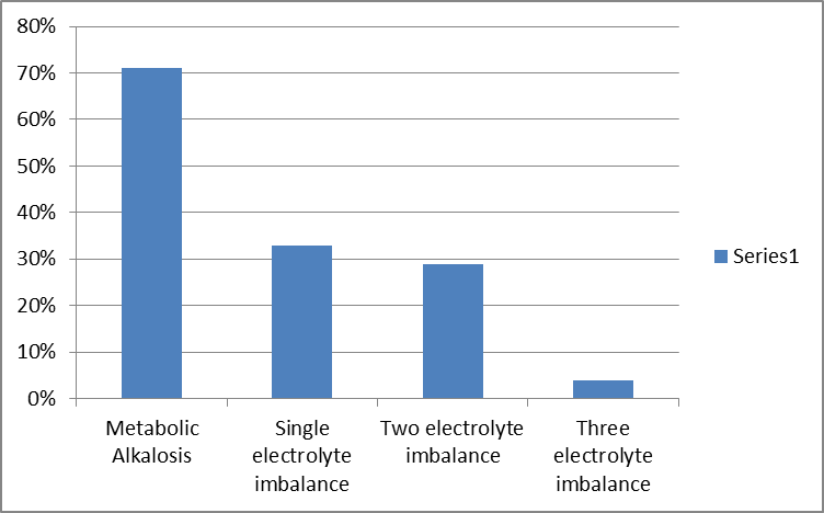
Figure 1: Distribution metabolic alkalosis and electrolyte imbalance graphically.
Outcome analysis:
All the patients in this study were surgically managed by pyloromyotomy following resuscitation with 100% good outcome without single mortality. The mean duration for resuscitation was 2.8 days ranging from 1 to 7 days. During this period fluid, electrolyte and metabolic derangements were corrected. Around 07 patients (29%) developed post operative complications. Post operative complications include surgical site infection, intraoperative mucosal perforation, Pneumonia, sepsis (blood culture positive with staphylococcus aureus), dyselectrolytemia, drop in haemoglobin.( Table 3) One patient with surgical site infection required minimal debridement and secondary suturing.
Among 24 neonates operated, 06 (25%) underwent laparoscopic surgery and 18(75%) underwent by open technique. The mean post operative hospital stay in laparascopy patients was 04 days as compared to 08 days in patients underwent surgery by open technique. (Table 4).
13 patients had prolonged post operative hospital stay as per predefined criteria in methodology. Among these 13 patients 09 had prolonged duration of symptoms, 08 with dyselectrolytemia, 06 showing higher sonologic pyloric length (around 20mm), 06 with metabolic alkalosis, 04 with post operative complications, 01 with deranged RFT (Renal function test).
Table 3: Distribution of patients with post operative complications
|
Number of patients (%) |
Post operative complication |
|
02 (8.3%) |
Surgical site infection |
|
01 (4%) |
Mucosal injury |
|
01 (4%) |
Pneumonia |
|
01 (4%) |
Sepsis |
|
01 (4%) |
Drop in Haemoglobin |
|
01 (4%) |
Dyselectrolytemia |
Table 4: Mean post operative hospital stay between open and Laparoscopic Pyloromyotomy
|
Surgery |
Number of patients |
Mean post operative stay |
P Value
|
|
Open |
18 (75%) |
08 Days |
|
|
Laparoscopic |
06 (25%) |
04 Days |
DISCUSSION:
Since Harald Hirschsprung first described IHPS in 1888. It has been one of the most common cause of gastric outlet obstruction in infant and the most common surgical emergency in a newborn [3]. The reason for male preponderance is not well understood though authors like Carter and Evans defined the disease as a “paradigm for the multifactorial, sex-modified threshold model of inheritance.[9]”. In our studies male neonates were affected more commonly than female neonates. These findings were in conformity with the studies conducted by A C. Paul et al [4], Salman Al-Ghazwany et al[5] and Amier. A.Ejrish et al [6].
In our study most of the neonates presented with nonbilious vomiting and dehydration with visible gastric peristalsis and palpable olive mass . These findings were similar to the studies conducted by Kumar A et al [7] and Phillipo L Chalya et al [8].
The clinical features of IHPS include forceful or projectile non-bilious vomiting, visible peristalsis and a palpable lump in right upper quadrant. In our study all the neonates with IHPS had projectile non-bilious vomiting and around 8% had visible peristalsis and palpable olive mass each. 3% had abdominal distention. And decreased urine output with refusal of feed was found in 1% infants. These findings were similar to the studies conducted by JD Ranells et al[9].
Hyponatremia, hypernatremia and hypokalemia was seen in 54%, 5% and 8.3% patients respectively. Similar findings were reported by NMadu PT, Spicer RD and JG Tutay [10,11,12].
The ultrasonography of abdomen is a sensitive and specific test for diagnosis of IHPS. In this study ultrasonographic finding of pyloric thickness of 3-5mm and length of 12-20mm is 54% and 46% presented with 5-7mm,21-30mm.These findings were similar to the findings of the authors like Meena Said et al, M Hussain et al and Sílvia Costa Dias et al [13,14,15].
In our study the most common IV fluid used in pre-operative period 5%D with ½ NS which was used in 50% neonates while rest of the infants received a combination of Isolyte-p and either Ringer’s lactate or DNS. This management was similar to Shwartz who used 5 % dextrose with 1⁄2 normal saline and Hisham Nazerwho used Ringer’s lactate to correct dehydration and metabolic disturbances [16,17].
In our study patients of IHPS 18(75%) neonates underwent classical Ramstedt's pyloromyotomy, and 6 (25%) neonates managed Laproscopically . They were managed postoperatively by IV Isolyte p and lactated ringer with prophylactic antibiotics. Feeding was started within 72 hours in all neonates. Similar post-operative feeding pattern were seen in study conducted by Carpenter RO, G Aspelund and Georgeson KE[18,19,20].surgical site inection(8.3%),mucosal injury(4%), pneumonia(4%),sepsis (4%), drop in haemoglobin (4%), dyselecrolytemia(4%), were the most common complications seen postoperatively. Similar complication rates were found in study done by Solowiejczyk M et al and Gibbs MK et al [21].
CONCLUSION:
Early recognition of IHPS, prompt correction of fluid and electrolyte imbalances, and early referral from peripheral centres are crucial to prevent morbidity.
REFERENCES:
1. Hirschsprung H. Falle von angeborener pyloric stenose. Jb Kinderheik. 1888;27:61
2. Ohshiro K, Puri P. Pathogenesis of infantile hypertrophic pyloric stenosis: recent progress. PediatrSurg Int. 1998 Apr;13(4):243-52.
3. Tan H, Roy P, Lakhoo K. Bilious vomiting in infantile hypertrophic pyloric stenosis. Afr J Paediatr Surg. 2007;4(2):101–2.
4. A.C Paul , MJ Alam IHPS Faridpur Med Coll.J.2011;6 (1); 01-04 .
5. Salman Al Ghawzwany , Infantile hypertrophic pyloric stenosis, Annals pediatric surgery 2012 vol 8 no 2 ; 39-42.
6. Amier A Ejrish IHPS, MEDICAL JOURNAL of BABYLON ; 2014; Vol 6 (3) 596-599.
7. Kumar A et al , study of clinical, biochemical evaluation and outcomes in hypertrophic pyloric stenosis , Int J ContempPediatr. 2016 may ; 3 (2) 473-476
8. Phillipo L Chalya, Mange Manyama, Neema M. Kayange , Infantile pyloric stenosis BMC Res Notes 2015 ; 8: 690 Mitchell LE, Risch N. The genetics of infantile hypertrophic pyloric stenosis A reanalysis. Am J Dis Child. 1993 Nov;147(11):1203-11.
9. Nmadu PT. Alterations in serum electrolytes in congenital hypertrophic pyloric stenosis: a study in Nigerian children. Ann Trop Paediatr 1992; 12:169.
10. Spicer RD. Infantile hypertrophic pyloric stenosis: a review. Br J Surg 1982; 69:128.
11. Tutay GJ, Capraro G, Spirko B, Garb J, Smithline H. Electrolyte profile ofpediatric patients with hypertrophic pyloric stenosis. PediatrEmerg Care. 2013 Apr;29(4):465-8.
12. Said M, Shaul DB, Fujimoto M, Radner G, Sydorak RM, Applebaum H. Ultrasound Measurements in Hypertrophic Pyloric Stenosis: Don’t Let the Numbers Fool You. The Permanente Journal. 2012;16(3):25-27.
13. Hussain M. Sonographic Diagnosis of Infantile Hypertrophic Pyloric stenosis Use of Simultaneous Grey-scale &Colour Doppler Examination. International Journal of Health Sciences. 2008;2(2):134140.
14. Costa Dias S, Swinson S, Torrão H, et al. Hypertrophic pyloric stenosis: tips and tricks for ultrasound diagnosis. Insights into Imaging. 2012;3(3):247-250.
15. Schwartz MZHypertrophic pyloric stenosis in paediatric surgery. 1111-17.
16. HishamNazer Allan C. Terminantsog good outcome in pyloric stenosis. JPS 2006:42(3):86-8.
17. Carpenter RO, Schaffer RL, Maeso CE, Sasan F, Nuchtern JG, Jaksic T, Harber FJ, Wesson DE, Brandt ML. Postoperative ad lib feeding for hypertrophic pyloric stenosis. J Pediatr Surg. 1999 Jun;34(6):959-61.
18. Aspelund G, Langer JC. Current management of hypertrophic pyloric stenosis. SeminPediatr Surg. 2007 Feb; 16(1):27-33. Review. PubMed PMID: 17210480.
19. Georgeson KE, Corbin TJ, Griffen JW, Breaux CW Jr. An analysis of feeding regimens after pyloromyotomy for hypertrophic pyloric stenosis. J Pediatr Surg. 1993 Nov;28(11):1478-80
20. Solowiejczyk M, Holtzman M, Michowitz M. Congenital hypertrophic pyloric stenosis: a long-term follow-up of 41 cases. Am Surg. 1980 Oct;46(10):567-71.
21. Gibbs MK, Van Herrden JA, Lynn HB. Congenital hypertrophic pyloric stenosis. Surgical experience. Mayo Clin Proc. 1975 Jun;50(6):312-6.