Co-existence of Psoriasis vulgaris and Bullous Pemphigoid
- Prachi Bhatt , Senior Resident, Department of Pathology, PDUMC, Rajkot.
- Priyanka Gangani , Senior Resident, Department of Pathology, PDUMC, Rajkot.
- Rohit Bhalara , Associate Professor Department of Pathology Rajkot
- Shruti Korvadiya , Assistant Professor Department of Pathology Rajkot.
- Manisha Popat , Tutor Department of Pathology Rajkot.
- Gauravi Dhruva , Professor and Head, Department of Pathology, PDUMC, Rajkot.
Article Information:
Abstract:
Background: Psoriasis vulgaris and bullous pemphigoid (BP) are chronic inflammatory dermatoses with distinct pathogenetic mechanisms. Their coexistence in a single patient is uncommon. Case Report: We report a 70-year-old male with a 15-year history of psoriasis vulgaris who presented with generalized pruritic bullous lesions of recent onset. Clinical examination revealed tense bullae and erosions over both psoriatic plaques and normal skin, along with oral mucosal involvement. Two skin punch biopsies were obtained from a bullous lesion and a psoriatic plaque. Histopathology from the bullous lesion demonstrated a subepidermal blister with a prominent eosinophil-rich inflammatory infiltrate, while the plaque biopsy showed classical features of psoriasis vulgaris. These findings confirmed the coexistence of psoriasis vulgaris and bullous pemphigoid. Conclusion: The association of BP with long-standing psoriasis, although rare, should be considered when new blistering lesions arise in psoriatic patients. Histopathological evaluation from representative sites is essential for accurate diagnosis.
Keywords:
Article :
INTRODUCTION:
Psoriasis is a chronic immune-mediated inflammatory disorder that predominantly affects the skin and joints. It is frequently associated with a broad spectrum of cutaneous and systemic comorbidities. Epidemiological studies have demonstrated that patients with psoriasis carry an increased risk of cardiovascular disease and metabolic abnormalities. Besides these systemic associations, various dermatological conditions may also occur in conjunction with psoriasis. However, the subsequent development of autoimmune blistering disorders in individuals with psoriasis is rare.
CASE REPORT:
Clinical History
A 70-year-old male presented with multiple fluid-filled lesions over the body for 20–25 days, associated with intense pruritus. The lesions initially appeared on the arm and subsequently involved other parts of the body. He also complained of painful oral ulcers with dysphagia. The patient had a known history of psoriasis vulgaris for 15 years.
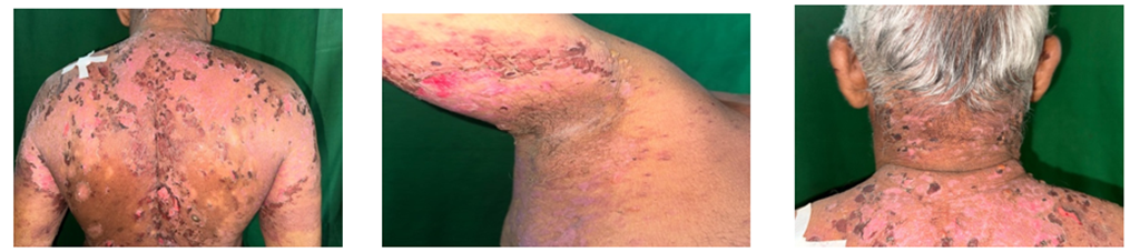
Cutaneous examination revealed multiple tense bullae and crusted erosions over the upper and lower limbs [Figure 1]. Oral examination showed erosions over the right buccal mucosa. Nikolsky’s sign and bulla spread sign were negative. In addition, well-defined hyperpigmented scaly plaques were present over the trunk, extremities, and scalp. Bullae were noted both on pre-existing psoriatic plaques and on clinically normal skin [Figure 2].
A clinical diagnosis of psoriasis vulgaris with suspected bullous pemphigoid was considered. Routine hematological and biochemical investigations were within normal limits.
Gross Findings
Two skin punch biopsy specimens were received in formalin.
Specimen A: From a bullous lesion on the upper back, measuring 0.5 × 0.5 × 0.3 cm, greyish-white, soft to firm.
Specimen B: From a plaque on the lower back, measuring 0.5 × 0.5 × 0.3 cm, greyish-white, soft to firm.
Microscopic Findings
Sections from Specimen A (bullous lesion) showed epidermis with mild spongiosis and scattered inflammatory cells. A focal subepidermal bulla containing eosinophilic proteinaceous material was identified. The underlying dermis exhibited a dense inflammatory infiltrate composed predominantly of eosinophils and neutrophils. Perivascular inflammatory infiltrates consisting mainly of eosinophils and neutrophils, along with occasional lymphocytes, were also present. Sections from Specimen B (plaque) demonstrated features consistent with psoriasis vulgaris. The epidermis showed hyperkeratosis with parakeratosis, psoriasiform acanthosis, and suprapapillary thinning. The dermal papillae revealed dilated and proliferating capillaries. The papillary dermis showed perivascular inflammatory infiltrates predominantly composed of lymphocytes with occasional neutrophils.
Histopathological Impression
The histomorphological features from the two biopsy sites are consistent with psoriasis vulgaris coexisting with bullous pemphigoid.
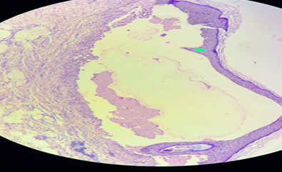
Subepidermal bullous lesion with secretory material inside.[ H&E,40x]
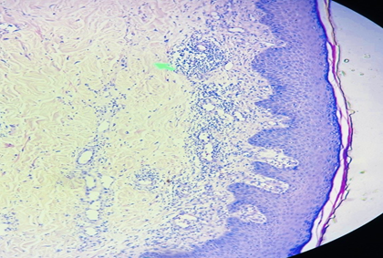
Blunting of rete ridges and dilated capillaries and lymphocyticinfiltration in papillary dermis.[ H&E,40x]
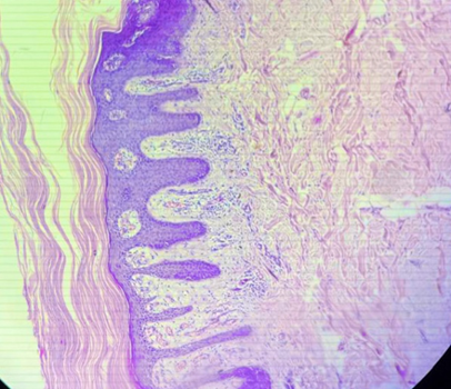
Regular acanthosis of epidermis with focal neutrophilic collection.[ H&E,40x]
DISCUSSION:
Psoriasis vulgaris and bullous pemphigoid (BP) are chronic inflammatory dermatoses with distinct pathogenetic mechanisms, yet their coexistence in the same patient has been described in a limited number of reports. Psoriasis is driven predominantly by T-cell–mediated inflammation and epidermal hyperproliferation, whereas BP is an autoantibody-mediated subepidermal blistering disorder directed against structural proteins of the basement membrane zone (BMZ). Despite these differences, epidemiological observations suggest that patients with psoriasis may have an increased predisposition to autoimmune blistering diseases [1–3].
Several bullous disorders have been reported in association with psoriasis, including pemphigus vulgaris, pemphigus foliaceus, linear IgA bullous dermatosis, cicatricial pemphigoid, and epidermolysis bullosa acquisita. Among these, BP appears to be the most frequently encountered entity [4–7]. Population-based studies indicate that the incidence of BP is higher in individuals with psoriasis compared with the general population, suggesting that the association may not be merely coincidental [4,5].
Clinically, BP occurring in psoriatic patients usually affects elderly individuals and often develops after a prolonged duration of psoriasis. In most reported cases, psoriasis precedes the onset of BP by several years or decades [8]. The present case follows a similar pattern, with bullous lesions appearing in a patient with long-standing psoriasis vulgaris.
The mechanism underlying this association remains uncertain and is likely multifactorial. One proposed explanation involves the role of antipsoriatic therapies, such as phototherapy or systemic agents, which may alter immune regulation or damage the dermoepidermal junction, thereby facilitating autoantibody formation [6]. However, the occurrence of BP in untreated patients argues against therapy as the sole causative factor [6,9].
Chronic inflammation in psoriasis may itself contribute to structural and immunological changes at the BMZ. Persistent infiltration by activated lymphocytes, increased cytokine activity, and enhanced antigen presentation can lead to disruption of the dermoepidermal junction, exposing normally concealed antigens such as BP180 and BP230. Subsequent autoantibody formation against these components can result in subepidermal blistering [10,13].
Ultrastructural studies have demonstrated abnormalities of the basal lamina in psoriatic skin, including focal discontinuity and duplication, which may increase susceptibility to blister formation once an autoimmune response develops [13]. Another widely accepted mechanism is epitope spreading, wherein ongoing tissue damage exposes previously hidden antigens, triggering a secondary autoimmune response directed against new targets [14]. This concept provides a plausible explanation for the emergence of BP in chronic inflammatory dermatoses such as psoriasis.
Genetic susceptibility and shared immune pathways may also play a role, as both diseases involve dysregulated immune activation and cytokine-mediated inflammation [11,12]. Recognition of this association is clinically important because the appearance of tense bullae in a patient with longstanding psoriasis should prompt evaluation for an autoimmune blistering disorder.
Histopathological examination remains central to diagnosis, supported by immunofluorescence studies where available [15,16]. In the present case, biopsy from a bullous lesion revealed a subepidermal blister with eosinophil-rich inflammation consistent with BP, while biopsy from a plaque showed classical features of psoriasis vulgaris, confirming the coexistence of both conditions.
CONCLUSION:
The coexistence of bullous pemphigoid in a patient with long-standing psoriasis vulgaris is uncommon and may be easily overlooked. This case underscores the need for careful evaluation when a known psoriatic patient develops new-onset blistering lesions. Early recognition, supported by histopathological examination, is essential to establish the correct diagnosis and guide appropriate treatment.
Acknowledgment
The authors acknowledge the Department of Dermatology and the technical staff of the Department of Pathology, P.D.U. Government Hospital, Rajkot, for their assistance in the clinical evaluation and processing of biopsy specimens.
Financial Support and Sponsorship
Nil.
Conflicts of Interest
There are no conflicts of interest.
Author Contributions
All authors contributed to the conception, clinical evaluation, histopathological analysis, drafting, and final approval of the manuscript. All authors agree to be accountable for all aspects of the work.
REFERENCES:
1. Griffiths CEM, Barker JNWN. Pathogenesis and clinical features of psoriasis. Lancet. 2007;370(9583):263-71. doi:10.1016/S0140-6736(07)61128-3
2. Nestle FO, Kaplan DH, Barker J. Psoriasis. N Engl J Med. 2009;361(5):496-509. doi:10.1056/NEJMra0804595
3. Parisi R, Symmons DPM, Griffiths CEM, Ashcroft DM. Global epidemiology of psoriasis. J Invest Dermatol. 2013;133(2):377-85. doi:10.1038/jid.2012.339
4. Kridin K, Bergman R. Association between bullous pemphigoid and psoriasis. J Am Acad Dermatol. 2017;77(2):370-372. doi:10.1016/j.jaad.2017.03.035
5. Langan SM, Groves RW, West J. The relationship between pemphigoid and psoriasis. Br J Dermatol. 2010;163(3):525-530. doi:10.1111/j.1365-2133.2010.09868.x
6. Stavropoulos PG, Soura E, Antoniou C. Drug-induced pemphigoid. J Eur Acad Dermatol Venereol. 2014;28(9):1133-1140. doi:10.1111/jdv.12366
7. Cozzani E, Gasparini G, Parodi A. Autoimmune blistering diseases and psoriasis. G Ital Dermatol Venereol. 2016;151(4):437-445. doi:10.23736/S0392-0488.16.05286-3
8. Kridin K, Ludwig RJ. The growing incidence of bullous pemphigoid. Front Med (Lausanne). 2018;5:220. doi:10.3389/fmed.2018.00220
9. Amber KT, Hertl M. Treatment options for bullous pemphigoid. Expert Opin Pharmacother. 2018;19(2):183-192. doi:10.1080/14656566.2017.1412403
10. Schmidt E, Zillikens D. Pemphigoid diseases. Lancet. 2013;381(9863):320-332. doi:10.1016/S0140-6736(12)61140-4
11. Borradori L, Van Beek N, Feliciani C, et al. Updated S2K guidelines for BP. J Eur Acad Dermatol Venereol. 2022;36(10):1689-1704. doi:10.1111/jdv.18274
12. Di Zenzo G, Della Torre R, Zambruno G, Borradori L. Bullous pemphigoid pathogenesis. Clin Rev Allergy Immunol. 2012;42(1):3-16. doi:10.1007/s12016-010-8210-4
13. Tsuruta D, Hashimoto T. Immunopathogenesis of bullous pemphigoid. J Dermatol Sci. 2012;66(1):1-8. doi:10.1016/j.jdermsci.2012.02.003
14. Vanderlugt CL, Miller SD. Epitope spreading in immune-mediated diseases. Nat Rev Immunol. 2002;2(2):85-95. doi:10.1038/nri724
15. Schmidt E, Zillikens D. Diagnosis of autoimmune blistering diseases. Dermatol Clin. 2011;29(3):427-438. doi:10.1016/j.det.2011.03.003
16. Chan LS, Ahmed AR, Anhalt GJ, et al. The first international consensus on BP diagnosis. Arch Dermatol. 2002;138(3):370-379. doi:10.1001/archderm.138.3.370