Expression of immunohistochemical markers ki-67 and p16 in cervical intraepithelial neoplasia and carcinoma cervix.
- Sweta Sinha , Department of Pathology, Shri Atal Bihari Vajpayee, Government Medical College, Chhainsa, Faridabad, Haryana, India.
- Sana Tafseer , Department of Pharmacology, Shri Atal Bihari Vajpayee, Government Medical College, Chhainsa, Faridabad, Haryana, India.
- Naghma Anjum , Department of Pathology, Shri Guru Gobind Singh Tricentenary University, Gurugram, Haryana, India.
Article Information:
Abstract:
Background: Cervical cancer remains a leading cause of cancer-related morbidity and mortality among women, particularly in low- and middle-income countries. Persistent infection with high-risk human papillomavirus (hrHPV) plays a central role in cervical carcinogenesis. While histopathology is the diagnostic gold standard, interobserver variability and limitations in distinguishing equivocal lesions necessitate the use of adjunct biomarkers such as p16 and Ki-67 to improve diagnostic accuracy. Aims and Objectives: To evaluate and correlate the expression of p16 and Ki-67 in cervical intraepithelial neoplasia (CIN) and carcinoma cervix. Materials and Methods: A cross-sectional study was conducted on 50 cervical biopsy and hysterectomy specimens diagnosed as CIN or carcinoma cervix. Routine histopathological evaluation was performed using haematoxylin and eosin staining. Immunohistochemistry (IHC) for p16 and Ki-67 was carried out on formalin-fixed paraffin-embedded tissues. Expression was graded based on staining intensity and proportion, and statistical analysis was performed using SPSS version 21, with p < 0.05 considered significant. Results: The mean age of patients was 36.3 ± 8.74 years, with the majority in the 31–40-year age group. CIN I was the most common lesion (40%), followed by carcinoma cervix (32%). Ki-67 expression showed a progressive increase with lesion severity, with high scores predominantly in CIN III and carcinoma. Similarly, p16 expression showed positive correlation with lesion grade, showing higher expression in high-grade lesions and carcinoma. Ki-67 demonstrated higher sensitivity (92.31%) and specificity (100%) compared to p16 (sensitivity 91.67%, specificity 80%). Combined use of both markers improved diagnostic accuracy. Conclusion: Ki-67 and p16 are valuable adjunct biomarkers in the evaluation of cervical lesions. Their combined use enhances diagnostic accuracy, particularly in distinguishing low-grade from high-grade lesions and in resolving equivocal cases.
Keywords:
Article :
INTRODUCTION:
Cervical cancer remains a major global public health concern and is among the most common malignancies affecting women worldwide. According to GLOBOCAN 2020 estimates, approximately 604,000 new cases and 342,000 deaths occur annually, making it the fourth most common cancer in women after breast, colorectal, and lung cancers.¹ A disproportionate burden of disease is observed in low- and middle-income countries (LMICs), which account for nearly 85–90% of both incidence and mortality. In India, cervical cancer constitutes a significant proportion of female malignancies and continues to be a leading cause of cancer-related morbidity and mortality.²
Persistent infection with high-risk human papillomavirus (hrHPV) is now well established as the necessary etiological factor in the development of cervical cancer. Among the numerous HPV types, approximately 15 are classified as high-risk, with types 16 and 18 accounting for the vast majority of cervical carcinoma cases.³,⁴ HPV infection is highly prevalent, with more than 80% of sexually active women acquiring the infection at some point in their lifetime. However, only a small proportion of these infections persist, and it is this persistence that plays a critical role in the progression to cervical intraepithelial neoplasia (CIN) and invasive carcinoma.⁵
Most HPV infections are transient, with nearly 90% clearing spontaneously within two years due to host immune responses.⁶ Nevertheless, in a subset of women, the virus may persist or remain latent within basal epithelial cells, with the potential for later reactivation and progression to malignancy.
Importantly, cervical cancer is largely preventable through effective screening and early intervention strategies. The introduction of cytological screening using the Papanicolaou (Pap) smear has significantly reduced the incidence and mortality of cervical cancer, particularly in developed countries.⁷ Despite its success, conventional cytology has inherent limitations, including suboptimal sensitivity, sampling errors, and inter- and intra-observer variability. The sensitivity of Pap smear for detecting high-grade squamous intraepithelial lesions (HSIL) has been reported to be moderate, leading to false-negative results. Furthermore, false-positive interpretations may occur due to reactive or inflammatory changes, resulting in unnecessary follow-up procedures and overtreatment.⁸
Histopathological examination of cervical biopsy specimens remains the gold standard for diagnosis; however, it is also subject to variability among observers, especially in borderline or equivocal lesions. These limitations highlight the need for adjunctive biomarkers that can enhance diagnostic accuracy and improve reproducibility in cervical cancer screening and diagnosis.⁸,⁹
At the molecular level, HPV-mediated carcinogenesis is driven primarily by the viral oncoproteins E6 and E7, which interfere with key tumour suppressor pathways. The E6 protein promotes degradation of p53, while E7 binds to and inactivates the retinoblastoma (Rb) protein, leading to dysregulation of the cell cycle. This inactivation results in increased expression of p16INK4a, a cyclin-dependent kinase inhibitor, due to disruption of the Rb pathway. Overexpression of p16INK4a is therefore considered a surrogate marker of oncogenic HPV activity. Additionally, Ki-67, a nuclear protein associated with cellular proliferation, is expressed in actively dividing cells and is frequently upregulated in dysplastic and neoplastic cervical epithelium.⁹,¹⁰
In view of these molecular alterations, immunohistochemical markers such as p16INK4a and Ki-67 have emerged as valuable adjuncts in the evaluation of cervical lesions. These markers can improve diagnostic precision, particularly in cases with ambiguous histopathological findings, and may aid in distinguishing reactive changes from true neoplastic transformation.
The present study was undertaken to evaluate the expression of p16INK4a and Ki-67 in cervical intraepithelial neoplasia and carcinoma using immunohistochemistry on formalin-fixed paraffin-embedded biopsy specimens, with the objective of assessing their role in enhancing diagnostic accuracy in equivocal cases.
AIMS AND OBJECTIVES
Aims:
• To correlate expression of p16 and ki-67in intraepithelial neoplasia and carcinoma cervix.
Objectives:
• Histological examination and grading of cervical intra-epithelial neoplasia and carcinoma cervix in cervical biopsies and hysterectomy specimens.
• To study and correlate ki-67 and p16 expression in premalignant lesions and carcinoma cervix.
MATERIALS AND METHODS:
STUDY PLACE:
The study was conducted in the Department of Pathology, Shree Guru Gobind Singh Tricentenary Hospital, Gurugram, on cervical biopsies and hysterectomy specimens received from the Department of Obstetrics and Gynecology.
STUDY DESIGN:
It was a cross-sectional study
DURATION OF STUDY:
One and a half year
SAMPLE SIZE:
50 cervical biopsies and hysterectomies of premalignant and malignant lesions was included the study aims to evaluate the degree of expression of ki-67 and p16 in cervical intraepithelial lesions and carcinoma cervix.
STUDY POPULATION
50 hysterectomies or cervical biopsy specimens diagnosed as CIN or carcinoma cervix was included in the study.
INCLUSION CRITERIA
Cervical biopsies/ hysterectomy specimens diagnosed as cervical intraepithelial neoplasia (CINI, CINII, CINIII) or cervical carcinoma on hemotoxylin and eosin staining was included.
EXCLUSION CRITERIA
1. ¬Patient with cervicitis
2. Mesenchymal lesions of cervix
3. Recurrence of malignancy
4. Patient under radiotherapy
CLINICAL DETAILS:
Routine clinical details and investigation was recorded as per proforma attached.
PROCEDURE:
Biopsies and hysterectomies received in the Department of Pathology were processed as per routine protocol. Tissue was fixed in 10% formalin. Routine 3-4 microns sections were cut and stained with Hematoxylin & Eosin. Cervical dysplasia was graded histologically. Cervical carcinoma was classified according to WHO classification. Cases diagnosed as CIN and carcinoma cervix were subjected to IHC for p16 and ki-67.
Immunohistochemical processing:
IHC was performed on 3-4m-thick sections taken on poly-L-lysine-coated slides. Retrieval of antigen was done by heating the sections in Tris-EDTA buffer at pH 6.0 using pressure cooker. Mouse Monoclonal antibody was done to bind with the primary antigen and was identified by adding secondary antibody conjugated with horse radish peroxidase – polymer and diaminobenzidine substrate. In our study, antigen of Ki-67 and p16 of DAKO laboratory products was used.
Immunohistochemistry Processing:
• 3µm thick sections were cut using new blade in microtome from the paraffin blocks.
• The floated sections were taken in slides coated with poly-L- lysine.
• They were incubated overnight at 60 c.
• Those slides were given to 2 changes of xylene, 5 minutes each for deparaffinization and transferred to absolute alcohol for 5 minutes. Then followed by 80% and 70% alcohol for 5 minutes to rehydrate the sections. Then washed in distilled water.
• Antigen retrieval was performed by using pressure cooker in Tris-EDTA buffer.
• The sections were cooled to room temperature and washed with distilled water
• Incubating the tissue sections with enough drops of 3% peroxide block in a humid chamber for 5minutes to remove endogenous peroxidase activity. Then sections were washed in TRIS wash buffer.
• Primary antibody was added then to cover the sections and incubation for 30 minutes was done.
• The tissue sections were then washed in TRIS wash buffer.
• Then amplifier for 15 minutes was done to enhance the activity of primary antibody and then washed in TRIS wash buffer.
• Secondary antibody was added and incubated for 20 minutes.
• Then sections were washed with TRIS wash buffer.
• DAB chromogen (1ml DAB buffer +1 drop DAB chromogen) was added and incubated for 4 minutes.
• Then washed with 2 changes of distilled water.
• Hematoxylin counterstaining was done for 30 seconds and then washed in tap water.
EVALUATION FOR p16:
Paraffin wax sections of 3-4 microns thickness was prepared on poly-L-lysine coated slides. Deparaffinised tissue sections were incubated with p16 primary antibody for one hour followed by blocking with DAKO, REALTM peroxidase solution. Sections were washed and incubated with secondary antibody for 30 minutes. After proper washing it was subjected to Di-amine benzene colorimetric reaction. For counter staining Harris Hematoxylin was used. Both cytoplasmic and nuclear staining was considered positivity and called as block positivity. Intensity of block positivity is graded from weak to strong as in following table 1,2 and 3.
EVALUATION FOR ki-67:
Paraffin wax sections of 3-4 microns thickness was prepared on poly-L-lysine coated slides. Deparaffinised tissue sections were incubated with ki-67 primary antibody for 60 minutes followed by blocking with DAKO, REALTM peroxidase solution. Now the sections were then washed and incubated with secondary antibody for half an hour. After proper washing, they were subjected to Di-amine benzene colorimetric reaction. Counterstaining was done using Harris hematoxylin. Ki-67stained nuclei were counted in 200 cells and immunoreactive score was expressed as % of total cell count.
Ki-67 and p16scoring:
INTENSITY OF STAINING:
|
Scoring |
Grading |
|
0 |
no staining |
|
1 |
weak staining |
|
2 |
moderate staining |
|
3 |
strong staining |
PROPORTION OF CELL STAINED
|
Scoring |
Grading |
|
0 |
No Staining |
|
1 |
Less than1% |
|
2 |
1-10% |
|
3 |
11-33% |
|
4 |
34-66% |
|
5 |
>66% |
Score will be calculated by adding points of intensity and proportions of cells stained
|
Scoring |
Grading |
|
0-2 |
Low |
|
3-5 |
Moderate |
|
6-8 |
Over expression |
STATISTICS:
SPSS statistical software version 21 used for statistical analysis. Continuous variables were presented as mean ± SD. Categorical variables was expressed as frequencies and percentages& chi-square also used. p value <0.05 will be considered significant.
RESULTS:
Table: 1. Age distribution
|
Age in years |
No of cases |
Percentage |
|
20 -30 years |
11 |
22.0 |
|
31 – 40 years |
27 |
54.0 |
|
41 – 50 years |
08 |
16.0 |
|
>50 years |
04 |
8.0 |
|
Total |
50 |
100.0 |
|
Mean & Sd |
36.30±8.74 |
|
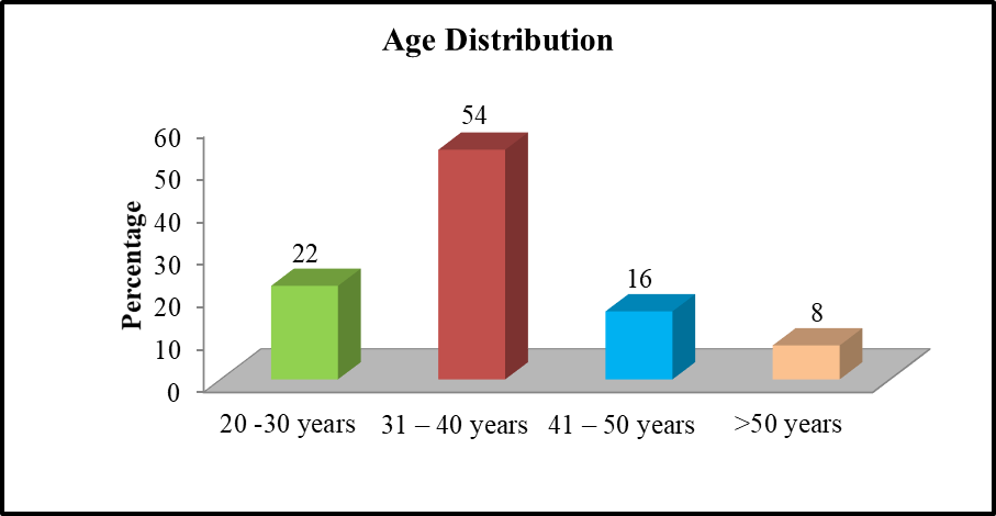
Graph: 1. Age distribution
The distribution of the age group consisted of 22% of patients in between 20 and 30 years of age, 54% of patients in between 31 to 40 years of age, 16% of patients in between 41 to 50 years of age, and 8% of patients over 50 years of age respectively. The mean age was 36.30 years.
Table: 2. Case Distribution among study Population (n=50)
|
Case Distribution |
No of cases |
Percentage |
|
CIN-I |
20 |
40.0 |
|
CIN-II |
9 |
18.0 |
|
CIN-III |
5 |
10.0 |
|
Carcinoma Cervix |
16 |
32.0 |
|
Total |
0 |
100.0 |
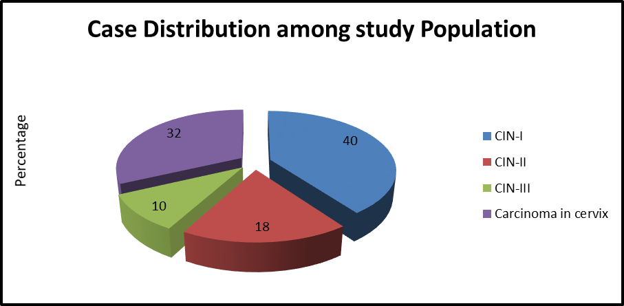
Graph: 2. Case Distribution among study Population (n=50)
Case distribution among study population we have found 40% cases were CIN-I, 18% cases were CIN-II, 10% Cases were CIN-III and 32% patients had Carcinoma in cervix, respectively.
Table: 3. Distribution of Carcinoma in cervix (n=16)
|
Age in Year |
No of cases |
Percentage |
|
Non keratinising squamous cell carcinoma |
10 |
62.5 |
|
Keratinising squamous cell carcinoma |
04 |
25.0 |
|
Adenocarcinoma |
02 |
12.5 |
|
Total |
16 |
100.0 |
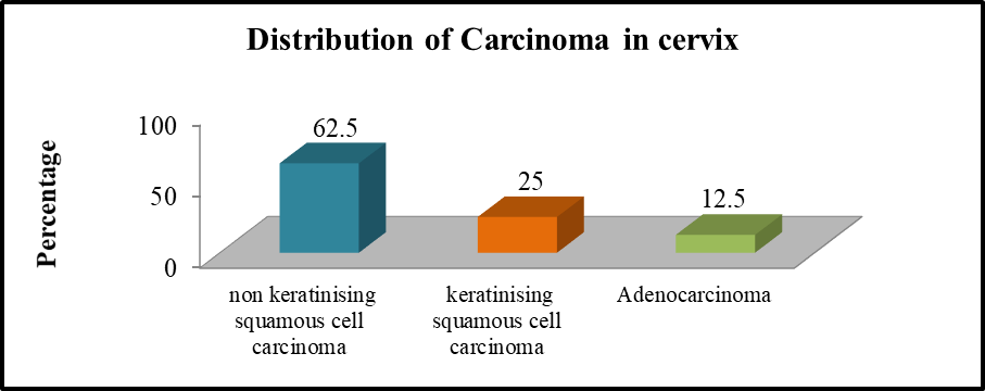
Graph: 3. Distribution of Carcinoma in cervix
Out of 16 Carcinoma in cervix cases 10(62.5%) were Non keratinising squamous cell carcinoma, 25% cases were Keratinising squamous cell carcinoma and only 2(12.5%) cases were Adenocarcinoma respectively.
Table:4. Grading of expression among Ki-67in the study cases
|
Case Distribution |
Total |
Grading of Ki67 |
|||
|
0 |
1 |
2 |
3 |
||
|
CIN-I |
20 |
5(25.0%) |
4(20.0%) |
9(45.0%) |
2(10.0%) |
|
CIN-II |
9 |
3(33.3%) |
3(33.3%) |
1(11.1%) |
2(22.2%) |
|
CIN-III |
5 |
0(0.0%) |
1(20.0%) |
2(40.0%) |
2(40.0%) |
|
Non-keratinising squamous cell carcinoma |
10 |
2(20.0%) |
1(10.0%) |
1(10.0%) |
6(60.0%) |
|
Keratinising squamous cell carcinoma |
04 |
0(0.0%) |
1(25.0%) |
1(25.0%) |
2(50.0%) |
|
Adenocarcinoma |
02 |
2(100.0%) |
0(0.0%) |
0(0.0%) |
0(0.0%) |
|
Total |
50 |
12(24.0%) |
10(20.0%) |
14(28.0%) |
14(28.0%) |
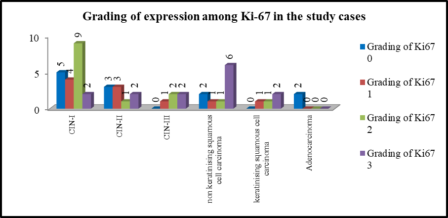
Graph:4. Grading of expression among Ki-67 in the study cases
Regarding grading of expression among Ki-67in the study population we found, among 20 patients of CIN-I, 5(25.0%) had grade ‘0’, 4(20.0%) had grade ‘1’, 9(45.0%) had grade ‘2’ and 2(10.0%) had grade ‘3’. Out of 9 CIN-II cases, 3(33.3%) each had grade ‘0’, & ‘1’, 1(11.1%) had grade ‘2’ and 2(22.2%) had grade ‘3. Among 5 patients of CIN-III, 1(20.0%) had grade ‘1’, 2(40.0%) each cases had grade ‘2’ and grade ‘3. On the other hand out of 10 Non-keratinising squamous cell carcinoma and 04 Keratinising squamous cell carcinoma cases maximum number of the patients were found in Grade-‘3’ i.e. 6(60%) & 2(50%) respectively. Among 2 Adenocarcinoma patients had grading ‘0’ of expression among Ki-67
Table:5. Grading of expression among p16 in the study cases
|
Case Distribution |
Total |
Grading of p16 |
|||
|
0 |
1 |
2 |
3 |
||
|
CIN-I |
20 |
15(75.0% |
3(15.0%) |
2(10.0%) |
0(0.0%) |
|
CIN-II |
9 |
6(66.7%) |
0 |
2(22.3%) |
1(11.1%) |
|
CIN-III |
5 |
3(60.0%) |
0 |
1(20.0%) |
1(20.0%) |
|
Non-keratinising squamous cell carcinoma |
10 |
2(20.0%) |
1(10.0%) |
2(20.0%) |
5(50.0%) |
|
Keratinizing squamous cell carcinoma |
04 |
1(25.0%) |
1(25.0%) |
1(25.0%) |
1(25.0%) |
|
Adenocarcinoma |
02 |
2(100.0%) |
0(0.0%) |
0(0.0%) |
0(0.0%) |
|
Total |
50 |
29(58.0%) |
5(10.0%) |
8(16.0%) |
8(16.0%) |
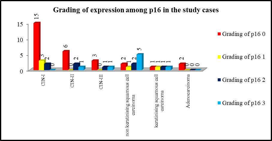
Graph:5. Grading of expression among p16 in the study cases
Regarding grading of expression among p16 in the study population we found, among 20 patients of CIN-I, 15(75.0%) had grade ‘0’, 3(15.0%) had grade ‘1’, 2(10.0%) had grade ‘2’ Out of 9 CIN-II cases, 6(66.7%) had grade ‘0’, and 2(22.3%) had grade ‘2. And 1(11.1%) had grade ‘3’Among 5 patients of CIN-III, 3(60%) had grade ‘0’, 1(20.0%) each cases had grade ‘2’ and grade ‘3. On the other hand, out of 10 Non-keratinising squamous cell carcinoma cases most of patients were belong to grade ‘3’ i.e. 5(50.0%) and 04 Keratinising squamous cell carcinoma cases 1 patent were present in each grade i.e. 25%. Among 2 Adenocarcinoma patients had grading’0’ of expression among p16.
Table:6. SCORE OF ki67 IN CERVICAL LESIONS:
|
Score ki 67in study case |
|||
|
Case Description |
LOW |
MODERATE |
HIGH |
|
CIN I |
5(25.0%) |
13(65.0%) |
2(10.0%) |
|
CIN II |
3(33.3%) |
4(44.5%) |
2(22.2%) |
|
CIN III |
0 |
3(60.0%) |
2(40.0%) |
|
Non-keratinising squamous cell carcinoma |
2(20.0%) |
2(20.0%) |
6(60.0%) |
|
Keratinizing squamous cell carcinoma |
0 |
2(50%) |
2(50.0%) |
|
ADENO CARCINOMA |
2(100.05) |
0 |
0 |
Graph:6. SCORE OF ki67 IN CERVICAL LESIONS
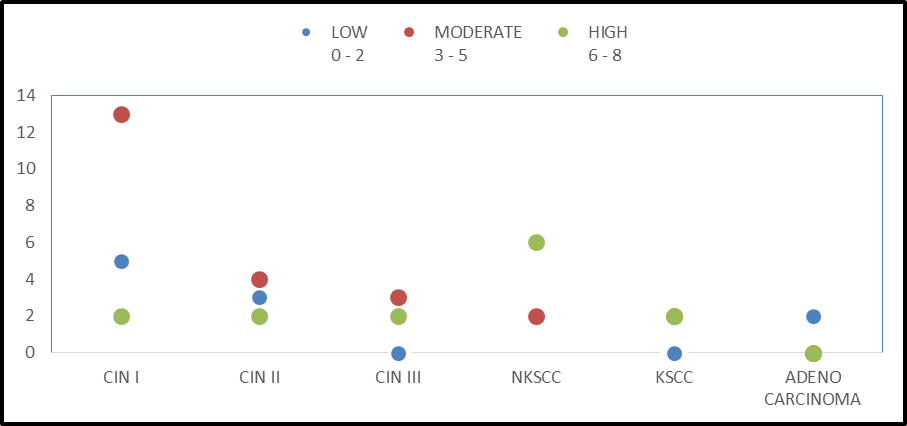
Regarding Scoring of ki-67 in the study population we found, among 20 patients of CIN-I, 5(25%) cases had Low score (0-2), 13(65.0%) cases Moderate score (3-5), & 2(10.0%) cases had High score (6-8). Out of 9 CIN-II, 3(33.3%) cases had Low score (0-2), 4(44.5%) cases Moderate score (3-5), & 2(22.2%) cases had High score (6-8). Among 5 patients of CIN-III, 3(60.0%) cases Moderate score (3-5), & 2(40.0%) cases had High score (6-8). On the other hand, out of 10 Non-keratinising squamous cell carcinoma cases most of patients were high ki67 moderate and high score i.e. 6(60.0%) and 04 Keratinising squamous cell carcinoma cases 2 each patents were present in each moderate and high score. Among 2(100.0%) Adenocarcinoma patients had low score (0-2) found in ki-67 respectively.
Table:7. SCORE OF p16 IN CERVICAL LESIONS:
|
Score P16 |
|||
|
Case Description |
LOW |
MODERATE |
HIGH |
|
CIN I |
15(75.0%) |
5(25.0%) |
0 |
|
CIN II |
6(66.7%) |
2(22.2%) |
1(11.1%) |
|
CIN III |
3(75.0%) |
1(25.0%) |
1(25.0%) |
|
Non-keratinising squamous cell carcinoma |
2(20.0%) |
3(30.0%) |
5(50.0%) |
|
Keratinizing squamous cell carcinoma |
0 |
2(66.7%) |
1(33.3%) |
|
ADENO CARCINOMA |
2(100.0%) |
0 |
0 |
Graph:7. SCORE OF p16 IN CERVICAL LESIONS:
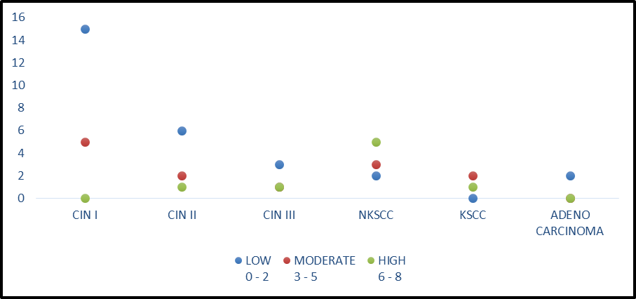
Regarding Scoring of p16 in the study population we found, among 20 patients of CIN-I, 15(75.0%) cases had Low score (0-2), 5(25.0%) cases Moderate score (3-5), there was no cases found of High score (6-8). Out of 9 CIN-II, 6(66.7%) cases had Low score (0-2), 2(22.2%) cases Moderate score (3-5), & 1(11.1%) cases had High score (6-8). Among 5 patients of CIN-III, 3(75.0%), 6(66.7%) cases had Low score (0-2), 1(25.0%) cases each Moderate & High score (3-5), & (6-8). On the other hand, out of 10 Non-keratinising squamous cell carcinoma cases 2(20.0%) cases had Low score (0-2), 3(30.0%) cases Moderate score (3-5), and 5(50.0%) cases had high score (6-8). Among 2, Adenocarcinoma patients had low score (0-2) found in p16 respectively.
Table:8. Ki-67 positivity in carcinoma cervix
|
Case Description |
Total |
positive |
Percentage |
|
Non-keratinising squamous cell carcinoma |
10 |
8 |
80.0 |
|
Keratinizing squamous cell carcinoma |
4 |
4 |
100.0 |
|
Adeno carcinoma |
2 |
0 |
0.0 |
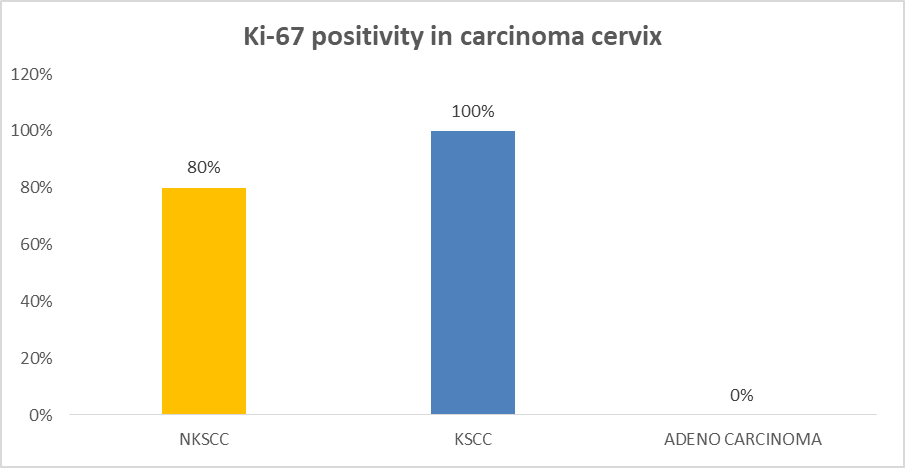
8 out of 10 cases were found positive in keratinising squamous cell carcinoma and 100% of positive cases found in Keratinizing squamous cell carcinoma.
Graph:8. Ki-67 positivity in carcinoma cervix
Table:9. p16 positivity in carcinoma cervix
|
Case Description |
Total |
positive |
Percentage |
|
Non-keratinising squamous cell carcinoma |
10 |
8 |
80.0 |
|
Keratinizing squamous cell carcinoma |
4 |
3 |
75.0 |
|
Adeno carcinoma |
2 |
0 |
0.0 |
8 out of 10(80%) cases were found positive in keratinising squamous cell carcinoma and 75% of positive cases found Keratinizing squamous cell carcinoma.
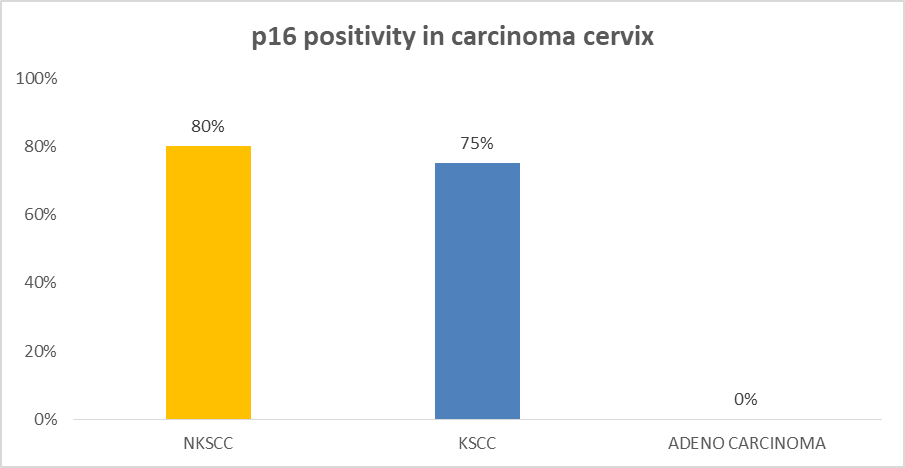
Graph:9. p16 positivity in carcinoma cervix
Table:10. Mean & SD value of ki 67 (cell proliferation marker) labeling index
|
Case Distribution |
ki 67 index |
|
|
Mean |
SD |
|
|
|
12.99 |
±0.14 |
|
CIN-II |
15.72 |
±0.15 |
|
CIN-III |
36.90 |
±0.15 |
|
Non keratinising squamous cell carcinoma |
46.28 |
±0.17 |
|
Keratinizing squamous cell carcinoma |
47.25 |
±1.47 |
|
Adenocarcinoma |
0.00 |
±0.0 |
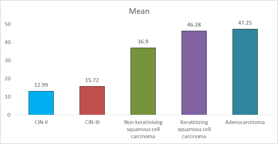
Graph:10. Mean value of ki67 (cell proliferation marker) labelling index
Mean & SD value of Ki-67 cell proliferation marker labeling index in CIN-I the mean & SD value was 12.99±0.14. Mean & SD value of CIN-II was 15.72±0.15, In CIN-III was 36.90±0.15, In Non keratinising squamous cell carcinoma, mean and SD value was 46.28±0.17, In Keratinising squamous cell carcinoma was 47.25±1.47 respectively.
Table:11. Comparison of mean of p16
|
Case Distribution |
p16index |
|
|
Mean |
SD |
|
|
CIN-I |
10.24 |
±0.33 |
|
CIN-II |
12.34 |
±0.15 |
|
CIN-III |
31.25 |
±0.15 |
|
Non keratinising squamous cell carcinoma |
39.28 |
±0.17 |
|
Keratinizing squamous cell carcinoma |
41.21 |
±0.99 |
|
Adenocarcinoma |
0 |
±0.0 |
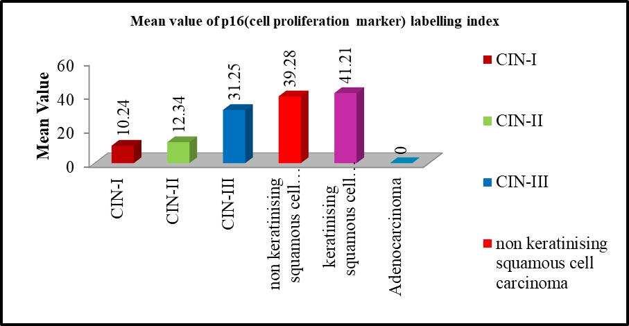
Graph: 11. Comparison of mean of p16
Mean & SD value of p16 in CIN-I the mean & SD value was 10.24±0.33. Mean & SD value of CIN-II was 12.34±0.65, In CIN-III was 31.25±0.45, In Non keratinising squamous cell carcinoma mean and SD value was 39.28±0.98, In Keratinising squamous cell carcinoma was 41.21±0.99 respectively.
Table: 12. Sensitivity & Specificity of Ki67
|
Statistic |
Value |
95% CI |
|
Sensitivity |
92.31% |
63.97% to 99.81% |
|
Specificity |
100.00% |
15.81% to 100.00% |
|
Positive Likelihood Ratio |
- |
- |
|
Negative Likelihood Ratio |
0.08 |
0.01 to 0.51 |
|
Disease prevalence (*) |
86.67% |
59.54% to 98.34% |
|
Positive Predictive Value (*) |
100.00% |
|
|
Negative Predictive Value (*) |
66.67% |
23.33% to 92.93% |
|
Accuracy (*) |
93.33% |
68.05% to 99.83% |
Table: 13. Sensitivity & Specificity of p16
|
Statistic |
Value |
95% CI |
|
Sensitivity |
91.67% |
61.52% to 99.79% |
|
Specificity |
80.00% |
28.36% to 99.49% |
|
Positive Likelihood Ratio |
4.58 |
0.79 to 26.68 |
|
Negative Likelihood Ratio |
0.10 |
0.02 to 0.72 |
|
Disease prevalence (*) |
70.59% |
44.04% to 89.69% |
|
Positive Predictive Value (*) |
91.67% |
65.40% to 98.46% |
|
Negative Predictive Value (*) |
80.00% |
36.80% to 96.49% |
|
Accuracy (*) |
88.24% |
63.56% to 98.54% |
Immunohistochemistry for p16 and Ki-67 in between ki67 was more Sensitive, specific and accuracy than p16 cell proliferation marker.
DISCUSSION:
Cervical cancer continues to be a leading cause of cancer-related mortality among women in India, underscoring the need for improved diagnostic and prognostic strategies. The natural history of cervical carcinogenesis, characterized by a prolonged pre-invasive phase progressing through cervical intraepithelial neoplasia (CIN), provides a valuable window for early detection and intervention. This has led to the exploration of adjunctive biomarkers that can enhance diagnostic precision beyond conventional histopathology.
In the present study, the mean age of patients was 36.3 years, with the majority belonging to the 31–40-year age group. This finding is consistent with previous studies, such as that by Kanthiya et al., which reported a comparable mean age of approximately 40 years, reflecting the higher prevalence of premalignant lesions in women of reproductive age.¹¹
The distribution of lesions in our study demonstrated that CIN I constituted the largest proportion (40%), followed by CIN II (18%), CIN III (10%), and invasive carcinoma (32%). Among carcinoma cases, non-keratinizing squamous cell carcinoma was the most frequent subtype, which aligns with the established predominance of squamous histology in cervical malignancies. Similar patterns have been described in earlier studies, although variations in lesion distribution may reflect differences in study population, screening practices, and diagnostic criteria.¹²–¹⁴
Immunohistochemical evaluation revealed a progressive increase in Ki-67 expression with increasing severity of cervical lesions. Ki-67, a well-established marker of cellular proliferation, showed low expression in CIN I and markedly higher expression in CIN III and invasive carcinoma. This trend is consistent with the biological behavior of cervical neoplasia, where increased proliferative activity correlates with disease progression. Previous studies have similarly demonstrated a stepwise increase in Ki-67 expression from low-grade lesions to high-grade dysplasia and carcinoma.¹⁵,¹⁶
However, the proportion of Ki-67 positivity observed in our study was relatively lower compared to some reports. This discrepancy may be attributed to differences in criteria used to define positivity. While some studies have considered both nuclear and cytoplasmic staining as positive, the present study adopted stricter criteria by including only nuclear staining in basal and parabasal layers. Studies employing similar stringent criteria have reported comparable or slightly higher expression rates, suggesting that methodological differences significantly influence reported outcomes.¹³,¹⁴
The expression of p16INK4a also demonstrated a positive correlation with the severity of cervical lesions. Overexpression of p16INK4a is a surrogate marker of oncogenic HPV activity, reflecting functional inactivation of the retinoblastoma (Rb) pathway by viral oncoproteins. In our study, higher expression was observed in CIN II/III and carcinoma compared to CIN I, supporting its role as a marker of high-grade lesions. These findings are in agreement with previous studies that have established p16 as a reliable biomarker for distinguishing high-grade dysplasia from low-grade or reactive changes.¹²,¹⁷
Nevertheless, p16 expression was also noted in a subset of low-grade lesions, which may be attributed to inflammatory changes, transient HPV infections, or possible underdiagnosis of higher-grade lesions on histopathology. Similar observations have been reported in the literature, highlighting the potential for false-positive staining and emphasizing the need for cautious interpretation, particularly in CIN I lesions.¹⁴
When evaluated together, p16 and Ki-67 demonstrated improved diagnostic performance. In our study, combined positivity showed higher sensitivity and specificity compared to either marker alone. Ki-67 exhibited the highest sensitivity and specificity individually, while the combined use of both markers further enhanced diagnostic accuracy. These findings are consistent with earlier studies, which have shown that dual immunostaining improves the identification of high-grade lesions and reduces interobserver variability.¹⁸
The results of the present study reinforce the utility of Ki-67 as a sensitive marker of cellular proliferation and p16INK4a as a specific marker of HPV-mediated oncogenic transformation. The combined application of these markers can significantly aid in the differentiation of low-grade from high-grade lesions and improve diagnostic confidence in equivocal cases.
However, certain limitations must be acknowledged. The relatively small sample size and potential selection bias may limit the generalizability of the findings. Additionally, variability in immunohistochemical interpretation and lack of standardized scoring systems may influence results. Future studies with larger cohorts and standardized methodologies are warranted to validate these findings and establish robust diagnostic criteria.
REFERENCES:
1. Sung H, Ferlay J, Siegel RL, et al. Global cancer statistics 2020: GLOBOCAN estimates of incidence and mortality worldwide for 36 cancers in 185 countries. CA Cancer J Clin. 2021;71(3):209–249.
2. Ferlay J, Soerjomataram I, Ervik M, et al. GLOBOCAN 2012 v1.0: Cancer incidence and mortality worldwide. Lyon: International Agency for Research on Cancer; 2013.
3. Bosch FX, Lorincz A, Muñoz N, Meijer CJLM, Shah KV. The causal relation between human papillomavirus and cervical cancer. J Clin Pathol. 2002;55:244–265.
4. IARC Working Group. Human papillomaviruses. IARC Monographs on the Evaluation of Carcinogenic Risks to Humans. Lyon: IARC; 2007.
5. Franco EL, Villa LL, Sobrinho JP, et al. Epidemiology of acquisition and clearance of cervical human papillomavirus infection. J Infect Dis. 1999;180:1415–1423.
6. Ho GYF, Bierman R, Beardsley L, Chang CJ, Burk RD. Natural history of cervicovaginal papillomavirus infection. N Engl J Med. 1998;338:423–428.
7. Kavatkar AN, Nagwanshi CA, Dabak SM. Study of a manual method of liquid-based cervical cytology. Indian J Pathol Microbiol. 2008;51:190–194.
8. Klaes R, Friedrich T, Spitkovsky D, et al. Overexpression of p16INK4a as a specific marker for dysplastic and neoplastic epithelial cells of the cervix uteri. Int J Cancer. 2001;92:276–284.
9. Agoff SN, Lin P, Morihara J, et al. p16INK4a expression correlates with degree of cervical neoplasia: Comparison with Ki-67 and HPV detection. Mod Pathol. 2003;16:665–673.
10. Izadi-Mood N, Asadi K, Shojaei H, et al. Diagnostic value of p16 expression in premalignant and malignant cervical lesions. J Res Med Sci. 2012;17:428–433.
11. Kanthiya K, Khunnarong K, Tangjitgamol S, et al. Expression of p16 and Ki-67 in cervical squamous intraepithelial lesions and cancer. Asian Pac J Cancer Prev. 2016;17(7):3201–3206.
12. Volgareva G, Zavalishina L, Andreeva Y, et al. Protein p16 as a marker of dysplastic and neoplastic alterations in cervical epithelial cells. BMC Cancer. 2004;4:58.
13. Wang SS, Trunk M, Schiffman M, et al. Validation of p16INK4a as a marker of oncogenic HPV infection in cervical biopsies. Cancer Epidemiol Biomarkers Prev. 2004;13:1355–1360.
14. Hariri J, Oster A. The negative predictive value of p16 in assessing CIN1 outcomes. Int J Gynecol Pathol. 2007;26:223–228.
15. Nam EJ, Kim JW, Hong JW, et al. Expression of p16INK4a and Ki-67 in relation to CIN grade and HPV infection. J Gynecol Oncol. 2008;19:162–168.
16. Kim SM, Lee JU, Lee DW, et al. Prognostic significance of p16, Ki-67, p63, and CK17 in CIN1. Korean J Obstet Gynecol. 2011;54:184–191.
17. Klaes R, Friedrich T, Spitkovsky D, et al. Overexpression of p16INK4a as a marker for cervical dysplasia. Int J Cancer. 2001;92:276–284.
18. van Niekerk D, Guillaud M, Matisic J, et al. p16 and MIB1 improve sensitivity and specificity in diagnosing HSIL. Gynecol Oncol. 2007;107:233–240.
19. Murphy N, Ring M, Heffron CC, et al. p16INK4a, CDC6, and MCM5 as predictive biomarkers. J Clin Pathol. 2005;58:525–534.
20. Queiroz C, Silva TC, Venancio AF. p16 expression as a prognostic marker in cervical lesions. Pathol Res Pract. 2006;202:77–83.
21. Conesa-Zamora P, Domenech-Peris A, Orantes-Casado FJ, et al. HPV-related biomarkers in precursor lesions. Am J Clin Pathol. 2009;132:378–390.
22. Cavalcante DM, Linhares LM, Pompeu ML, et al. Utility of p16INK4a and Ki-67 in identifying HSIL. Indian J Med Microbiol. 2012;55:339–342.
23. Jackson JA, Kapur U, Ersahin C. Utility of p16, Ki-67, and HPV testing in CIN diagnosis. Int J Surg Pathol. 2012;20:146–153.