COMPARTIVE STUDY BETWEEN ENDOSCOPIC TRANSCANAL MYRINGOPLASTY USING CARTILAGE GRAFT V/S SILASTIC SHEET IN SAFE CHRONIC SUPPURATIVE OTITIS MEDIA.
- Rohit Maheshwari , Senior Resident, Department of Otorhinolaryngology, Shyam Shah Medical College, Rewa.
- Surendra Singh Moupachi , Professor & Head of Department, Department of Otorhinolaryngology, Shyam Shah Medical College, Rewa.
- Hareesh Dev , Junior Resident (JR-3), Department of Otorhinolaryngology, Shyam Shah Medical College, Rewa.
- Saksham Kumar. , Junior Resident (JR-2), Department of Otorhinolaryngology, Shyam Shah Medical College, Rewa.
Article Information:
Abstract:
Background: Chronic suppurative otitis media (CSOM) is a common middle ear disease characterized by tympanic membrane perforation, persistent ear discharge, and hearing impairment. It remains a significant health concern, particularly in developing countries. Endoscopic myringoplasty has emerged as a minimally invasive technique with improved visualization and better cosmetic outcomes. Aim: To compare the efficacy of cartilage graft and silastic sheet in endoscopic transcanal myringoplasty with respect to graft uptake and hearing improvement. Materials and Methods: This prospective comparative study was conducted in Department of Otorhinolaryngology, Shyam Shah Medical College, Rewa, Op Shyam Shah Medical College, Rewa over a period of 6 months. A total of 60 patients with inactive CSOM were included and divided into two groups: Group A (silastic sheet) and Group B (cartilage graft), with 30 patients in each group. All patients underwent endoscopic transcanal myringoplasty. Preoperative assessment included clinical evaluation, otoendoscopy, and pure tone audiometry (PTA). Postoperative follow-up was done at 1, 3, 6, and 12 weeks to evaluate graft status and hearing outcomes. Results: The mean age was comparable between the groups. Hearing loss was present in 58.3% of patients. Postoperative PTA showed significant improvement in both groups, with greater improvement observed in the cartilage graft group (p < 0.001). Graft uptake was 100% in both groups up to 6 weeks. At 12 weeks, intact tympanic membrane was observed in 90% of patients in the cartilage group compared to 40% in the silastic sheet group (p = 0.001). Conclusion: Cartilage graft is more effective than silastic sheet in endoscopic myringoplasty, showing better graft uptake and hearing outcomes. It provides superior long-term stability and can be considered the preferred graft material.
Keywords:
Article :
INTRODUCTION:
Chronic suppurative otitis media (CSOM) is a persistent inflammatory and infectious condition of the middle ear characterized by tympanic membrane perforation, chronic mucosal inflammation, and possible ossicular involvement. [1] It remains a significant health problem, particularly in developing countries, due to its high prevalence and association with recurrent infections, poor hygiene, and limited access to healthcare. Clinically, CSOM commonly presents with hearing loss, persistent ear discharge, tinnitus, and ear pain, all of which can adversely affect quality of life and daily functioning. [2]
The primary goals in the management of CSOM are eradication of infection, closure of tympanic membrane perforation, and restoration of hearing. [3] Myringoplasty, defined as the surgical repair of the tympanic membrane without ossicular reconstruction, has long been considered the gold standard for treating tympanic membrane defects. Since the 1950s, microscopic myringoplasty (MM) has been widely used, employing operative microscopes and approaches such as postauricular, endaural, and transcanal techniques. [4] The microscopic approach offers advantages such as binocular vision and improved depth perception, facilitating precise surgical manipulation. However, it is limited by restricted visualization of certain middle ear areas due to the narrow external auditory canal. [5]
To address these limitations, endoscopic myringoplasty (EM) has gained increasing acceptance since the late 1990s. This technique provides superior visualization of hidden regions of the middle ear and allows a minimally invasive transcanal approach without the need for external incisions or canalplasty. [8–11] Additionally, EM is associated with reduced operative time, better cosmetic outcomes, and lower postoperative morbidity compared to conventional microscopic techniques. Despite these advantages, comparative data on the outcomes of EM and MM remain limited, especially in regions with a high burden of CSOM. [9–11]
The success of myringoplasty is significantly influenced by the choice of graft material. Various grafts such as temporalis fascia, cartilage, perichondrium, fat, and synthetic substitutes have been used. [13] Cartilage grafts are particularly valued for their rigidity, resistance to retraction, and long-term stability, especially in high-risk cases. [13,14] Concerns regarding their impact on hearing have not been substantiated clinically. [15] Silastic sheets, as synthetic materials, act as inert barriers preventing adhesions and maintaining middle ear space, thus aiding healing and sound conduction. [16]
However, limited evidence exists comparing cartilage grafts and silastic sheets in endoscopic myringoplasty. Therefore, this study aims to evaluate and compare their efficacy in terms of graft uptake, hearing improvement, and surgical outcomes in patients with CSOM.
MATERIALS AND METHODS:
This hospital-based prospective comparative study was conducted in the Department of Otorhinolaryngology, Shyam Shah Medical College, Rewa, over a period of 6 months (October 2025 to March 2026) after obtaining approval from the Institutional Ethics Committee. Prior to enrollment, written informed consent was obtained from all patients in their vernacular language.
A total of 60 patients of either sex aged more than 15 years, presenting with ear discharge and diagnosed with inactive chronic suppurative otitis media (CSOM), were included in the study. Sample size was calculated using a prevalence rate of 4% with 95% confidence interval and 5% precision, yielding a sample size of 59, which was rounded to 60 (30 patients in each group)
Inclusion Criteria
§ Patients of either sex
§ Age >12 years
§ Patients with small to medium central perforation
§ Diagnosed cases of inactive CSOM
§ Patients willing to provide informed consent
Exclusion Criteria
§ Patients refusing consent
§ Patients with large or subtotal perforation
§ Patients requiring ossiculoplasty
§ Patients with unsafe CSOM
§ Patients below 12 years of age
Methodology
After obtaining informed consent, eligible patients were enrolled and divided into two groups:
§ Group A: Endoscopic transcanal myringoplasty using silastic sheet
§ Group B: Endoscopic transcanal myringoplasty using cartilage graft
Baseline data including demographic profile, clinical history, and symptom duration were recorded using a prestructured proforma. All patients underwent thorough ENT examination, and diagnosis was confirmed using otoendoscopy, X-ray mastoid, and pure tone audiometry (PTA). In cases with active discharge, appropriate antibiotic therapy was given, and surgery was performed after achieving a dry ear for at least 3 weeks.
Surgical procedures were performed under standard aseptic precautions using a 0° endoscope. In Group A, silastic sheet was placed using the underlay technique, whereas in Group B, tragal cartilage graft was harvested and placed similarly. Middle ear structures were assessed intraoperatively, and external auditory canal was packed with medicated materials following the procedure.
Follow-Up
Patients were followed up at 1st, 3rd, 6th, and 12th week postoperatively to assess graft uptake and complications. Postoperative PTA was conducted at 6 and 12 weeks to evaluate hearing improvement.
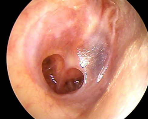
Figure 1. Tympanic membrane with small/medium size central perforation.
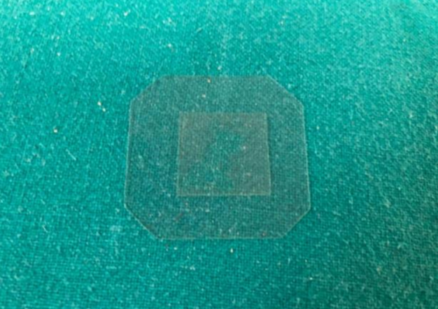
Figure 2. Silastic sheet
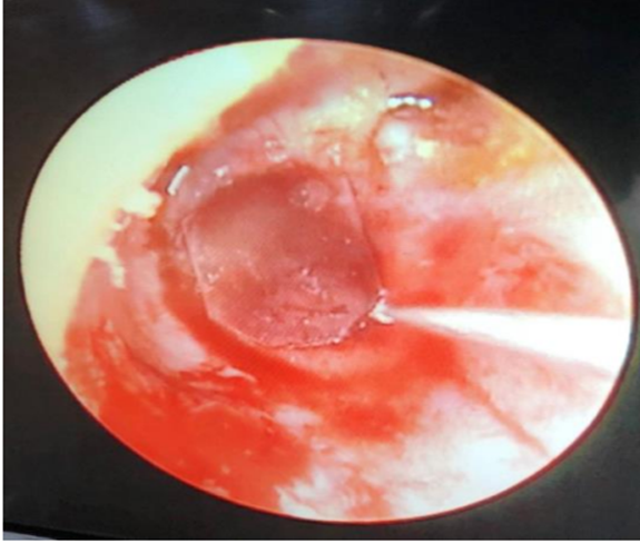
Figure 3. Postop repair of tympanic membrane perforation done with silastic sheet
Statistical Analysis
Data were entered into Microsoft Excel and analyzed using SPSS version 22.0. Continuous variables were expressed as mean ± standard deviation, and categorical variables as frequency and percentage. Comparative analysis was performed using Chi-square test and unpaired t-test. A p-value <0.05 was considered statistically significant
RESULTS:
The present study included 60 patients with chronic suppurative otitis media undergoing endoscopic transcanal myringoplasty using either silastic sheet (Group A) or cartilage graft (Group B). The mean age of patients in Group A was 28.23 ± 9.82 years and in Group B was 24.73 ± 8.27 years. The difference between the groups was statistically non-significant (p = 0.141). Male patients constituted the majority (65%), with a significantly higher proportion in Group B compared to Group A (83.3% vs. 46.7%) (χ² = 8.864, p = 0.003). [Table 1]
Table 1: Demographic Characteristics
|
Variable |
Category |
Group A (n=30) |
Group B (n=30) |
Total |
p-value |
|
Age (years) |
Mean ± SD |
28.23 ± 9.82 |
24.73 ± 8.27 |
— |
0.141 |
|
Gender |
Male |
14 (46.7%) |
25 (83.3%) |
39 (65%) |
0.003* |
|
|
Female |
16 (53.3%) |
5 (16.7%) |
21 (35%) |
Regarding clinical presentation, discharge from the right ear (53.3%) was slightly more common than the left ear (46.7%), with no significant intergroup difference (p = 0.121). Duration of discharge showed a statistically significant difference between groups (χ² = 24.833, p < 0.001), with Group A having more cases in the 3–12 months category, whereas Group B had a higher proportion since childhood. Similarly, duration since last episode of discharge also differed significantly (χ² = 37.333, p < 0.001).
Table 2: Clinical Profile
|
Variable |
Category |
Group A |
Group B |
Total |
p-value |
|
Chief complaint |
Right ear discharge |
13 (43.3%) |
19 (63.3%) |
32 (53.3%) |
0.121 |
|
|
Left ear discharge |
17 (56.7%) |
11 (36.7%) |
28 (46.7%) |
|
|
Duration of discharge |
3–12 months |
18 (60%) |
1 (3.3%) |
19 (31.7%) |
<0.001* |
|
|
>12 months |
9 (30%) |
13 (43.3%) |
22 (36.7%) |
|
|
|
Since childhood |
3 (10%) |
16 (53.3%) |
19 (31.7%) |
|
|
Last episode |
<1 month |
10 (33.3%) |
2 (6.7%) |
12 (20%) |
<0.001* |
|
|
1–6 months |
20 (66.7%) |
5 (16.7%) |
25 (41.7%) |
|
|
|
>6 months–1 year |
0 |
23 (76.7%) |
23 (38.3%) |
|
With respect to symptoms and risk factors, hearing loss was present in 58.3% of patients, while pain was reported in only 8.3%, with no significant difference between groups. However, history of trauma (χ² = 4.286, p = 0.038) and upper respiratory tract infection (χ² = 7.937, p = 0.005) were significantly more common in Group A. Itching and tinnitus were common but not statistically significant.
Table 3: Symptoms and Risk Factors
|
Variable |
Category |
Group A (n=30) |
Group B (n=30) |
Total (n=60) |
p-value |
|
Pain |
Present |
3 (10.0%) |
2 (6.7%) |
5 (8.3%) |
0.640 |
|
Absent |
27 (90.0%) |
28 (93.3%) |
55 (91.7%) |
|
|
|
Hearing loss |
Present |
16 (53.3%) |
19 (63.3%) |
35 (58.3%) |
0.432 |
|
Absent |
14 (46.7%) |
11 (36.7%) |
25 (41.7%) |
|
|
|
Trauma |
Present |
20 (66.7%) |
12 (40.0%) |
32 (53.3%) |
0.038* |
|
Absent |
10 (33.3%) |
18 (60.0%) |
28 (46.7%) |
|
|
|
Upper RTI |
Present |
14 (46.7%) |
4 (13.3%) |
18 (30.0%) |
0.005* |
|
Absent |
16 (53.3%) |
26 (86.7%) |
42 (70.0%) |
|
|
|
Itching |
Present |
23 (76.7%) |
27 (90.0%) |
50 (83.3%) |
0.166 |
|
Absent |
7 (23.3%) |
3 (10.0%) |
10 (16.7%) |
|
|
|
Tinnitus |
Present |
25 (83.3%) |
28 (93.3%) |
53 (88.3%) |
0.228 |
|
Absent |
5 (16.7%) |
2 (6.7%) |
7 (11.7%) |
|
On otoscopic examination, all patients had central perforation with normal pars tensa and ossicles. The most common site was anteroinferior quadrant (66.7%), with no significant difference between groups (p = 0.273). Radiological findings showed mastoid sclerosis in most cases without significant variation.
Table 4: Otoscopic and Radiological Findings
|
Variable |
Category |
Group A |
Group B |
Total |
p-value |
|
Location of perforation |
Anteroinferior |
22 (73.3%) |
18 (60%) |
40 (66.7%) |
0.273 |
|
Posteroinferior |
8 (26.7%) |
12 (40%) |
20 (33.3%) |
||
|
Radiology |
Right mastoid sclerosis |
15 (50%) |
20 (66.7%) |
35 (58.3%) |
0.190 |
|
Left mastoid sclerosis |
15 (50%) |
10 (33.3%) |
25 (41.7%) |
Pure tone audiometry revealed comparable preoperative hearing levels between groups (p = 0.095). However, postoperative hearing improvement was significantly better in Group B (p < 0.001). Graft uptake was 100% up to 6 weeks in both groups. At 12 weeks, Group B showed significantly higher intact tympanic membrane rates (90% vs. 40%) (χ² = 17.969, p = 0.001).
Table 5: Audiological Outcome and Graft Status
|
Variable |
Category |
Group A |
Group B |
p-value |
|
PTA (dB HL) |
Preoperative |
28 (26–30) |
33 (26–33) |
0.095 |
|
|
Postoperative |
25 (22–28) |
21 (20–22) |
<0.001* |
|
Graft status (12 weeks) |
TM intact |
12 (40%) |
27 (90%) |
0.001* |
|
|
Graft in-situ |
4 (13.3%) |
1 (3.3%) |
|
|
|
Graft displaced |
1 (3.3%) |
1 (3.3%) |
|
|
|
Small perforation |
9 (30%) |
1 (3.3%) |
|
|
|
Medium perforation |
4 (13.3%) |
0 |
DISCUSSION:
Myringoplasty remains a well-established surgical procedure aimed at repairing tympanic membrane perforations in patients with intact ossicular chain function, adequate cochlear reserve, and satisfactory Eustachian tube function. The evolution of this procedure dates back to Berthold (1878) [17], with further refinements by Wullstein (1956) [18] and Zollner (1955) [19]. Despite advances in surgical techniques, the choice of graft material continues to influence postoperative outcomes significantly.
In the present study, both groups were comparable in terms of baseline characteristics, thereby ensuring minimal selection bias. The mean age of patients in Group A and Group B was 28.23 ± 9.82 years and 24.73 ± 8.27 years respectively, with no statistically significant difference. This is consistent with findings reported by S. Mahdiani et al., where a broad age distribution was observed among patients undergoing myringoplasty [20]. Similarly, studies conducted in India have demonstrated that CSOM is more prevalent in younger age groups, particularly between 1–20 years, followed by adults up to 60 years [21].
A male predominance (65%) was observed in the present study, which aligns with previous literature suggesting higher prevalence of CSOM among males [22,23]. This gender distribution has been attributed to increased exposure to environmental and occupational risk factors in males.
Regarding clinical presentation, otorrhea was the most common symptom, with right ear involvement slightly more frequent than the left. The majority of patients presented with insidious onset, gradual progression, and profuse, non-foul-smelling discharge. These findings are in agreement with studies by Mahdiani et al. and other international reports, where otorrhea was identified as the predominant presenting complaint [24,25]. Hearing loss was present in 58.3% of patients, which is comparable to previous studies reporting hearing impairment in approximately 40–60% of CSOM cases.
In the present study, significant differences were observed in duration of discharge and duration since last episode between the groups. Group A had more recent and shorter duration of symptoms, whereas Group B included a higher proportion of long-standing cases. However, other clinical parameters such as pain, tinnitus, and itching did not differ significantly between the groups, suggesting comparable disease severity at baseline.
On otoscopic examination, all patients demonstrated central perforation with normal pars tensa and ossicles, ensuring uniformity in disease profile. The most common location was anteroinferior quadrant, consistent with previous studies. No statistically significant difference in perforation characteristics was observed between the groups, indicating that surgical outcomes were primarily influenced by graft material rather than anatomical variability.
Preoperative hearing assessment using tuning fork tests (Rinne and Weber) did not show significant differences between the groups. Similarly, radiological findings revealed comparable mastoid sclerosis patterns. Preoperative pure tone audiometry (PTA) also demonstrated no significant difference, confirming that both groups were comparable in terms of baseline hearing status.
Postoperative outcomes, however, revealed important differences. Although both groups showed significant improvement in hearing, Group B (cartilage graft) demonstrated superior postoperative hearing gain compared to Group A (silastic sheet) (p < 0.001). This finding is consistent with literature suggesting that cartilage grafts provide better structural support and long-term stability without significantly compromising acoustic properties.
Graft uptake remained 100% in both groups up to 6 weeks, indicating good early surgical outcomes. However, at 12 weeks, Group B showed a significantly higher rate of intact tympanic membrane (90%) compared to Group A (40%) (p = 0.001). In contrast, residual perforations were more common in the silastic sheet group. These findings highlight the superior efficacy of cartilage grafts in maintaining long-term graft integrity.
Müller et al. (2022) reported that silastic sheeting may offer certain advantages in maintaining middle ear space and preventing adhesions, particularly in staged procedures [26]. However, the present study suggests that while silastic sheets may be beneficial as supportive materials, they are less effective than cartilage grafts in achieving long-term graft uptake and stability.
Despite these findings, certain limitations must be acknowledged. The sample size was relatively small, and the follow-up period was limited to 12 weeks, which may not fully reflect long-term outcomes. Additionally, factors such as Eustachian tube function, middle ear ventilation, and patient-related variables like smoking were not assessed. Furthermore, variability in reporting standards across studies makes direct comparison challenging, as noted in systematic reviews by Mor et al. Standardized outcome measures are essential for improving comparability and guiding clinical decision-making.
CONCLUSION:
Cartilage graft appears to be more effective than silastic sheet in endoscopic myringoplasty for CSOM, showing higher graft uptake rates and better improvement in hearing. While both techniques are safe and yield good early postoperative results, cartilage graft provides greater long-term stability and reliability. Silastic sheet may serve as a supportive option but is less consistent as a primary graft material. Overall, cartilage graft stands out as a preferable choice for achieving better surgical outcomes. Further studies with larger sample sizes and longer follow-up periods are required to strengthen these observations.
REFERENCES:
1. Monasta L, Ronfani L, Marchetti F, Montico M, Vecchi Brumatti L, Bavcar A, et al. Burden of disease caused by otitis media: systematic review and global estimates. PLoS One. 2012;7(4):e36226.
2. Reiss M, Reiss G. Suppurative chronic otitis media: etiology, diagnosis and therapy. Med Monatsschr Pharm. 2010;33(1):11–16.
3. Plinkert P, Löwenheim H. Trends and perspectives in minimally invasive surgery in otorhinolaryngology-head and neck surgery. Laryngoscope. 1997;107(11 Pt 1):1483–1489.
4. Shakya D, KC A, Nepal A. A comparative study of endoscopic versus microscopic cartilage type I tympanoplasty. Int Arch Otorhinolaryngol. 2020;24:e80–e85.
5. Choi N, Noh Y, Park W, Lee JJ, Yook S, Choi JE, et al. Comparison of endoscopic tympanoplasty to microscopic tympanoplasty. Clin Exp Otorhinolaryngol. 2017;10:44–49.
6. Kang HS, Ahn SK, Jeon SY, Hur DG, Kim JP, Park JJ, et al. Sensation recovery of auricle following chronic ear surgery by retroauricular incision. Eur Arch Otorhinolaryngol. 2012;269:101–106.
7. Kozin ED, Gulati S, Kaplan AB, Lehmann AE, Remenschneider AK, Kaplan BA, et al. Systematic review of endoscopic middle ear surgery outcomes. Laryngoscope. 2015;125:1205–1214.
8. Lee S, Cho HH. Transcanal endoscopic tympanoplasty for pediatric patients under 15 years of age with chronic otitis media. Clin Exp Otorhinolaryngol. 2020;13:41–46.
9. Badr-el-Dine M. Value of ear endoscopy in cholesteatoma surgery. Otol Neurotol. 2002;23:631–635.
10. Thomassin JM, Korchia D, Doris JM. Endoscopic-guided otosurgery in the prevention of residual cholesteatomas. Laryngoscope. 1993;103:939–943.
11. Puria S. Middle ear mechanics in research and otosurgery. In: Hüttenbrink KB, editor. Proceedings of the International Workshop on Middle Ear Mechanics and Otosurgery; 1996 Sep 19–22; Dresden, Germany. Dresden: Bibliothek der HNO-Universitätsklinik; 1997.
12. Iacovou E, Vlastarakos PV, Papacharalampous G, Kyrodimos E, Nikolopoulos TP. Is cartilage better than temporalis muscle fascia in type I tympanoplasty? Implications for current surgical practice. Eur Arch Otorhinolaryngol. 2013;270:2803–13.
13. Yang T, Wu X, Peng X, Zhang Y, Xie S, Sun H. Comparison of cartilage graft and fascia in type I tympanoplasty: systematic review and meta-analysis. Acta Otolaryngol. 2016;136:1085–90.
14. Barake R, El Natout T, Bassim M, El Natout MA. Loop underlay tympanoplasty for anterior, subtotal and total tympanic membrane perforations: a retrospective review. J Otolaryngol Head Neck Surg. 2019;48:12.
15. Fayad J.N. & House J.W. (2004) Silastic sheeting in the middle ear. Ear Nose Throat J. 83, 732
16. Courteney-Harris RG, Ford GR, Ganiwalla TM, et al. Closure of tympanic membrane perforation after the removal of Goode-type tympanostomy tubes: the use of silastic sheeting. J Laryngol Otol. 1992;106:960–962.
17. Berthold E (1878) Ueber myringoplastik. Wier Med Bull 1:627
18. Zollner F. The principals of plastic surgery of the sound conducting apparatus. J Laryngol Otol. 1955;69:637.
19. Wullstein H. The restoration of the function of the middle ear in chronic otitis media. Ann Otol Rhinol Laryngo. 1971;l(80):210–2
20. Mahdiani S, Lasminingrum L, Anugrah D. Management evaluation of patients with chronic suppurative otitis media: A retrospective study. Ann Med Surg (Lond). 2021 Jun 11;67:102492.
21. M.C. Basavaraj, P. Jyothi, Chronic suppurative otitis media (CSOM): etiological agents and antibiotic sensitivity pattern of the isolates, J. Med. 16 (2) (2015) 79
22. Hunt L, Mulwafu W, Knott V, Ndamala CB, Naunje AW, Dewhurst S, et al. Prevalence of paediatric chronic suppurative otitis media and hearing impairment in rural Malawi: a cross-sectional survey. PLoS One. 2017;12(12).
23. Jensen RG, Koch A, Homøe P. The risk of hearing loss in a population with a high prevalence of chronic suppurative otitis media. Int J Pediatr Otorhinolaryngol. 2013;77(9):1530–1535.
24. Muftah S, Mackenzie I, Faragher B, Brabin B. Prevalence of chronic suppurative otitis media (CSOM) and associated hearing impairment among school-aged children in Yemen. Oman Med J. 2015;30(5):358–365.
25. J. Kim, G.H. Jung, S.Y. Park, W.S. Lee, Facial nerve paralysis due to chronic otitis media: prognosis in restoration of facial function after surgical intervention, Yonsei Med. J. 53 (3) (2012) 642–648,
26. Müller M, Honegger F, Podvinec M, et al. Silastic sheeting in staged ear surgery: Is there still a role for this procedure? Eur Arch Otorhinolaryngol. 2022;279:1203–1210.