Venous Pseudoaneurysm of a Left Brachiocephalic Arteriovenous Fistula in a Patient on Maintenance Hemodialysis.
- Divya Kandula , 3rd Year PG, Department of Cardiothoracic and Vascular Surgery, Osmania General Hospital, Hyderabad, Telangana, India.
- Yashwanth Gundala , 1st Year PG, Department of Cardiothoracic and Vascular Surgery, Osmania General Hospital, Hyderabad, Telangana, India.
- Krishna Kishore B , Assistant Professor, Department of Cardiothoracic and Vascular Surgery, Osmania General Hospital, Hyderabad, Telangana, India.
Article Information:
Abstract:
Objectives: To highlight the clinical presentation, management, and outcome of a rare venous pseudoaneurysm complicating a brachiocephalic arteriovenous fistula in a patient on maintenance hemodialysis. Materials and Methods: A 38-year-old woman with chronic kidney disease on long-term hemodialysis via a left brachiocephalic fistula presented with a painless, progressively enlarging swelling over the medial aspect of the left upper arm for four months. Clinical examination revealed a soft, non-tender, pulsatile mass measuring approximately 3 × 4 cm. CT angiography confirmed a pseudoaneurysm arising from the left cephalic vein with a normal brachial artery. Due to difficulty in cannulation, surgical exploration and excision of the lesion were undertaken. Results: Intraoperative findings confirmed a venous pseudoaneurysm originating from the cephalic vein. Postoperatively, thrombosis of the cephalic vein occurred, rendering the fistula non-functional for further dialysis. The patient otherwise had an uneventful recovery and was discharged on the fifth postoperative day. Conclusion: Venous pseudoaneurysm is a rare but significant complication of arteriovenous fistula, often associated with repeated cannulation trauma. Early diagnosis and timely surgical intervention are important; however, preservation of fistula function may be challenging, particularly in advanced cases.
Keywords:
Article :
INTRODUCTION:
Native arteriovenous fistula remains the most widely accepted vascular access for patients who require long-term hemodialysis. It is generally preferred because it offers better durability, fewer infections, and improved long-term outcomes when compared with grafts and central venous catheters [1-6].
However, the repeated use of fistulas for dialysis is associated with several complications, including stenosis, thrombosis, infection, aneurysm formation, and pseudoaneurysm development [2,6,7]. A pseudoaneurysm is different from a true aneurysm. In a true aneurysm, all layers of the vessel wall are involved in the dilatation, whereas a pseudoaneurysm results from a defect in the vessel wall with persistent blood leakage into the surrounding tissue, producing a contained hematoma without a true endothelial lining [2,8]. In patients on hemodialysis, pseudoaneurysms usually develop because of repeated puncture at the same site, trauma during cannulation, infection, or structural weakness of the vessel wall in a high-flow access circuit [2,4,9].
Venous pseudoaneurysm involving the outflow vein of a brachiocephalic fistula is uncommon. Even though it is rare, it has important clinical implications because it may cause pain, progressive enlargement, skin thinning, difficulty in cannulation, thrombosis, hemorrhage, and eventual loss of vascular access [2,4,10].
The diagnosis is usually established by clinical examination and confirmed by duplex ultrasonography or CT angiography, which also helps define the exact anatomy before intervention [2,11]. Management depends on the size of the lesion, site of origin, presence of infection, risk of rupture, availability of endovascular expertise, and the possibility of preserving the existing fistula. Treatment options described in the literature include ultrasound-guided compression, thrombin injection in selected cases, covered stent placement, aneurysmorrhaphy, surgical excision with repair, or fistula ligation when salvage is not possible [2,9,11-13].
In this report, we present a case of venous pseudoaneurysm arising from the cephalic vein at the venous end of a left brachiocephalic fistula in a young woman on maintenance hemodialysis, managed by surgical excision.
MATERIALS AND METHODS:
Study design and setting
This work was prepared as a hospital-based case report in the Department of Cardiothoracic and Vascular Surgery, Osmania General Hospital, Hyderabad. The report was developed from the clinical records, imaging findings, operative notes, and postoperative follow-up details of patients with dialysis access-related vascular complications managed in our unit.
Study period
The cases were observed during the period from 2023 to 2025. During this time, ten patients with suspected aneurysmal or pseudoaneurysmal complications related to arteriovenous fistula access were evaluated in the department.
Case selection
Among the ten cases seen during the study period, one patient was selected for detailed presentation because of the unusual nature of the lesion. The present report focuses on a venous pseudoaneurysm arising from the venous end of a left brachiocephalic arteriovenous fistula in a patient on maintenance hemodialysis. This case was chosen because venous pseudoaneurysm at this site is uncommon and has important implications for dialysis access preservation and surgical decision-making.
Clinical evaluation
The selected patient underwent detailed clinical assessment at the time of presentation. A careful history was taken with emphasis on the duration and progression of swelling, dialysis access history, prior cannulation pattern, and the presence or absence of pain, bleeding, infection, or difficulty during dialysis. General and local examination findings were recorded. The swelling was assessed for its site, size, pulsatility, tenderness, and relation to the existing fistula.
Radiological assessment
To define the nature of the swelling and its vascular origin, CT angiography of the left upper limb was performed. Imaging was used to identify the size of the lesion, its relation to the cephalic vein and brachial artery, and to confirm whether the lesion was venous or arterial in origin. The radiological findings were correlated with the clinical examination before planning treatment.
Preoperative workup
Routine laboratory investigations were reviewed as part of preoperative assessment. These included the standard blood tests required for anaesthetic and surgical fitness. The patient was then evaluated for operative management based on the symptoms, imaging findings, and the effect of the lesion on the functioning dialysis access.
Surgical management
As the swelling had become clinically significant and was causing difficulty in cannulation during hemodialysis, surgical exploration was planned. The procedure was carried out under general anaesthesia. Intraoperative findings, including the exact vessel involved, approximate size of the lesion, and operative steps performed, were documented. Excision of the pseudoaneurysm was undertaken as the definitive treatment in this patient.
Postoperative follow-up
The patient was monitored during the postoperative period for immediate complications, fistula status, wound condition, and suitability of the vascular access for continued dialysis. Particular attention was given to the development of thrombosis, bleeding, or loss of fistula function. The short-term outcome until discharge was recorded.
Data source and documentation
The case report was prepared using anonymised information obtained from the patient’s clinical notes, imaging records, operative findings, and inpatient postoperative course. Although ten related cases were seen during the study period, only this single case is described in detail in order to highlight its uncommon presentation and management.
Ethical considerations
Patient identity was not disclosed at any stage of reporting. All clinical details were presented in a de-identified manner to maintain confidentiality. Images and case-related information were included only for academic and scientific documentation.
RESULTS:
Patient profile
The patient was a 38-year-old woman with chronic kidney disease who was on maintenance hemodialysis. She had bilateral renal failure and had been undergoing dialysis through a left brachiocephalic arteriovenous fistula for nearly one year. The fistula had been created with the anastomosis at the cubital region.
Presenting complaint
She came with a swelling over the medial aspect of the left upper arm that had been increasing gradually over the previous four months. The swelling was painless throughout this period and there was no history of acute bleeding or local tenderness at presentation.
Clinical examination findings
On local examination, the lesion was found to be soft in consistency, non-tender, and pulsatile. Its approximate size was 3 × 4 cm. Multiple puncture marks were visible over the swelling, suggesting repeated cannulation at the same site or in the surrounding segment of the fistula. The clinical appearance raised suspicion of an access-related vascular complication.
Radiological findings
CT angiography of the left upper limb showed a pseudoaneurysm measuring about 3 × 4 cm arising from the left cephalic vein. The brachial artery appeared normal, which supported the view that the lesion was venous in origin rather than arterial. The imaging findings were in favour of a venous pseudoaneurysm involving the outflow segment of the left brachiocephalic fistula.
Preoperative assessment
Routine laboratory investigations were within acceptable limits for surgery. As the lesion had started interfering with dialysis cannulation, operative treatment was considered necessary. Based on the clinical and radiological findings, the patient was planned for surgical exploration and excision of the pseudoaneurysm.
Intraoperative findings
Surgery was performed under general anaesthesia. A skin incision of about 5 cm was placed over the swelling. During exploration, the brachial artery and cephalic vein were identified clearly. A pseudoaneurysm measuring nearly 3 × 4 cm was seen arising from the cephalic vein. The operative findings matched the CT angiography findings. Excision of the pseudoaneurysm was carried out.
Postoperative outcome
During the postoperative period, thrombosis of the cephalic vein was noted. Following this, the fistula could no longer be used for dialysis. No other immediate postoperative complication was documented. The patient remained haemodynamically stable and was discharged on the fifth postoperative day.
Overall outcome of the case
This case showed that venous pseudoaneurysm of a brachiocephalic fistula may present as a painless enlarging swelling and can become clinically important once cannulation becomes difficult. Although the lesion was managed surgically, preservation of the access was not possible because of postoperative venous thrombosis.
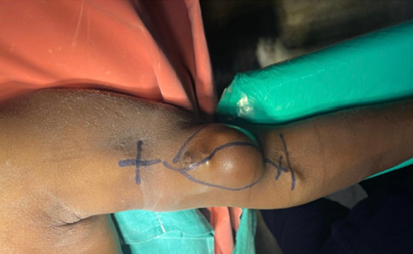
Figure 1: Preoperative clinical photograph showing a localized, smooth, rounded swelling over the medial aspect of the left upper arm at the venous end of the left brachiocephalic arteriovenous fistula. The lesion was soft, non-tender, and clinically suggestive of a venous pseudoaneurysm in a patient on maintenance hemodialysis.
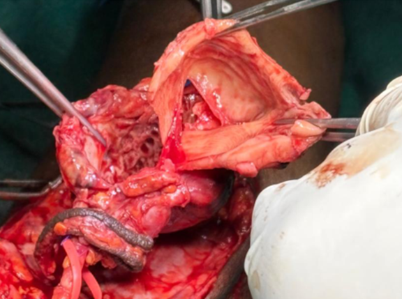 Figure 2: Intraoperative photograph showing the venous pseudoaneurysm arising from the cephalic vein during surgical exploration and excision
DISCUSSION:
This case demonstrates an uncommon but clinically relevant complication of dialysis access. Although aneurysmal changes are seen in longstanding fistulas, venous pseudoaneurysm involving the outflow cephalic vein is reported far less often than arterial or anastomotic lesions [2,10,14]. The most likely mechanism in the present patient was repeated cannulation trauma in a venous segment exposed to increased flow and pressure after creation of the brachiocephalic fistula. The multiple puncture marks observed over the swelling and the absence of documented signs of infection support a traumatic rather than infective origin.
Pseudoaneurysm formation in an arteriovenous fistula is clinically important because the lesion may remain painless for a prolonged period while progressively enlarging. Patients may first seek care only when the lesion interferes with dialysis cannulation or when cosmetic deformity becomes obvious. If left untreated, pseudoaneurysms may predispose to skin thinning, rupture, thrombosis, infection, hemorrhage, and loss of access [2,4,9]. In the present case, difficulty in cannulation was the practical indication for intervention.
Imaging has a key role in the evaluation of suspected access complications. Duplex ultrasonography is often the initial modality because it is readily available, non-invasive, and useful for identifying flow abnormalities, mural defects, and associated stenosis. CT angiography is valuable in selected cases where detailed anatomical delineation is needed before intervention [2,11]. In this patient, CT angiography clearly demonstrated the pseudoaneurysm arising from the cephalic vein and confirmed that the brachial artery was normal, helping establish the lesion as venous in origin.
Treatment must be individualized. Small and stable lesions may be monitored in carefully selected patients, whereas symptomatic, enlarging, infected, or difficult-to-cannulate lesions usually require active management. Reported treatment options include ultrasound-guided compression, thrombin injection in selected pseudoaneurysms, covered stent placement, aneurysmorrhaphy, excision with venous repair, and fistula ligation when salvage is not feasible [9,11-13]. Each option has its own limitations. Endovascular techniques may preserve access in some patients, but cost, expertise, lesion anatomy, and future cannulation issues may restrict their use.
In our patient, surgical excision was chosen because the lesion was established, symptomatic from a dialysis-access perspective, and being managed in a setting where open surgical treatment was feasible. However, the postoperative thrombosis of the cephalic vein led to loss of fistula function. This highlights an important clinical reality: even when the pseudoaneurysm itself is treated successfully, preservation of long-term access may not always be possible, especially when the lesion is large or when the underlying vein has already been structurally compromised [10,12,14].
Prevention remains an important component of access care. Proper cannulation technique, rotation of puncture sites, avoidance of repeated needling at a single point, and regular surveillance of vascular access may reduce the risk of aneurysm and pseudoaneurysm formation [1,2,15]. Early referral for vascular assessment when a new swelling is noted may improve the possibility of fistula salvage before thrombosis or extensive venous wall damage occurs.
CONCLUSION:
Venous pseudoaneurysm at the venous end of a brachiocephalic arteriovenous fistula is an uncommon but important complication in patients on maintenance hemodialysis. Repeated cannulation injury in an arterialized cephalic vein was the most likely mechanism in this case. CT angiography helped define the lesion, and surgical excision provided local control. However, postoperative thrombosis led to loss of fistula function, showing that access preservation may remain difficult once the lesion becomes advanced. Careful cannulation technique, regular access surveillance, and early intervention are essential for reducing morbidity and improving the chances of fistula salvage.
REFERENCES:
1. Lok CE, Huber TS, Lee T, Shenoy S, Yevzlin AS, Abreo K, et al. KDOQI clinical practice guideline for vascular access: 2019 update. Am J Kidney Dis. 2020;75(4 Suppl 2):S1-S164. doi:10.1053/j.ajkd.2019.12.001.
2. Agha RA, Franchi T, Sohrabi C, Mathew G, Kerwan A; SCARE Group. The SCARE 2020 guideline: updating consensus Surgical CAse REport guidelines. Int J Surg. 2020;84:226-230. doi:10.1016/j.ijsu.2020.10.034.
3. Mudoni A, Cornacchiari M, Gallieni M, Guastoni C, McGrogan D, Logias F, et al. Aneurysms and pseudoaneurysms in dialysis access. Clin Kidney J. 2015;8(4):363-367. doi:10.1093/ckj/sfv042.
4. Padberg FT Jr, Calligaro KD, Sidawy AN. Complications of arteriovenous hemodialysis access: recognition and management. J Vasc Surg. 2008;48(5 Suppl):55S-80S. doi:10.1016/j.jvs.2008.08.067.
5. Balaz P, Rokosny S, Bafrnec J, Whitley A, O'Neill S. Repair of aneurysmal arteriovenous fistulae: a systematic review and meta-analysis. Eur J Vasc Endovasc Surg. 2020;59(4):614-623. doi:10.1016/j.ejvs.2019.07.033.
6. Zibari GB, Rohr MS, Landreneau MD, Bridges RM, DeVault GA, Petty FH, et al. Complications from permanent hemodialysis vascular access. Surgery. 1988;104(4):681-686.
7. Schatz IJ, Fine G. Venous aneurysms. N Engl J Med. 1962;266:1310-1312. doi:10.1056/NEJM196206212662504.
8. Kresowik TF, Khoury MD, Miller BV, Winniford MD, Shamma AR, Sharp WJ, et al. A prospective study of the incidence and natural history of femoral vascular complications after percutaneous transluminal coronary angioplasty. J Vasc Surg. 1991;13(2):328-333. doi:10.1067/mva.1991.26786.
9. Najibi S, Bush RL, Terramani TT, Chaikof EL, Gunnoud AB, Lumsden AB, et al. Covered stent exclusion of dialysis access pseudoaneurysms. J Surg Res. 2002;106(1):15-19. doi:10.1006/jsre.2002.6389.
10. Chaudhary A, Acharya S, Karmacharya RM, Lamsal A, Pokharel S, Bhatt S. Venous pseudoaneurysm of left brachiocephalic fistula treated by excision and aneurysmorrhaphy: a case report. Int J Surg Case Rep. 2022;98:107496. doi:10.1016/j.ijscr.2022.107496.
11. Une D, Nakanishi K, Shimizu S, Nakai M, Kato G. Cephalic vein pseudoaneurysm after blunt trauma in a patient with arteriovenous fistula for hemodialysis. Circ J. 2014;78:232-234. doi:10.1253/circj.CJ-13-1156.
12. Rahil MA, Patel MS, Patel B, McCann M. Successful treatment of a venous pseudoaneurysm in a complicated arteriovenous fistula using ultrasound-guided manual compression. Hemodial Int. 2018;22(3):E63-E66. doi:10.1111/hdi.12621.
13. Fotiadis N, Koukounaras J, Tsetis D, Ioannidis A, Gkrinia E, Brountzos E. Endovascular repair of symptomatic hemodialysis access graft pseudoaneurysms. J Vasc Access. 2014;15(1):5-11. doi:10.5301/jva.5000161.
14. Belli S, Parlakgumus A, Colakoglu T, Ezer A, Toktas O, Yildirim S, et al. Surgical treatment modalities for complicated aneurysms and pseudoaneurysms of arteriovenous fistulas. J Vasc Access. 2012;13(4):438-445. doi:10.5301/jva.5000077.
15. Plonski A, Plonski AF, Glowinski J. Surgical management, prevention and outcomes for aneurysms of arteriovenous dialysis fistulas: a case series study and review. Int J Environ Res Public Health. 2023;20(13):6256. doi:10.3390/ijerph20136256.