Beyond Morphology: Precision Diagnosis and Sub-Classification of Acute Myeloid Leukemia Using Multiparameter Flow Cytometry
- Syeda Muneza Nausheen , Assistant Professor, GMCH Aurangabad (CSN), India.
- Anil R. Joshi , Professor, Department of Pathology, GMCH Aurangabad (CSN), India.
Article Information:
Abstract:
Acute myeloid leukemia (AML) is a biologically heterogeneous hematological malignancy requiring precise classification to guide therapy. Conventional morphology and cytochemistry have well-recognized limitations, including subjectivity and inter-observer variability. Multiparameter flow cytometry (MFC) has emerged as a reliable adjunct for objective immunophenotypic characterization. This study evaluates the diagnostic role of MFC in AML subclassification in a resource-limited tertiary care setting, with emphasis on lineage-specific and aberrant antigen expression. Methods: This cross-sectional study included 30 newly diagnosed acute leukemia cases over 18 months. Initial classification used French-American-British (FAB) criteria, applied in the context of limited molecular diagnostic availability at our center. All cases underwent multiparameter flow cytometry using a 10-color Beckman Coulter Navios EX platform with a standardized antibody panel. Antigen positivity was defined as expression in ≥20% of blast cells (≥10% for CD34 and cytoplasmic markers). Data were analyzed using descriptive statistics; frequencies and proportions are reported. Results: Of 30 cases, AML-M2 was the most frequent subtype (47%), followed by AML-M1 (17%) and AML-M4 (13%). MFC confirmed myeloid lineage in all cases via universal CD33 expression (100%) and near-universal CD13 expression (93%). CD34 was absent in all AML-M3 and AML-M5 cases, consistent with established immunophenotypic profiles. MFC altered or refined the morphological classification in 6 cases (20%), including reclassification of 2 cases initially classified as AML-M0 and confirmation of lineage in 1 case of biphenotypic (mixed phenotype) acute leukemia. Aberrant antigen expression was detected in 14 cases (47%), with CD56 the most frequent (30%), followed by CD7 (13%). One case showed extramedullary leukemic infiltration of the appendix. Conclusion: MFC changed or refined the diagnosis in 20% of cases, demonstrating its added value beyond morphology alone. In resource-limited settings, MFC serves as an objective and practical diagnostic adjunct. Prospective studies with larger cohorts and molecular correlation are needed for comprehensive risk stratification per WHO 2022 and ELN 2022 guidelines.
Keywords:
Article :
INTRODUCTION:
Acute myeloid leukemia (AML) is a clonal hematological malignancy arising from immature myeloid precursor cells, characterized by impaired differentiation and accumulation in the bone marrow and peripheral blood [1]. It is biologically heterogeneous, driven by diverse genetic mutations and clonal evolution patterns that profoundly influence prognosis and treatment response [6].
Classification of AML has evolved substantially. The 5th edition of the World Health Organization (WHO) classification and the International Consensus Classification (ICC) now integrate morphologic, immunophenotypic, cytogenetic, and molecular features into a unified diagnostic framework [1,2]. The European LeukemiaNet (ELN) 2022 recommendations provide risk-stratified management guidance grounded in molecular profiling [3]. These advances have shifted AML classification toward molecularly defined entities, relegating purely morphological approaches to a supplementary role in well-resourced centers.
In practice, however, many centers in low- and middle-income countries — including tertiary hospitals in India — operate without routine access to fluorescence in situ hybridization (FISH) or next-generation sequencing (NGS). In these settings, the French-American-British (FAB) classification remains the operational standard, supported by morphology and cytochemistry. Multiparameter flow cytometry (MFC) offers a practical bridge in such contexts, providing objective, rapid immunophenotypic characterization that complements morphological assessment [8].
The EuroFlow consortium has established standardized antibody panels and instrument protocols that significantly improve the reproducibility of flow cytometric immunophenotyping across laboratories [9,10]. In AML, MFC enables reliable lineage assignment, identification of maturation arrest, detection of aberrant antigen co-expression, and — increasingly — the monitoring of measurable residual disease (MRD) following therapy [11–15].
Several studies from India have confirmed the utility of MFC in AML diagnosis, reporting immunophenotypic profiles broadly comparable to global data [16,17]. More recent work has highlighted the spectrum of aberrant antigen expression and the heterogeneity of AML in the Indian population [18,19]. Clinico-hematological data from tertiary centers further underscore the importance of integrating laboratory and clinical findings for optimal management [20].
Against this background, the present study was designed to evaluate the role of MFC in the diagnosis and subclassification of AML at a tertiary care center operating without routine molecular diagnostics. A specific objective was to document cases where MFC altered or refined the morphological impression, thereby quantifying its added diagnostic value.
MATERIALS AND METHODS:
Study Design and Setting
This was a cross-sectional study conducted over 18 months in the Department of Pathology at a tertiary care center. Ethical approval was obtained from the Institutional Ethics Committee. Written informed consent was waived off as we did not collect any samples but used only the samples received for diagnosis. Patient confidentiality was maintained throughout.
Study Population
Thirty newly diagnosed cases of acute leukemia were enrolled. Inclusion criteria were: (a) clinical suspicion of acute leukemia with supporting peripheral blood or bone marrow findings; (b) patients of any age or sex; and (c) adequate sample cellularity for both morphological and flow cytometric analysis. Exclusion criteria were: (a) prior chemotherapy or treatment for leukemia; (b) inadequate sample cellularity; and (c) previously diagnosed chronic leukemias or other hematological malignancies.
Morphological and Cytochemical Assessment
Peripheral blood smears and bone marrow aspirates were collected under aseptic conditions and stained with Leishman stain. Differential counts were performed on a minimum of 200 nucleated cells on peripheral blood smears and 500 cells on bone marrow aspirates. Initial leukemia classification was performed according to FAB criteria. This approach was adopted at our center given the absence of routine FISH and molecular diagnostics. We acknowledge that FAB classification does not align with WHO 2022 or ICC 2022 standards; however, it provides a reproducible operational framework in resource-constrained settings. All morphological assessments were reviewed by two senior pathologists; discordant cases were resolved by consensus.
Cytochemical staining for myeloperoxidase (MPO) was performed in all cases to distinguish myeloid from lymphoid lineage. (Figures 2-7).
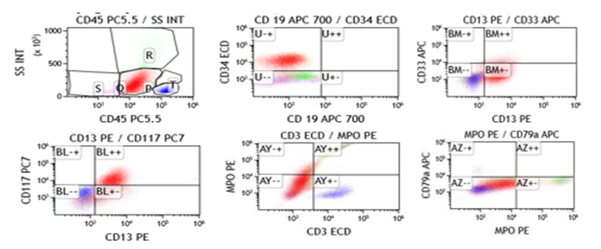
Figure 1: Flowcytometry graphs showing sequential gating of blasts(in red) in a case of AML-M1. (green:granulocytes, blue :lymphocytes).
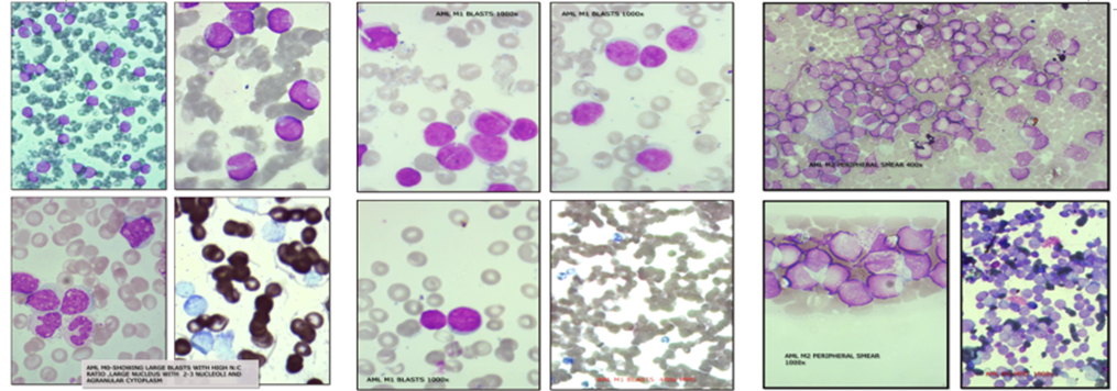
Figure 2 Figure 3 Figure 4
Figure 2: Peripheral smear showing large agranular myeloblasts (AML-M0) with high nuclear-to-cytoplasmic ratio and prominent nucleoli. MPO cytochemistry negative (1000×).
Figure 3: AML-M1 blasts with high nuclear-to-cytoplasmic ratio and prominent nucleoli. MPO-positive cytoplasmic staining confirms myeloid lineage (1000×).
Figure 4: AML-M2 blasts with partial maturation. MPO-positive staining is seen in differentiating cells (400×/1000×).
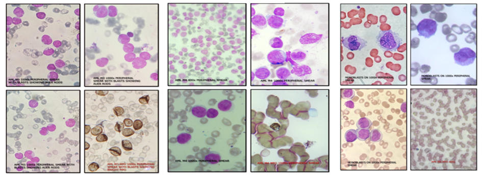
Figure 5 Figure 6 Figure 7
Figure 5: AML-M3 (acute promyelocytic leukemia): hypergranular promyelocytes with Auer rods. Strong MPO positivity. HLA-DR and CD34 negative by MFC (1000×).
Figure 6: AML-M4 blasts showing myelomonocytic differentiation. MPO positive; CD64 and CD14 expressed by MFC (400×/1000×).
Figure 7: AML-M5 monoblasts: abundant cytoplasm, folded/convoluted nuclei. MPO weak/negative; CD64-positive by MFC (1000×).
Multipara meter Flow Cytometry
Immunophenotyping was performed on peripheral blood or bone marrow aspirates using a 10-color Beckman Coulter Navios EX flow cytometer with bulk-lyse staining. A minimum of 10,000 events were recorded per sample. Data were stored as list-mode files and analyzed using dedicated flow cytometry software.
The antibody panel included: myeloid markers (CD13, CD33, CD117, intracellular MPO); monocytic markers (CD14, CD16, CD64); progenitor and immaturity markers (CD34, CD45, HLA-DR); and lymphoid or aberrant markers (CD3, CD7, CD10, CD19, CD20, CD56, cytoplasmic CD3, cytoplasmic CD79a). Blast identification used a CD45 versus side scatter gating strategy; blasts characteristically showed low side scatter and dim CD45 expression.
Antigen positivity was defined as expression in ≥20% of blast cells. For CD34 and cytoplasmic markers, a threshold of ≥10% was applied, consistent with established flow cytometry practice [8,11]. HLA-DR negativity in the context of CD33-bright, CD34-negative blasts was used as a supportive criterion for AML-M3 (acute promyelocytic leukemia, APL).
For cases classified as biphenotypic or lineage-ambiguous by morphology, MPAL criteria were applied per WHO 2022: B-lymphoid lineage was defined by CD19 strong positivity or expression of ≥2 of CD19 (weak), CD10, cytoplasmic CD79a; T-lymphoid lineage by cytoplasmic CD3 or surface CD3; myeloid lineage by intracellular MPO or monocytic differentiation (CD11c, CD14, CD64, lysozyme). Lineage scoring used EGIL criteria as a secondary reference.
Quality control included daily instrument calibration, internal controls, Levy-Jennings chart monitoring, and reagent standardization.
Diagnostic Impact Assessment
To quantify the added diagnostic value of MFC, cases were classified first by morphology and cytochemistry alone, and then re-evaluated after incorporating MFC findings. Any change in FAB subtype, lineage assignment, or detection of MPAL was recorded as a diagnostic impact event. This comparison was performed prospectively as part of the study workflow.
Statistical Analysis
Data were compiled using standard spreadsheet software and analyzed using descriptive methods. Results are expressed as frequencies and proportions (rounded to one decimal place). Given the small sample size (n=30), no inferential statistics were performed. Confidence intervals are not reported given this exploratory nature; findings should be interpreted as descriptive. No power calculation was performed for this pilot study; sample size was determined by consecutive enrollment over the study period.
RESULTS:
Demographics and Clinical Features
Thirty cases were included (15 male, 15 female; M:F ratio 1:1). Age ranged from childhood to late adulthood, with the highest proportion in the 31–45 year group (33%). Table 1 shows the demographic distribution.
Table 1: Demographic Profile of Study Population (n=30)
|
Variable |
n |
% |
|
Male |
15 |
50.0 |
|
Female |
15 |
50.0 |
|
Age 0–15 years |
6 |
20.0 |
|
Age 16–30 years |
5 |
16.7 |
|
Age 31–45 years |
10 |
33.3 |
|
Age 46–60 years |
6 |
20.0 |
|
Age 61–70 years |
3 |
10.0 |
Generalized weakness was the most common presenting symptom (93%), followed by fever (77%). Pallor was the predominant physical finding (93%); hepatomegaly and splenomegaly were present in 57% and 27% of cases, respectively. Severe anemia (hemoglobin <8 g/dL) was documented in 80% and thrombocytopenia in 97% of cases (Table 2).
Table 2: Clinical and Hematological Findings (n=30)
|
Parameter |
n |
% |
|
Generalized weakness |
28 |
93.3 |
|
Fever |
23 |
76.7 |
|
Pallor |
28 |
93.3 |
|
Hepatomegaly |
17 |
56.7 |
|
Splenomegaly |
8 |
26.7 |
|
Hemoglobin <8 g/dL |
24 |
80.0 |
|
Thrombocytopenia |
29 |
96.7 |
FAB Subtype Distribution
Following morphological assessment and MFC, cases were classified by FAB criteria. AML-M2 was the most common subtype (47%), followed by M1 (17%) and M4 (13%). AML-M3 accounted for 10% of cases. One case (3%) was classified as mixed phenotype acute leukemia (MPAL) following MFC evaluation; this case was initially classified as AML-M0 by morphology (Table 3).
Table 3: FAB Subtype Distribution (n=30)
|
FAB Subtype |
n |
% |
|
AML-M0 |
2 |
6.7 |
|
AML-M1 |
5 |
16.7 |
|
AML-M2 |
14 |
46.7 |
|
AML-M3 |
3 |
10.0 |
|
AML-M4 |
4 |
13.3 |
|
AML-M5 |
1 |
3.3 |
|
MPAL (WHO 2022) |
1 |
3.3 |
Immunophenotypic Findings
CD33 was expressed in all 30 cases (100%), confirming its role as a highly sensitive pan-myeloid marker. CD13 was expressed in 93% of cases. CD117 expression was found in 77% of cases, with higher frequency in less-differentiated subtypes (M0, M1, M2). CD34 was expressed in 53% of cases; notably, it was absent in all AML-M3 and AML-M5 cases, consistent with their well-characterized immunophenotypic profiles. HLA-DR negativity was confirmed in all three M3 cases (see Table 4).
Monocytic differentiation markers showed selective expression. CD64 was positive in 23% of cases, predominantly in M4 and M5. CD16 was expressed in 13%, also associated with monocytic subtypes.
Aberrant antigen expression was detected in 14 cases (47%). CD56 was the most common aberrant marker (30%), with the highest frequency in AML-M2. CD7 was expressed in 13% of cases, particularly in M0 and M1 subtypes(Figure 1). In the MPAL case, co-expression of myeloid (intracellular MPO, CD33) and T-lymphoid (cytoplasmic CD3) markers satisfied WHO 2022 MPAL criteria.
Table 4: Immunophenotypic Marker Expression (n=30)
|
Marker |
Positive Cases |
% |
Predominant Subtype(s) |
|
CD33 |
30 |
100.0 |
All subtypes |
|
CD13 |
28 |
93.3 |
All subtypes |
|
CD117 |
23 |
76.7 |
M0, M1, M2 |
|
CD34 |
16 |
53.3 |
M0, M1, M2 (absent in M3, M5) |
|
HLA-DR (negative) |
3 |
10.0 |
M3 (all 3 cases) |
|
CD64 |
7 |
23.3 |
M4, M5 |
|
CD16 |
4 |
13.3 |
M4, M5 |
|
CD56 (aberrant) |
9 |
30.0 |
M2 predominantly |
|
CD7 (aberrant) |
4 |
13.3 |
M0, M1 |
|
MPAL markers (cCD3+MPO) |
1 |
3.3 |
MPAL case |
Diagnostic Impact of MFC
MFC altered or refined the morphological diagnosis in 6 of 30 cases (20%). Specific changes included: (a) reclassification of 2 AML-M0 cases — one confirmed as AML-M0 with aberrant CD7 expression (altering clinical risk assessment), one reclassified as MPAL based on co-expression of cytoplasmic CD3 and intracellular MPO per WHO 2022 criteria; (b) confirmation of AML-M3 in 3 morphologically suspected cases via CD34-negative, HLA-DR-negative, CD33-bright phenotype, reinforcing ATRA eligibility pending PML-RARA molecular confirmation; and (c) identification of aberrant CD56 expression in AML-M2 cases not apparent on morphology, with prognostic implications.
Note: PML-RARA FISH/RT-PCR confirmation was not available at our center for the three AML-M3 cases. ATRA therapy was initiated based on morphological and immunophenotypic findings. This is acknowledged as a limitation; our center is in the process of establishing molecular diagnostic capacity.
Case Note: Extramedullary Leukemic Infiltration
One case — a 4-year-old female presenting with acute abdomen — was incidentally found to have leukemic infiltration of the appendix and adjacent tissues on histopathological examination following emergency appendectomy. Blast cells involved the mucosa, muscularis propria, and serosa (Figure 8). This preceded bone marrow diagnosis of AML-M2 by 72 hours. MFC confirmed myeloid lineage. Extramedullary AML (chloroma/myeloid sarcoma) presenting as acute appendicitis is rare and may represent the initial manifestation of AML. This case highlights the importance of flow cytometry in rapidly characterizing blast populations encountered in unexpected anatomical sites.
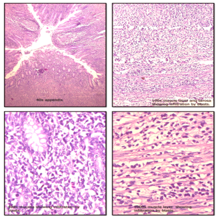
Figure 8. Extramedullary AML — histopathological sections of the appendix showing leukemic infiltration by myeloid blast cells involving the mucosa, muscularis propria, and serosa (40×–400×). Myeloid lineage confirmed by MFC.
DISCUSSION:
This study evaluated the diagnostic contribution of MFC in 30 cases of AML in a tertiary center where molecular diagnostic infrastructure is limited. The key finding is that MFC changed or refined the morphological diagnosis in 20% of cases, providing objective evidence of its added value in this setting.
AML-M2 was the predominant subtype (47%), consistent with prior reports from Indian tertiary centers and with the finding that t(8;21) — the cytogenetic correlate of M2 — is among the most common recurrent AML abnormalities in South Asian populations [16,18]. The relatively high M2 frequency may also reflect diagnostic ascertainment: M2 blasts have distinct morphological features that facilitate recognition even without molecular testing. Studies from Tata Memorial Hospital and other large Indian centers have reported similar M2 predominance [16,20].
CD33 positivity in 100% and CD13 in 93% of cases reaffirm these markers as robust myeloid lineage indicators, consistent with established literature [11,12]. The utility of CD117 as a marker of early myeloid differentiation, particularly in M0, M1, and M2, aligns with its role as a receptor tyrosine kinase expressed on myeloid progenitors [13]. CD34 negativity in all M3 and M5 cases is immunophenotypically characteristic: APL blasts typically lack CD34 and HLA-DR, a pattern that serves as a critical diagnostic flag when morphological assessment of Auer rods may be insufficient[5,11].
Aberrant antigen expression was detected in 47% of cases in this series. CD56 was the most common aberrant marker (30%), predominantly in AML-M2. Prior studies have reported CD56 aberrant expression in 15–30% of AML cases [15,19], placing our finding within the expected range. CD56 expression in AML is clinically significant: multiple studies have associated it with shorter overall survival, increased extramedullary disease, and resistance to standard induction chemotherapy [15]. Its detection therefore has direct prognostic implications, even in the absence of molecular profiling. CD7 aberrant expression (13%) in minimally differentiated and myeloblastic subtypes is similarly well-documented and may reflect origin from an early lymphomyeloid progenitor [12,14].
The single MPAL case warrants particular attention. Initially classified morphologically as AML-M0 given the absence of clear myeloid differentiation, MFC revealed co-expression of intracellular MPO and cytoplasmic CD3, satisfying WHO 2022 criteria for T/myeloid MPAL. This is a clinically critical distinction: MPAL requires a different therapeutic approach — typically AML-type or ALL-type induction, sometimes with allogeneic stem cell transplantation — compared to standard AML therapy [1,2]. This case alone illustrates why MFC is indispensable in diagnostically ambiguous presentations. The reclassification of this case from AML-M0 to MPAL by MFC is one of the clearest demonstrations of added diagnostic value in this series.
The extramedullary case is also noteworthy. Myeloid sarcoma presenting as acute appendicitis is uncommon, with fewer than 50 cases reported in the English literature to date. In resource-limited settings, the absence of rapid MFC may delay leukemia diagnosis when extramedullary involvement precedes bone marrow presentation. This case demonstrates the value of immunophenotyping in unexpected tissue specimens.
From a broader perspective, our findings support the continued role of MFC as a practical diagnostic bridge in settings where WHO 2022 molecular classification is not yet achievable. We used FAB criteria operationally because cytogenetic and molecular data were unavailable; this is a real-world constraint shared by many tertiary hospitals in India and other middle-income countries. We do not claim equivalence with WHO 2022 classification; rather, we demonstrate that within the FAB framework, MFC adds measurable diagnostic value. Future integration of FISH for key cytogenetic abnormalities (t(8;21), inv(16), t(15;17)) and targeted molecular panels would substantially improve classification accuracy and risk stratification at our center.
Limitations of this study must be acknowledged. The sample size of 30 cases limits generalizability and prevents meaningful subgroup analysis for rare subtypes (M0, M5, MPAL). No power calculation was performed; this was a consecutive enrollment pilot study. The absence of cytogenetic and molecular data precludes WHO 2022 classification and full ELN risk stratification. PML-RARA molecular confirmation was not available for the three M3 cases. Patient follow-up and outcome data were not collected, precluding survival analysis. These limitations are the subject of a planned follow-up study incorporating FISH, targeted molecular profiling, and prospective outcome tracking.
CONCLUSION:
morphological classification in 20% of cases. Key contributions included identification of MPAL, confirmation of AML-M3 immunophenotype supporting ATRA eligibility, and detection of prognostically significant aberrant markers — particularly CD56. In resource-limited settings, MFC provides objective, reproducible lineage characterization that morphology and cytochemistry alone cannot achieve.
The high frequency of aberrant antigen expression (47%) underscores the immunophenotypic heterogeneity of AML and the inadequacy of morphological subclassification in isolation. The case of appendicular myeloid sarcoma illustrates the diagnostic value of MFC when leukemia presents at atypical sites.
These findings support integrating MFC into the routine diagnostic workflow for acute leukemias at tertiary centers, even where molecular infrastructure is limited. Prospective studies with larger cohorts, molecular correlation, and outcome data are needed to validate these observations and to support the transition toward WHO 2022-aligned classification at our center.
REFERENCES:
1. Khoury JD, Solary E, Abla O, et al. The 5th edition of the World Health Organization classification of haematolymphoid tumours: myeloid and histiocytic/dendritic neoplasms. Leukemia. 2022;36(7):1703–1719.
2. Arber DA, Orazi A, Hasserjian RP, et al. International Consensus Classification of myeloid neoplasms and acute leukemias. Blood. 2022;140(11):1200–1228.
3. Döhner H, Wei AH, Appelbaum FR, et al. Diagnosis and management of AML in adults: 2022 ELN recommendations from an international expert panel. Blood. 2022;140(12):1345–1377.
4. Short NJ, Rytting ME, Cortes JE. Acute myeloid leukaemia. Lancet. 2018;392(10147):593–606.
5. Estey EH. Acute myeloid leukemia: 2021 update on risk-stratification and management. Am J Hematol. 2020;95(11):1368–1398.
6. Papaemmanuil E, Gerstung M, Bullinger L, et al. Genomic classification and prognosis in acute myeloid leukemia. N Engl J Med. 2016;374(23):2209–2221.
7. Heuser M, Ofran Y, Boissel N, et al. Acute myeloid leukaemia in adult patients: ESMO Clinical Practice Guidelines for diagnosis, treatment and follow-up. Ann Oncol. 2020;31(6):697–712.
8. Craig FE, Foon KA. Flow cytometric immunophenotyping for hematologic neoplasms. Blood. 2008;111(8):3941–3967.
9. van Dongen JJM, Lhermitte L, Böttcher S, et al. EuroFlow antibody panels for standardized n-dimensional flow cytometric immunophenotyping of normal, reactive and malignant leukocytes. Leukemia. 2012;26(9):1908–1975.
10. Kalina T, Flores-Montero J, van der Velden VHJ, et al. EuroFlow standardization of flow cytometer instrument settings and immunophenotyping protocols. Leukemia. 2012;26(9):1986–2010.
11. Chen X, Cherian S. Acute myeloid leukemia immunophenotyping by flow cytometric analysis. Clin Lab Med. 2017;37(4):753–769.
12. Lucas F, Hergott CB. Advances in acute myeloid leukemia classification, prognostication and monitoring by flow cytometry. Clin Lab Med. 2023;43:377–398.
13. Ally F, Lucas F, Roshal M. Acute myeloid leukemia: diagnosis and evaluation by flow cytometry. Cancers (Basel). 2024;16(22):3855.
14. Saft L. The role of flow cytometry in the classification of myeloid disorders. Pathologie. 2023;44(3):164–175.
15. Buccisano F, Hourigan CS, Walter RB. The prognostic significance of measurable ("minimal") residual disease in acute myeloid leukemia. Curr Hematol Malig Rep. 2017;12(6):547–556.
16. Ghosh S, Shinde SC, Kumaran GS, et al. Haematologic and immunophenotypic profile of acute myeloid leukemia: an experience of Tata Memorial Hospital. Indian J Cancer. 2003;40(2):71–76.
17. Saxena R, Anand H. Flow cytometry in acute leukemia. Indian J Hematol Blood Transfus. 2008;24(4):146–150.
18. Gupta N, Pawar R, Banerjee S, et al. Spectrum and immunophenotypic profile of acute leukemia: a tertiary center flow cytometry experience. Mediterr J Hematol Infect Dis. 2019;11(1):e2019017.
19. Venugopalan RK, Singh N, Anthony ML, et al. Leukemia-associated aberrant immunophenotype: a flow cytometry-based experience of 110 cases from a tertiary care center in Northern India. J Cancer Res Ther. 2023;19(5):1335–1339.
20. Vemprala A, Gajendra S, Gupta R, et al. Clinico-hematological profile of acute myeloid leukemia: experience from a tertiary care cancer center in North India. Cureus. 2023;15(12):e50869.