A STUDY ON PLATELET COUNT / SPLENIC DIAMETER RATIO AS A NON-INVASIVE PREDICTOR OF OESOPHAGEAL VARICES IN HEPATIC CIRRHOSIS.
- NITIN GUPTA , Resident, Department of Medicine, S.P. Medical College Bikaner.
- VIJAY KUMAR TUNDWAL , Professor, Department of Medicine, S.P. Medical College Bikaner.
- PRIYANKA SUNDA , Assistant Professor, Department of Medicine, S.P. Medical College Bikaner.
- RITWIK AGARWAL , Assistant Professor, Department of Medicine, S.P. Medical College Bikaner.
- DIVYANSHI TUNDWAL , MBBS
- SARANSHI SINGH , Resident, department of Anaesthesia, S.P. Medical College Bikaner.
- B L MEENA , Professor, Department of Medicine, S.P. Medical College Bikaner.
Article Information:
Abstract:
Background: Cirrhosis is a common disease which has significant morbidity and it is one of the common cause for premature mortality. The spectrum is from acute infection mostly asymptomatic to a chronic hepatitis B which leads to complications such as cirrhosis and hepatocellular carcinoma. Following exposure to hepatitis B 15-20% develop cirrhosis over 5-20 years. Aim: To assess the validity of platelet count/splenic diameter ratio as a non invasive predictor of oesophageal varices in Hepatic cirrhosis according to endoscopic grading of varices. Methods: This was a analytical cross-sectional study conducted at S.P. Medical College & P.B.M. Associated Group of Hospitals, Bikaner during July 2023 to June 2024. One hundred ten patients were randomly selected from IPD, Dept of Medicine. Results: Mean age of patients was 38.46±9.63 years, while male to female ratio was 1.82:1. Out of these 110 patients, 72.7% were alcoholic, 7.27% cases had hepatitis B, 6.36% patients had hepatitis C. Out of 11 cases of grade 1 varices, 0, 1 and 10 cases had Hb <7gm/dl, 7-11gm/dl and >11gm/dl respectively. Out of 9 cases of grade 2 varices, 2,3,4 cases had Hb <7gm/dl, 7-11gm/dl >11gm/dl respectively. Out of 16 cases of grade 3, varices, 14,1 and 1 cases had Hb <7gm/dl, 7-11gm/dl and >11gm/dl respectively. Out of 38 cases of grade 4 varices, 30, 6 and 2 cases had Hb <7gm/dl, 7-11gm/dl and >11gm/dl respectively. Conclusion : We found that higher grades of esophageal varices exists with lower platelet and larger spleen size. Lower the platelet count and spleen size ratio, higher the incidence of varices and higher the grades.
Keywords:
Article :
INTRODUCTION:
Cirrhosis is a common disease which has significant morbidity and is one of the common causes for premature mortality. Portal hypertension is principal complication of hepatic cirrhosis. It can occur at any age. World-wide the most common causes are viral hepatitis and consumption of excessive alcohol1.
Among the viral hepatitis the most common are hepatitis B and C viruses. 300 million people are affected by viral hepatitis B. The spectrum is from acute infection mostly asymptomatic to a chronic hepatitis B which leads to complications such as cirrhosis and hepatocellular carcinoma. Following exposure to hepatitis B 15-20% develop cirrhosis over 5-20 years. The incidence of cirrhosis is directly proportional to the positivity of HBeAg. The patients infected with Hepatitis C are unaware of the disease and diagnosed only when they progress to chronic liver disease more than 3/4th of patients succumb to chronic infection and later leads to inadequate clearance of virus2.
The next most common cause is chronic liver disease is alcohol. The death due to alcoholic liver disease is on raising trend with over 556800 deaths/year. The mean age at presentation is also falling. When the alcohol consumption is > 160g/day for 8 years they are more to develop cirrhosis. The 3rd most common cause of chronic liver disease is Non-alcoholic fatty liver disease (NAFLD). It mostly affects the affluent societies. The prevalence increases with raise of obesity. The patients with Diabetes and Metabolic syndrome are more prone to develop NASH3. The major complication of cirrhosis is portal hypertension which in turn leads to variceal bleeding which accounts for 35-40% mortality4.
Aim: To assess the validity of platelet count/splenic diameter ratio as a non invasive predictor of oesophageal varices in Hepatic cirrhosis according to endoscopic grading of varices.
MATERIALS AND METHODS:
This was a analytical cross-sectional study conducted at S.P. Medical College & P.B.M. Associated Group of Hospitals, Bikaner during July 2023 to June 2024. One hundred ten patients were randomly selected from IPD, Dept of Medicine, S.P. Medical College & P.B.M. Associated Group of Hospitals, Bikaner.
Sample size – 110 subjects.
Sample size was calculated on OpenEpi.com version 3 using the formula:
Sample Size Formula
N=(Z 1-@/2)2*sensitivity (1-sensitivity)/d 2 *prevalence where,
N= sample size
Z= standard score at 95% level of significance = 1.96
p=prevalence of esophageal varices = 0.802 Sensitivity=0.84
d=(absolute error) = 10% of prevalence
N = 1 1 0
Data Collection:
After noting the preliminary data of the subject, a complete history was taken and clinical examination was done including history of active or previous haemetemesis, melena, bleeding PR, alcoholism, blood transfusion, intake of hepatotoxic drug, STD, IV drug abuse, jaundice, anemia, edema, stigmata of CLD, dilated abdominal veins, ascites, splenomegaly and encephalopathy. All patients underwent biochemical tests, like liver function tests, complete blood counts, renal function tests, prothrombin time, ultrasonography of the abdomen to confirm the presence of cirrhosis and to record the spleen bipolar diameter, portal vein size, ascites and presence of collaterals.
UGIE was done for the presence and size of EV was determined and recorded for each patient. Platelet count, spleen diameter was calculated in all patients as platelet count/spleen diameter ratio was evaluated. A previously designed proforma was used to collect the demographic and clinical details of the patients.
Statistical methods:
Appropriate statistical analysis were applied as and when required using Epi info software from CDC.
RESULTS:
In our study out of 110 cases 40.91% cases were seen in the age group of 31- 40 years age followed by 32.73% cases in 18-30 years age and 10.91% cases were in 41–50 years age whereas 6.36% cases in >60 years followed by 9.09% cases in 51– 60 years age group. The mean age of study population was 38.46 ± 9.63 years.
Table 1 Distribution of cases according to age group (years)
|
Age Group (Years) |
No. of cases |
Percentage |
|
18-30 |
36 |
32.7 |
|
31-40 |
45 |
40.9 |
|
41-50 |
12 |
10.9 |
|
51-60 |
10 |
9.1 |
|
>60 |
7 |
6.4 |
|
Total |
110 |
|
|
Mean |
38.46 |
|
|
SD |
9.63 |
|
64.55% cases were male whereas 35.55% cases were female in our study with male to female ratio was 1.82:1. 72.7% cases were alcoholic, 7.27% cases had hepatitis B, 6.36% cases had hepatitis C and 13.6% had other etiology.
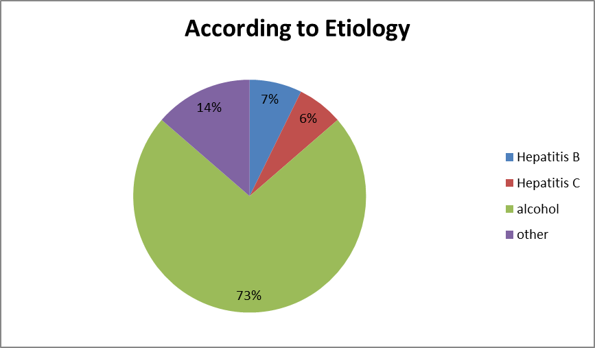
Graph 1. Distribution of cases according to etiology
Out of 11 cases of grade 1 varices, 0 case had Hb <7 gm/dl, 1 case had Hb 7-11gm/dl and 10 cases had Hb >11gm/dl. Out of 9 cases of grade 2 varices, 2 cases had Hb <7gm/dl, 3 cases had Hb 7-11gm/dl and 4 cases had Hb >11gm/dl. Out of 16 cases of grade 3 varices, 14 cases had Hb <7gm/dl, 1 case had Hb 7-11gm/dl and 1 case had Hb >11 gm/dl. Out of 38 cases of grade 4 varices, 30 cases had Hb <7gm/dl, 6 cases had Hb 7-11gm/dl and 2 cases had Hb >11gm/dl. This shows that more the grade of esophageal varices, lesser will be the Hb of patient.
Table 2. Distribution of cases according to grading of esophageal varices with haemoglobin status
|
Grade of Varices |
Haemoglobin level (gm/dl) |
|||
|
<7 |
7-11 |
>11 |
Total |
|
|
1 |
0 |
1 |
10 |
11 |
|
2 |
2 |
3 |
4 |
9 |
|
3 |
14 |
1 |
1 |
16 |
|
4 |
30 |
6 |
2 |
38 |
|
Total |
46 |
11 |
17 |
74 |
In present study, 7.27% cases had 1-2 mg% serum bilirubin and 12.73% cases had 2.1 – 3 mg% serum bilirubin whereas 23.64% cases had 3.1 – 4 mg% serum bilirubin followed by 56.36% cases had >4 mg% serum bilirubin.
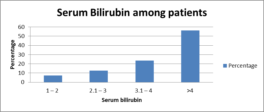
Graph 2. Distribution of Serum Bilirubin among patients in our study
In our findings, 22 cases had <60000 platelets followed by 18 cases had 80 – 60000 and 9 cases had >12.5 * 103. P<0.05 signifies that lesser the no. of platelets, more will be the chance of having esophageal varices.
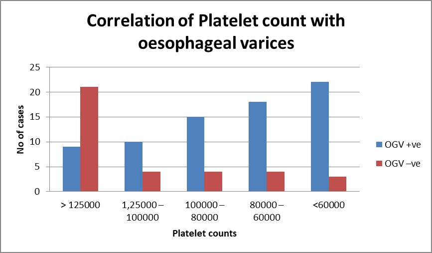
Graph 3. Correlation of Platelet count with oesophageal varices
Out of 74 cases of esophageal varices, maximum 9 cases had spleen diameter <150 mm and 65 cases had >150 mm.
Table 3. Distribution of esophageal varices in patients according to spleen diameter
|
SPLEEN DIAMETER(mm) |
OGV +ve |
OGV -ve |
|
< 150 |
9 |
21 |
|
> 150 |
65 |
15 |
|
Total |
74 |
36 |
|
P value |
0.0001** |
|
Out of 74 cases of esophageal varices, maximum 65 cases had Platelet count and Spleen diameter ratio <909 and 9 cases had >909. (p<0.05) Out of 65 cases of esophageal varices which had ratio of platelet count / splenic diameter <909, 37 cases were in grade 4, 15 cases in grade 3, 8 cases had grade 2 and 5 cases had grade 1 esophageal varices. Out of 9 cases of EV which had ratio of platelet count/splenic diameter >909, 6 cases were in grade 1, 1 case had grade 2, 1 case had grade 3 and 1 case had grade 4 esophageal varices.
Table 4. Correlation of grading of esophageal varices and ratio of platelet count / splenic diameter
|
Grade |
RATIO |
Total |
|
|
< 909 |
> 909 |
||
|
1 |
5 |
6 |
11 |
|
2 |
8 |
1 |
9 |
|
3 |
15 |
1 |
16 |
|
4 |
37 |
1 |
38 |
|
Total |
65 |
9 |
74 |
DISCUSSION:
Many studies were conducted based on platelet count- spleen diameter ratio as a non-invasive predictor of oesophageal varices in patients with cirrhosis. This was a hospital based analytical cross-sectional study conducted on 110 cases of liver cirrhosis presenting to the IPD of Medicine Department, at S.P. Medical College and P.B.M. Associated Group of Hospitals, Bikaner (Raj.).
In our study out of 110 cases 40.91% cases were seen in the age group of 31- 40 years age followed by 32.73% cases in 18-30 years age and 10.91% cases were in 41–50 years whereas 6.36% cases in >60 years followed by 9.09% cases in 51 – 60 years age group. The mean age of study population was 38.46 ± 9.63 years with age range of 18 – 70 years. Similarly, El Makarem5 the mean age was 48 years (range 36-60 years). Also, Mandar et al6 observed patient’s age range from 26 to 70 years. Maximum number of patients were in age group of 31-50 years (58.57%), followed by 51-70 years (35.74%). This is almost similar to study done by Cherian et al7 and Sarangapani et al8. Results of above studies were quite similar with our study.
In our study, out of 110 cases 64.55% cases were male whereas 35.55% cases were female in our study. Out of 110 cases, 63.64% cases were rural whereas 36.36% cases were urban. El Makarem5 found that 115 men and 60 women were included in the study and Mandar et al6 observed, out of 70 patients, 64 (91.43%) were male and 6 (8.57%) were females. Male preponderance was seen.
In our study, 72.7% cases were alcoholic, 7.27% cases had hepatitis B followed by 6.36% cases had hepatitis C and 13.6% cases had other etiology. Similarly, Mandar et al6 majority of patients had alcohol in5d1uced liver disease (90.00%) and remaining were due to hepatotrophic virus infection (10.00%), 63(90%) patients had cause of alcoholism, 7 (10%) were due to hepatotrophic virus infection of which 4 (5.71%) were HbsAg related and 3 (4.28%) HCV related. This is almost similar to study by Cherian et al8 and Baig et al9. However, this is in sharp contrast to study by Berger et al10 and Sarangapani et al7 who observed hepatotropic viruses as main cause of cirrhosis of liver in their study as compared to alcohol as cause.
In our study, out of 11 cases of grade 1 varices, 0 case had Hb <7 gm/dl, 1 case had Hb 7- 11gm/dl and 10 cases had Hb >11gm/dl. Out of 9 cases of grade 2 varices, 2 cases had Hb <7gm/dl, 3 cases had Hb 7-11gm/dl and 4 cases had Hb >11gm/dl. Out of 16 cases of grade 3 varices, 14 cases had Hb <7gm/dl, 1 case had Hb 7-11gm/dl and 1 case had Hb >11 gm/dl. Out of 38 cases of grade 4 varices, 30 cases had Hb <7gm/dl, 6 cases had Hb 7-11gm/dl and 2 cases had Hb >11gm/dl. This shows that more the grade of esophageal varices, lesser will be the Hb of patient. Konrad et al11 and Lur et al12 also had similar results as that of our study.
In our study, out of 110 cases 7.27% cases had 1-2 mg% serum bilirubin and 12.73% cases had 2.1 – 3 mg% serum bilirubin whereas 23.64% cases had 3.1 – 4 mg% serum bilirubin followed by 56.36% cases had >4 mg% serum bilirubin.
In our study, out of 74 cases of esophageal varices maximum 9 cases had spleen diameter <150 mm and 65 cases had >150 mm. Similarly El Makarem5 had bipolar spleen diameter (mm) of 159.4 ± 24.2. Also Mandar et al6 observed, 63 (90.00%) patients having splenomegaly also they observed 36 (51.43%) patients having spleen diameter ranging from 126-150 mm, 18 (25.71%) patients with diameter more than 150 mm and 16 (22.86%) patients having range below or equal to 125 mm.
In our study, out of 74 cases of esophageal varices maximum 22 cases had <60000 platelets followed by 18 had 80 – 60000 and 9 cases had >12.5 * 103. (p<0.05) Similarly El Makarem5 had Platelet count (No/cmm) of 119480.9 ± 38725.7 in cases with oesophageal varices. Also Mandar et al6 35.71% patients belonged to platelet ranging below 100000. 44.28% patients belonged to platelet range from 100000–200000 and 20.00% patients had platelet range above 200000.
In contrast to study by Abbasi et al13 in which there was inverse correlation between platelet count with grading of varices (r=0.321, p<0.001) which means that the severity of thrombocytopenia increased as the grading of esophageal varices increased.
Out of 65 cases of esophageal varices which had ratio of platelet count / splenic diameter <909, 37 cases were in grade 4, 15 cases in grade 3, 8 cases had grade 2 and 5 cases had grade 1 cases with esophageal varices. Out of 9 cases of EV which had ratio of platelet count/splenic diameter >909, 6 cases were in grade 1, 1 case had grade 2, 1 case had grade 3 and 1 case had grade 4 esophageal varices. Similar study was done by Schwarzenberger et al10 and Abbasi et al,13 Navgeet mathur et al,14 Mona A. Abu El Makarem,5 Soichiro Shibata,15 patients with EVs were older; had lower platelet counts; higher Child-Pugh scores; developed hepatic encephalopathy, ascites, and jaundice more frequently; and had lower prothrombin concentrations, lower platelet count/bipolar spleen diameter ratios, and higher bipolar spleen diameters.
CONCLUSION:
Our study found that higher grades of esophageal varices exists with lower platelet and larger spleen size. Lower the platelet count and spleen size ratio, higher the incidence of varices and higher the grades.
A cut off value of platelet count/ splenic diameter ratio of ≤ 909 when applied in order to take in to consideration the decrease in platelet count due to hypersplenism; it was found to be a good predictor of presence and grade of esophageal varices.
The use of platelet counts and platelet count/ splenic diameter ratio in appropriate subgroups of cirrhotic patients for screening and follow up of esophageal varices can substantially reduce the cost of healthcare and discomfort for patients as well as reduce burden on endoscopy units.
REFERENCES:
1. Gupta TK, Chen L, Groszmann RJ. Pathophysiology of portal hypertension. Clin. Liver Dis. 1997; 1:1-12.
2. De Franchis R, Primignani M. Natural history of portal hypertension inpatients with cirrhosis. Clin. Liver Disease 2001; 5:645-63.
3. Luketic VA, Sanyal AJ. Oesophageal varices. I clinical presentation, medical therapy and endoscopic therapy. Gastroenterology clinics of North America 2000:29:337-85.
4. Rigo GP, Merghi A, Chalen NJ, Mastronardi M, Codoluppi PL, Ferrari Aet al. A prospective study of the ability of the three endoscopic classifications to predict haemorrhage from oesophageal varices. Gastro intest Endos. 1992;38:425-9.
5. El Makarem MAA. Platelet count/bipolar spleen diameter ratio for the prediction of esophageal varices: The special Egyptian situation Noninvasive prediction of esophageal varices PMID: 22087154 PMCID: PMC3206702
6. Waghralkar MG, Vijay S. Correlation of platelet count/splenic diameter ratio for the diagnosis of esophageal varices in liver. Int J Res Med Sci 2021;9(6):1609-15.
7. Sarangapani A, Shanmugam C, Kalyanasundaram M, Rangachari B, Thangavelu P, Subbarayan KJ. Noninvasive prediction of large esophageal varices in chronic liver disease patients. Saudi J Gastroenterol. 2010;16(1):38-42.
8. Cherian JV, Deepak N, Ponnusamy PR, Somasundaram A, Jayanthi V. Noninvasive predictors of esophageal varices. Saudi J Gastroenterol. 2011;17(1):64-8.
9. Baig WW, Nagaraja MV, Varma M, et al. Platelet count to spleen diameter ratio for the diagnosis of esophageal varices: Is it feasible? Can J Gastroenterol. 2008;22(10):825-8.
10. Schwarzenberger E, Meyer T, Golla V, et al. Utilization of Platelet Count/Spleen Diameter Ratio in Predicting the Presence of Esophageal Varices in Patients With Cirrhosis. J Clin Gastroenterol. 2010;44:146-50.
11. Konrad H. Die hepatogene Anämie [Hepatogenic anemia]. Z Gesamte Inn Med. 1983;38(3):78-81.
12. Korolko IUR, Sarycheva TG, Kotelńikov VM, Kozinets GI, Zherebtsov LA. Anemic syndrome in chronic hepatitis and liver cirrhosis. Klin Med (Mosk). 1993;71(5):45-8.
13. Abbasi A, Butt N, Bhutto AR, Munir SM. Correlation of thrombocytopenia with grading of esophageal varices in chronic liver disease patients. J Coll Physicians Surg Pak. 2010;20(6):369-72.
14. Mathur N, Patel H, Mangalia R, Mehta A, Mathur M, Verma A, Patyal A. Correlation of non-invasive parameters with upper gastrointestinal endoscopy findings for presence and grading of gastroesophageal varices in liver cirrhosis patients. J Family Med Prim Care. 2024 Apr;13(4):1347-1353. doi: 10.4103/jfmpc.jfmpc_702_23. Epub 2024 Apr 22. PMID: 38827664; PMCID: PMC11141970.
15. Shibata S, Umemura T, Yamazaki T, Fujimori N, Ichikawa Y, Kimura T, Joshita S, Komatsu M, Matsumoto A, Tanaka E. Liver stiffness-spleen size-to-platelet ratio risk score identifies esophageal varices in Japanese patients with chronic hepatitis C. Hepatol Res. 2016 Aug;46(9):884-9. doi: 10.1111/hepr.12631. Epub 2016 Jan 11. PMID: 26662612.