INITIAL EXPRESSION OF MMP 9 AND TIMP 1 IN ACUTE SURGICAL WOUNDS AND THEIR RELATION TO WOUND HEALING.CRITICAL RESOLUTION OF VIABLE HEALING.
- Poorna Chandra Reddy Danduri. , Assistant Professor, Department of General Surgery, Sri Balaji Medical College Hospital and Research Institute.
- C S PRAVEEN , Associate Professor, Department of Pathology, PES Medical College and Research Institute.
- BHARI PRIYA , Assistant Professor, Department of DVL, Sri Balaji Medical College Hospital and Research Institute.
- M.Deepa , Associate Professor, Department of Biochemistry, Sri Balaji Medical College Hospital and Research Institute.
- Bokkisam Suneel , Assistant Professor, Department of Biochemistry, Narayana Medical College, Nellore.
Article Information:
Abstract:
Background: Wounds can be classified based on etiology, anatomical location, duration (acute or chronic), method of closure, and tissue characteristics. Acute wounds, such as surgical wounds, are characterized by orderly and timely healing through a sequence of overlapping processes including inflammation, proliferation, and remodeling. Matrix metalloproteinases (MMPs), particularly MMP-9, and their inhibitors like tissue inhibitor of metalloproteinase-1 (TIMP-1), play a crucial role in extracellular matrix remodeling, inflammation regulation, and wound healing. Dysregulation of these factors may influence healing outcomes. Objectives: To evaluate the initial expression of MMP-9 and TIMP-1 in acute surgical wounds and to study their relationship with wound healing. Materials and Methods: This prospective observational study was conducted at Kamineni Hospitals, LB Nagar, Hyderabad. A total of 50 patients of all age groups and both sexes undergoing open abdominal surgeries under the Department of General Surgery were included. Wound biopsies measuring 5 × 5 mm were obtained from the wound margins, including skin and subcutaneous tissue, at two different time intervals. The expression of MMP-9 and TIMP-1 was analyzed in relation to wound healing characteristics. Results: MMP-9 expression was observed to be elevated during the early inflammatory phase, facilitating extracellular matrix degradation, cell migration, and re-epithelialization. TIMP-1 expression played a regulatory role by balancing MMP activity. A coordinated expression of MMP-9 and TIMP-1 was associated with effective wound healing, while any imbalance may contribute to delayed or impaired healing. Conclusion: MMP-9 and TIMP-1 are critical regulators of wound healing in acute surgical wounds. Their balanced expression is essential for proper extracellular matrix remodeling and successful tissue repair. Monitoring these biomarkers may help in predicting wound healing outcomes and guiding therapeutic interventions.
Keywords:
Article :
INTRODUCTION:
A wound may be described in many ways—by its aetiology, anatomical location, by whether it is acute or chronic, by the method of closure, by its presenting symptoms or indeed by the appearance of the predominant tissue types in the wound bed. All definitions serve a critical purpose in the assessment and appropriate management of the wound through to symptom resolution or, if viable, healing.
Acute Wounds
Acute wounds are defined as disruptions in the integrity of the skin and underlying tissues that progress through the healing process in a timely and uneventful manner. The acute surgical wound is an example of a healthy wound in which healing can be maximized.
Causes of Acute Wounds
Many actions can cause an acute wound, including:
Rough surfaces scraping and rubbing against the skin
Sharp pointed objects, such as a nail, poking or jabbing into body tissue
Sharp edges or blades, such as a knife, cutting the skin cleanly
Hard blows by any objects, tearing the tissue roughly by sheer force.
Types of Acute Wounds
There are two main types of acute wounds: surgical and traumatic.
1. Surgical Wounds
These are incisions made purposefully by a healthcare professional and are cut precisely, creating clean edges around the wound. Surgical wounds may be closed (with stitches, staples or adhesive) or left open to heal.
Surgical Wound Classification
It was developed initially by the American College of Surgeons and adapted in 1985 by the Centers for Disease Control and Prevention. It divides surgical wounds into four classes:
Class I / Clean wounds: an uninfected surgical wound in which no inflammation is encountered and the respiratory, alimentary, genital, or uninfected urinary tracts are not entered. In addition, clean wounds are primarily closed and, if necessary, drained with closed drainage. Surgical wound incisions that are made after non-penetrating (i.e., blunt) trauma should be included in this category if they meet the criteria.
Class II / Clean-contaminated wounds: a surgical wound in which the respiratory, alimentary, genital, or urinary tract is entered under controlled conditions and without unusual contamination. Specifically, surgical procedures involving the biliary tract, appendix, vagina, and oropharynx are included in this category, provided no evidence of infection is encountered and no major break in technique occurs.
Class III / Contaminated wounds: open, fresh, accidental wounds. In addition, surgical procedures in which a major break in sterile technique occurs (e.g., open cardiac massage) or there is gross spillage from the gastrointestinal tract and incisions in which acute, non-purulent inflammation is encountered are included in this category.
Class IV / Dirty or infected wounds: old traumatic wounds with retained or devitalized tissue and those that involve existing clinical infection or perforated viscera. This definition suggests that the organisms causing postoperative infection were present in the wound before the surgical procedure.
2. Traumatic Wounds
These are injuries to the skin and underlying tissue caused by a force of some nature. They are classified by the object that caused the force.
Abrasion – A rough surface scrapes or rubs the skin, causing trauma and tearing the tissue (e.g., knees scraping against asphalt).
Puncture – A pointed object pokes into the tissue, sometimes causing deep multi-layered trauma (e.g., stepping on a nail).
Laceration – A sharp object delivers a hard blow to the tissue, resulting in a tear that can be jagged and irregular (e.g., bumping a leg on a table).
Incision – A straight-edged cut to the skin caused by a sharp blade (e.g., cutting a finger with a knife).
Wound Healing – Historical Aspects
The earliest recording of wound healing is in cave drawings in Spain dating back 20–30,000 years ago.
From the earliest recorded history, it is clear that the Assyrians and Egyptians knew about healing, not just from an observational point of view but also in terms of practical management.
John Hunter in 1763 AD recognized the “salutary” effects of inflammation over wound healing, described the phenomenon of wound healing and observed the factors that promote and delay wound healing.
The process by which new blood capillaries grow into a wound space after injury is known as angiogenesis. Wound angiogenesis is an important part of the proliferative phase of healing; in fact, the term “granulation tissue” was used by John Hunter in 1787 to describe the appearance of the prominent blood vessels of the initial connective tissue formed in the wound space. Healing of any skin wound, other than the most superficial, cannot occur without angiogenesis. Not only does any damaged vasculature need to be repaired, but the increased local cell activity necessary for healing requires an increased supply of nutrients from the bloodstream.
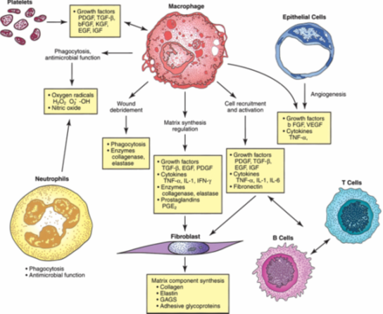
Figure1:Interaction of cellular and humoral factors in wound healing.
Acute (Normal) Wound Healing
Healing in acute wounds occurs as a sequential cascade of overlapping processes that requires the coordinated completion of a variety of cellular activities including phagocytosis, chemotaxis, mitogenesis, collagen synthesis and the synthesis of other matrix components. These activities do not occur in a haphazard manner but rather in a carefully regulated and systematic cascade that correlates with the appearance of different cell types in the wound during various stages of the healing process.
These processes involve four overlapping but well-defined phases: haemostasis, inflammation, proliferation, and remodeling.
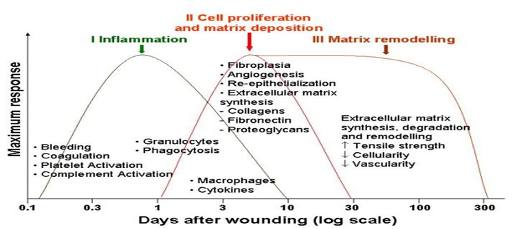
Figure2-Phases of repairing acute wound healing.
Haemostasis
Tissue injury is characterised by microvascular injury and therefore extravasation of blood into the wound. Injured vessels constrict rapidly and the coagulation cascade is activated to limit blood loss, leading to clot formation and platelet aggregation.
The clot, comprising of fibrin, fibronectin, vitronectin, von Willebrand factor and thrombospondin, provides the provisional matrix for cellular migration (10). The platelets trapped in the clot are essential for haemostasis as well as for a normal inflammatory response.
The alpha granules of the platelets contain growth factors, including platelet-derived growth factor (PDGF), insulin-like growth factor-1 (IGF-1), epidermal growth factor (EGF), and transforming growth factor-beta (TGF-β). These proteins initiate the wound healing cascade by attracting and activating fibroblasts, endothelial cells and macrophages.
The platelets also contain dense bodies that store vasoactive amines such as serotonin that increase microvascular permeability. This leads to the exudation of fluid into the extravascular space and results in tissue oedema, although this feature is more prominent during the inflammatory phase.
Early Inflammatory Phase
The next phase of healing is inflammation, which begins with the activation of complement and the initiation of the classical molecular cascade that leads to infiltration of the wound with granulocytes or polymorphonuclear leucocytes (PMNLs).
These cells are attracted to the wound site within 24 to 48 hours of injury by a number of agents, including complement components such as C5a, platelets, formyl-methionyl peptide products from bacteria and TGF-β.
Within a short time, the PMNLs begin to adhere to the endothelial cells in the adjacent blood vessels through a process called margination and start to move through the vessel wall, a process known as diapedesis.
Once in the wound environment, they phagocytose bacteria and other foreign particles, killing them by releasing degrading enzymes and oxygen-derived free radical species. PMNL activity usually ceases within a few days of wounding once contaminating bacteria have been cleared.
Redundant cells are cleared away from the wound by extrusion to the wound surface as slough or phagocytosis by macrophages. The main function of PMNLs is to prevent infection, so they contribute little to the normal wound healing process beyond this stage.
Late Inflammatory Phase
Blood monocytes undergo a phenotypic change on arrival at the wound site to become tissue macrophages. Monocytes are attracted to the wound by a variety of chemoattractants, including complement, clotting components, immunoglobulin G (IgG) fragments, collagen and elastin breakdown products, and cytokines such as leukotriene B4, platelet factor IV, PDGF and TGF-β.
Macrophages are the most important cells present in the later stages of the inflammatory process (48–72 hours) and appear to act as the key regulatory cells for repair. They release further cytokines and growth factors into the wound, recruiting fibroblasts, keratinocytes and endothelial cells to repair the damaged blood vessels (11).
Macrophages are also capable of releasing proteolytic enzymes such as collagenase that can debride tissue. The depletion of circulating monocytes and tissue macrophages causes severe alterations in wound healing, leading to poor wound debridement, delayed fibroblast proliferation, inadequate angiogenesis and poor fibrosis.
Additional growth factors such as transforming growth factor-alpha (TGF-α), heparin-binding epidermal growth factor (HB-EGF), and basic fibroblast growth factor (bFGF) are secreted by the PMNLs and macrophages, which further stimulate the inflammatory response.
Proliferative Phase
The proliferative phase starts at about day three and lasts for two weeks after wounding. It is characterised by the replacement of the provisional fibrin/fibronectin matrix with newly formed granulation tissue.
Fibroblast Migration
Fibroblasts and myofibroblasts appear in the wound between 2 and 4 days after wounding. Following injury, they are stimulated to migrate into the wound defect, proliferate and produce the matrix proteins fibronectin, hyaluronan (HA) and later collagen and proteoglycans.
Collagen Synthesis
Collagens, which are synthesised by fibroblasts, provide strength and integrity for all tissues in the body and therefore play a particularly vital role in wound repair.
Angiogenesis
The process of forming new blood vessels occurs concurrently during all stages of the healing process. TGF-β and PDGF, secreted by the platelets during the haemostatic phase, attract macrophages and granulocytes and promote angiogenesis.
Macrophages, in particular, play a key role in angiogenesis by releasing a number of other angiogenic substances including tumour necrosis factor-α and bFGF. Angiogenic capillary sprouts invade the fibrin/fibronectin-rich wound clot and organise into a microvascular network throughout the granulation tissue within a few days (14).
As collagen accumulates in the granulation tissue to produce scar tissue, the density of blood vessels diminishes.
Granulation Tissue Formation
Granulation tissue is so called because of the pink granular appearance of numerous capillaries that invade the wound stroma. Each “granule” contains a loop of capillaries and therefore bleeds easily if traumatised.
Granulation tissue is made up mainly of proliferating fibroblasts, capillaries and tissue macrophages in a matrix of collagen, glycosaminoglycans (GAGs) including HA, and the glycoproteins fibronectin and tenascin (15).
The extracellular matrix (ECM) is the largest component of the dermal skin layer, and the synthesis of ECM is a key feature of wound healing, especially when there has been a significant loss of tissue that precludes closure by primary intention. Proteins contained in the ECM of normal skin are important in the healing of acute and chronic wounds.
The ECM is composed of a variety of polysaccharides, water and collagen proteins which give the skin remarkable properties (19, 20). On a weight basis, the tensile (breaking) strength of normal skin approaches that of steel, yet skin also has substantial elasticity and compressibility.
These properties are due to the combination of two main classes of ECM molecules, which are secreted by fibroblasts and epidermal cells.
Matrix Metalloproteinases (MMPs)
Matrix metalloproteinases (MMPs) are calcium-dependent, zinc-containing endopeptidases (21). The MMPs belong to a larger family of proteases known as the metzincin superfamily.
These enzymes are capable of degrading all kinds of extracellular matrix proteins, but also can process a number of bioactive molecules. They are known to be involved in the cleavage of cell surface receptors, the release of apoptotic ligands and chemokine/cytokine inactivation (22).
MMPs are also thought to play a major role in cell behaviours such as cell proliferation, migration (adhesion/dispersion), differentiation, angiogenesis, apoptosis, and host defence.
MMPs were described initially by Jerome Gross and Charles Lapiere (1962), who observed enzymatic activity (collagen triple helix degradation) during tadpole tail metamorphosis by placing a tadpole tail in a collagen matrix plate (23). Therefore, the enzyme was named interstitial collagenase.
The MMPs have a common domain structure. The three common domains are the pro-peptide, the catalytic domain, and the haemopexin-like C-terminal domain, which is linked to the catalytic domain by a flexible hinge region.
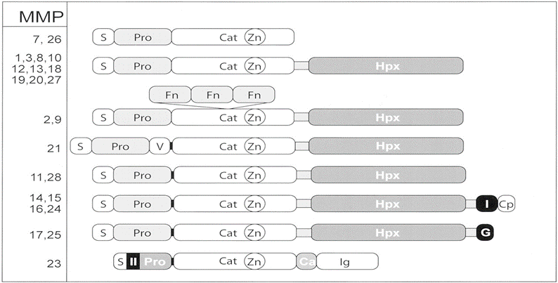
Domain structure of MMPs
The domain organization of MMPs is as indicated: S, signal peptide; Pro, propeptide; Cat, catalytic domain; Zn, active-site zinc; Hpx, hemopexin domain; Fn, fibronectin domain; V, vitronectin insert; I, type I transmembrane domain; II, type II transmembrane domain; G, GPI anchor; Cp, cytoplasmic domain; Ca, cysteine array region; and Ig, immunoglobulin (IgG)-like domain. A furin cleavage site is depicted as a black band between the propeptide and catalytic domain.
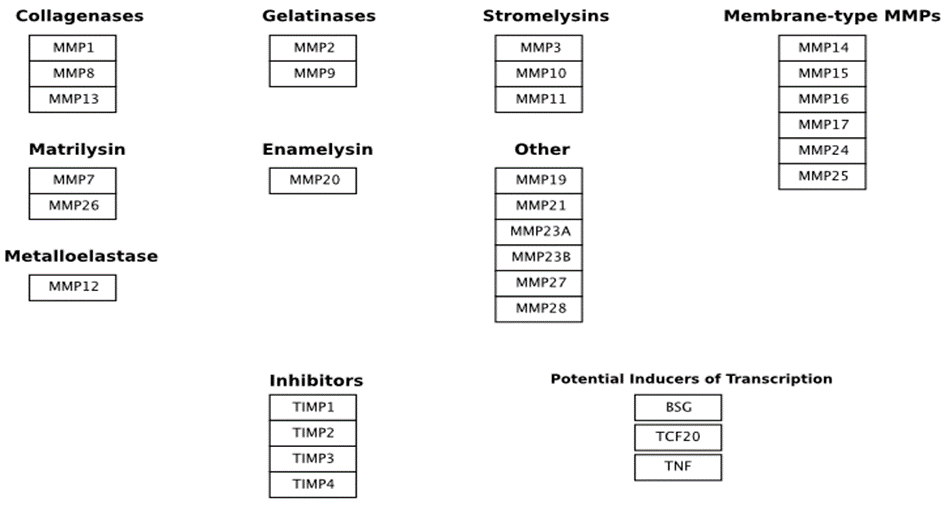
Activation
All MMPs are synthesized in the latent form (zymogen). They are secreted as proenzymes and require extracellular activation. MMPs can be activated by proteinases or in vitro by chemical agents (26).
Function
Besides wound healing, MMPs play an important role in tissue remodeling associated with various physiological or pathological processes such as morphogenesis, angiogenesis, tissue repair, cirrhosis, arthritis, and metastasis. MMP-2 and MMP-9 are thought to be important in metastasis.
MATERIALS AND METHODS:
Study Site
Kamineni Hospitals, LB Nagar, Hyderabad
Study Population
Patients of all ages and both sexes admitted under the Department of General Surgery, Kamineni Hospitals, Hyderabad, and undergoing open abdominal surgeries.
Study Design
This is a prospective observational study.
Sample Size: 50 subjects
This is a prospective observational study. The primary purpose of this study is to observe the initial expression of MMP-9 and TIMP-1 in acute wounds and their relation to wound healing. No sample size has been formally calculated.
Based on admissions over the past 4 years, approximately 120 cases fulfilling inclusion criteria were noted. The expected sample size was between 50–60. During the study period, a total of 62 patients fulfilling inclusion criteria were admitted and operated. After excluding deaths and patients lost to follow-up, 50 patients were included in the study.
Inclusion Criteria
All patients undergoing open abdominal surgeries (elective and emergency).
Exclusion Criteria
Traumatic wounds
Patients referred from outside centres with abdominal wound dehiscence or non-healing abdominal wounds
Methodology
Sample Collection
Wound biopsy was taken from the wound margin including skin and subcutaneous tissue measuring 5 × 5 mm at two intervals:
a. Immediately after giving skin incision
b. Before closure of incision (after surgical procedure)
Method of Staining
Staining of MMP-9 and TIMP-1 was done by immunohistochemistry.
MMP-9 and TIMP-1 Antibodies
Company: Leica Biosystems Newcastle Ltd., United Kingdom
Supplier: Labindia Instruments Pvt. Ltd., Hyderabad, Andhra Pradesh
0.1 ml Novocastra™ Lyophilized Mouse Monoclonal Antibody Matrix Metalloproteinase-9 (NCL-MMP9-439) and NCL-TIMP1-485
Novolink™ Mini Polymer Detection System (RE7290-K)
Immunohistochemistry scoring system for MMP-9 and TIMP-1 was used Procedure.
Method of Staining
Biopsies were sent in a formalin fixative and were paraffin-embedded. Endogenous peroxidase activity was neutralized using a peroxidase block. This was followed by application of the Novocastra™ protein block to reduce non-specific binding of the primary antibody and polymer.
The sections were subsequently incubated with optimally diluted primary antibody. This was washed with TBS (50 mM Tris-buffered saline) buffer for 5 minutes. Post-primary (rabbit anti-mouse IgG) was then used to detect mouse antibodies.
The Novolink™ polymer recognizes rabbit immunoglobulins; it detects the post-primary and any tissue-bound rabbit primary antibodies. Sections were further incubated with the substrate/chromogen, 3,3′-diaminobenzidine (DAB), prepared from DAB chromogen and Novolink™ DAB substrate buffer (polymer). This was washed with TBS buffer for 5 minutes.
The sections were incubated with peroxidase stain, producing a visible brown precipitate at the antigen site. Sections were washed under running tap water, counterstained with hematoxylin, and coverslipped.
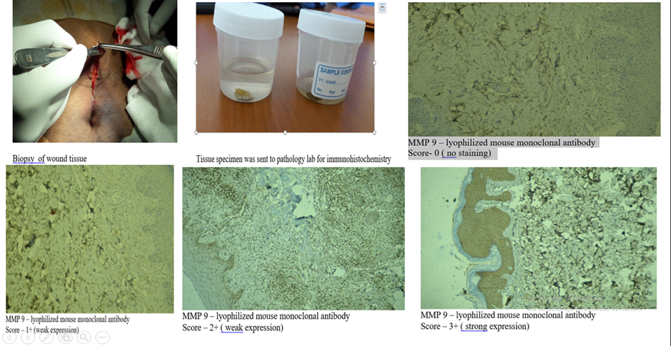
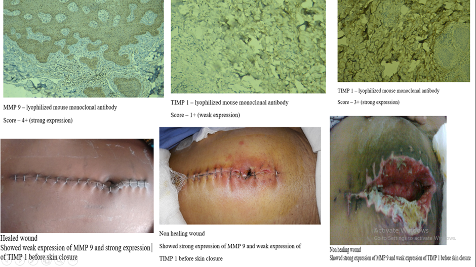
Results were interpreted using a light microscope, and staining was graded as weak or strong by comparison with controls. The analysis was performed by a pathologist, and scoring was assigned as follows.
|
SCORE |
STAINING |
|
0 |
No |
|
1+ |
Weak |
|
2+ |
Mild |
|
3+ |
Moderate |
|
4+ |
Strong |
MMP-9 and TIMP-1 tissue expression is graded as weak (0, 1+, 2+) and strong (3+, 4+).
Wound status at the 7th postoperative day (POD) is graded clinically as healed or non-healed using the Southampton wound grading score (53).
Southampton Wound Grading Score
Grade 0: Normal healing
Grade I: Normal healing with mild bruising
Ia: Some bruising
Ib: Considerable bruising
Ic: Mild erythema
Grade II: Erythema plus other signs of inflammation
IIa: At one point
IIb: Around sutures
IIc: Along wound
IId: Around wound
Grade III: Clear or hemoserous discharge
IIIa: At one point (<2 cm)
IIIb: Along wound (>2 cm)
IIIc: Large volume
IIId: Prolonged (>3 days)
Major complications
Grade IV: Pus
IVa: At one point only (<2 cm)
IVb: Along wound (>2 cm)
Grade V: Deep or severe wound infection with or without tissue breakdown
Healed: Southampton grades 0 and I
Non-healed: Southampton grades II, III, IV, and V
Statistical Methodology
Comparison between variables was made using odds ratio (OR) for TIMP, MMPs vs wound status.
The odds ratio (OR), standard error, and 95% confidence interval calculated (Altman, 1991)
P-value calculated (Sheskin, 2004).
Diabetes mellitus (DM) was categorized into diabetic and non-diabetic. MMP tissue expression was graded as weak (0, 1+, 2+) and strong (3+, 4+), and wound status was classified as healed or non-healed.
BMI ≥ 30 considered obese
Diabetes: diabetic/non-diabetic
Expression: weak vs strong
Outcome: healed vs non-healed
Wound Status at 7th Postoperative Day (POD)
RESULTS:
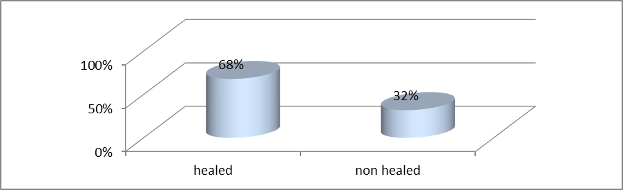
Graph-Wound status at 7th POD in 50 patients
At the 7th POD, 68% of patients had normally healed wounds, while 32% showed non-healing wounds.
Expression of MMP-9 and Wound Status (n = 50)
Table 1: After Skin Incision
|
MMP9 |
Healedwounds |
Nonhealedwounds |
|
Strong |
13 |
6 |
|
Weak |
21 |
10 |
|
Odd ratio |
1.0317 |
|
95%CI: |
0.3028to3.5155 |
|
statistic |
0.050 |
|
Significance level |
P=0.9601 |
MMP-9 expression was strong in 38% of healed wounds and 38% of non-healed wounds. MMP-9 expression was weak in 62% of healed wounds and 62% of non-healed wounds. The pattern of MMP-9 expression was statistically insignificant.
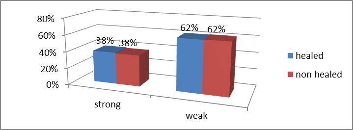
Graph1- After Skin Incision
Table 2: Before Skin Closure
|
MMP9 |
Healedwounds |
Nonhealedwounds |
|
Strong |
4 |
12 |
|
Weak |
30 |
4 |
|
Oddsratio |
0.0444 |
|
95%CI: |
0.0095to0.2071 |
|
zstatistic |
3.965 |
|
Significancelevel |
P=0.0001 |
Before skin closure, MMP-9 expression was strong in 12% of healed wounds and 75% of non-healed wounds. MMP-9 expression was weak in 88% of healed wounds and 25% of non-healed wounds.
The pattern of MMP-9 expression showed a significant association, being predominantly weak in healed wounds and strong in non-healed wounds.
.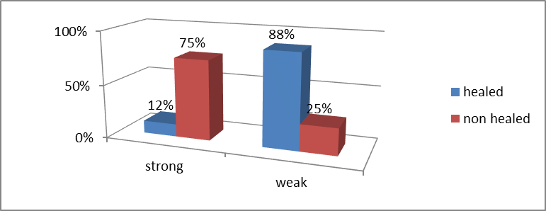
Graph2-Before skin closure (n=50).
Table 3: In Diabetic Patients After Skin Incision
|
MMP9 |
Healed |
Nonhealed |
|
Strong |
5 |
4 |
|
Weak |
9 |
6 |
|
Oddsratio |
0.8333 |
|
95%CI: |
0.1565to4.4360 |
|
zstatistic |
0.214 |
|
Significancelevel |
P=0.8308 |
In diabetic patients after skin incision, MMP-9 expression was strong in 35.70% of healed wounds and 40% of non-healed wounds.
MMP-9 expression was weak in 64.30% of healed wounds and 60% of non-healed wounds. The pattern of MMP-9 expression was statistically insignificant.
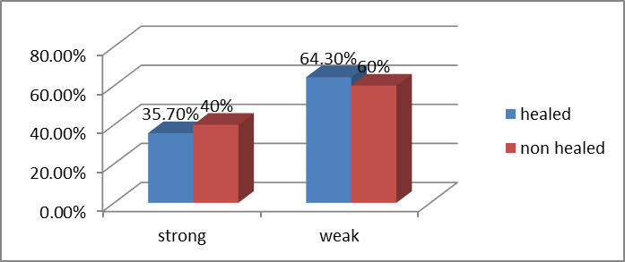
Graph 3- MMP 9 in diabetics after skin incision
Table 4: In Diabetic Patients Before Skin Closure.
|
MMP9 |
Healed |
Nonhealed |
|
Strong |
2 |
2 |
|
Weak |
9 |
7 |
|
Odds ratio |
0.7778 |
|
95%CI: |
0.0866to6.9832 |
|
Z statistic |
0.224 |
|
Significance level |
P=0.8224 |
MMP-9 expression was strong in 35.70% of healed wounds and 80% of non-healed wounds. MMP-9 expression was weak in 65.30% of healed wounds and 20% of non-healed wounds.
The pattern of MMP-9 expression was significantly weak in healed wounds and strong in non-healed wounds.
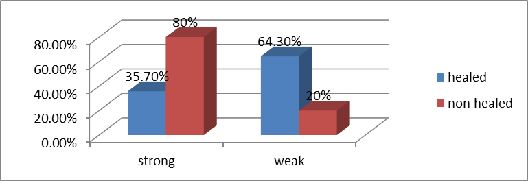
Graph4- MMP 9 in diabetics before skin closure
Table 5: In Obese Patients After Skin Incision
|
MMP9 |
Healedwounds |
Nonhealedwounds |
|
Strong |
5 |
8 |
|
Weak |
9 |
2 |
|
Oddsratio |
0.1389 |
|
95%CI: |
0.0208to0.9252 |
|
zstatistic |
2.040 |
|
Significancelevel |
P=0.0413 |
MMP-9 expression was strong in 18% of healed wounds and 22% of non-healed wounds.
MMP-9 expression was weak in 82% of healed wounds and 78% of non-healed wounds.
MMP-9 expression was statistically insignificant.
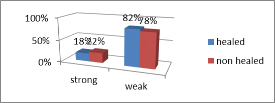
Graph5 –MMP 9 in obese patients (after skin incision)
Table 6: In Obese Patients Before Skin Closure
|
MMP9 |
Healedwounds |
Nonhealedwounds |
|
Strong |
2 |
7 |
|
Weak |
9 |
2 |
|
Oddsratio |
0.0635 |
|
95%CI: |
0.0071to0.5701 |
|
zstatistic |
2.462 |
|
Significancelevel |
P=0.0138 |
MMP-9 expression was weak in 82% of healed wounds and 22% of non-healed wounds. MMP-9 expression was strong in 18% of healed wounds and 78% of non-healed wounds.
The pattern of MMP-9 expression was significantly weak in healed wounds and strong in non-healed wounds.
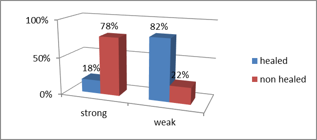
Graph6- MMP9 in Obesepatients (beforeskinclosure)
Expression of TIMP 1and wound status
Table 7: TIMP-1 Expression After Skin Incision
|
TIMP1 |
Healedwounds |
Nonhealedwounds |
|
Weak |
20 |
6 |
|
Strong |
14 |
10 |
|
Oddsratio |
2.3810 |
|
95%CI: |
0.7022to8.0730 |
|
zstatistic |
1.393 |
|
Significancelevel |
P=0.1638 |
TIMP-1 expression was weak in 59% of healed wounds and 38% of non-healed wounds. TIMP-1 expression was strong in 41% of healed wounds and 62% of non-healed wounds.
The pattern of TIMP-1 expression was statistically insignificant.
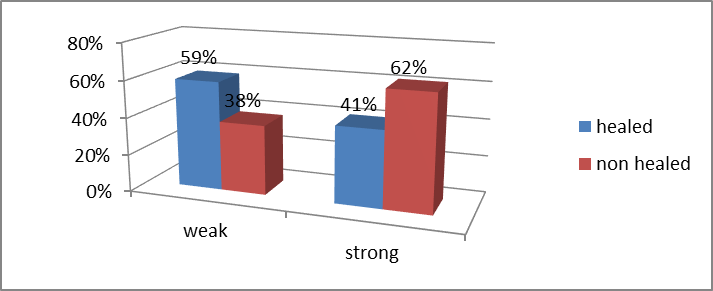
Graph7- After skin incision n=50
Table 8: TIMP-1 Expression Before Skin Closure
|
TIMP1 |
Healedwounds |
Nonhealedwounds |
|
Strong |
28 |
3 |
|
Weak |
6 |
13 |
|
Oddsratio |
0.0495 |
|
95%CI: |
0.0107to0.2293 |
|
zstatistic |
3.841 |
|
Significancelevel |
P=0.0001 |
TIMP-1 expression was weak in 18% of healed wounds and 81% of non-healed wounds. TIMP-1 expression was strong in 82% of healed wounds and 19% of non-healed wounds.
The pattern of TIMP-1 expression was significantly strong in healed wounds and weak in non-healed wounds.
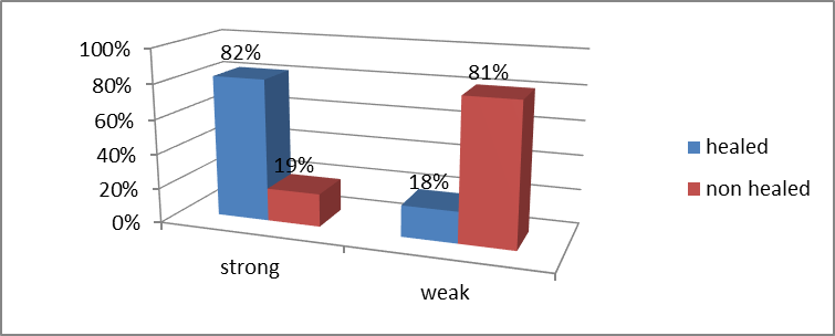
Graph8- TIMP1 Before skin closure n=50
Table 9: TIMP-1 Expression in Diabetic Patients After Skin Incision.
|
TIMP1 |
Healed |
Nonhealed |
|
Strong |
5 |
4 |
|
Weak |
9 |
6 |
|
Oddsratio |
0.8333 |
|
95%CI: |
0.1565to4.4360 |
|
zstatistic |
0.214 |
|
Significancelevel |
P=0.8308 |
TIMP-1 expression was strong in 35.70% of healed wounds and 40% of non-healed wounds. TIMP-1 expression was weak in 64.30% of healed wounds and 60% of non-healed wounds.
TIMP-1 expression was statistically insignificant.
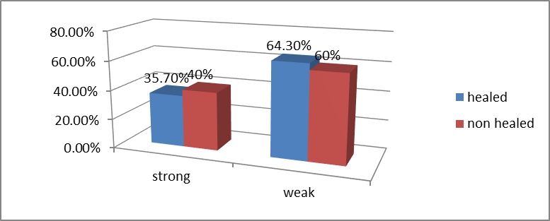
Graph9-TIMP1 in Diabetics (after skin incision)
Table 10: TIMP-1 Expression in Diabetic Patients Before Skin Closure
|
TIMP1 |
Healedwounds |
Nonhealedwounds |
|
Strong |
11 |
3 |
|
Weak |
3 |
7 |
|
Oddsratio |
8.5556 |
|
95%CI: |
1.3320to54.9513 |
|
zstatistic |
2.262 |
|
Significancelevel |
P=0.0237 |
TIMP-1 expression was strong in 78.50% of healed wounds and 30% of non-healed wounds. TIMP-1 expression was weak in 21.50% of healed wounds and 70% of non-healed wounds.
The pattern of TIMP-1 expression was significantly higher in healed wounds and weak in non-healed wounds.
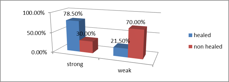
Graph10- TIMP1 in Diabetics (before skin closure)
Table 11: TIMP-1 Expression in Obese Patients After Skin Incision
|
TIMP1 |
Healed |
Nonhealed |
|
Strong |
3 |
2 |
|
Weak |
8 |
7 |
|
Oddsratio |
1.3125 |
|
95%CI: |
0.1678to10.2647 |
|
zstatistic |
0.259 |
|
Significancelevel |
P=0.7955 |
TIMP-1 expression was strong in 27% of healed wounds and 22% of non-healed wounds. TIMP-1 expression was weak in 73% of healed wounds and 78% of non-healed wounds. TIMP-1 expression was statistically insignificant.
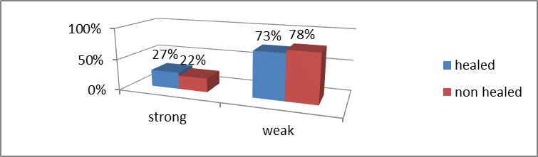
Graph11- TIMP1 in obese patients(after skin incision)
Table 12: TIMP-1 Expression in Obese Patients Before Skin Closure
|
TIMP1 |
Healedwounds |
Nonhealedwounds |
|
Strong |
7 |
1 |
|
Weak |
4 |
8 |
|
Oddsratio |
14.0000 |
|
95%CI: |
1.2515to156.6171 |
|
zstatistic |
2.142 |
|
Significancelevel |
P=0.0322 |
TIMP-1 expression was strong in 64% of healed wounds and 11% of non-healed wounds. TIMP-1 expression was weak in 36% of healed wounds and 89% of non-healed wounds.
The pattern of TIMP-1 expression was significantly strong in healed wounds and weak in non-healing wounds.
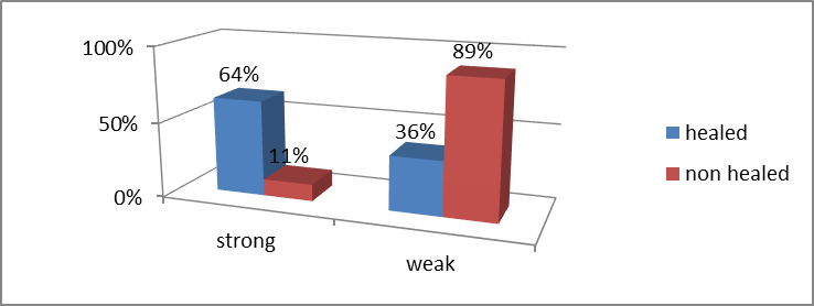
Graph12- TIMP1 in Obese patients (before skin closure)
DISCUSSION:
Role of MMPs in Inflammation
Inflammation is a key phase of wound healing that is required to protect against infection. Inflammatory cells are well known to express MMPs; however, epithelial and stromal cells in wounded tissue have also been demonstrated to express multiple MMPs, including MMP-1, MMP-2, MMP-3, MMP-7, MMP-9, MMP-10, and MMP-28 (27).
Many of these MMPs can regulate chemokine activity either by direct proteolysis or by affecting the formation of chemokine gradients. Among all MMPs, MMP-1, MMP-3, and MMP-9 appear to have the most widespread ability to regulate chemokine signalling.
Metalloproteinases have multiple roles in the regulation of inflammation, including control of chemokine activity, establishment of chemotactic gradients, and extravasation of leukocytes from blood into injured tissue. Additional evidence comes from MMP-deficient mice, where experimental challenge often results in altered inflammatory responses. These studies suggest that a major function of MMPs is the regulation of inflammation (6).
MMPs in Re-epithelialization
One major component of the repair phase of wound healing is re-epithelialization, which is the regrowth of epithelium over a denuded surface. This process requires cells at the wound margin to loosen their cell–cell and cell–ECM contacts and migrate across the wound (7).
Multiple MMPs are involved in this process, including MMP-1, MMP-3, MMP-7, MMP-9, MMP-10, MMP-14, and MMP-28.
MMP-9 (gelatinase B) has been strongly implicated in re-epithelialization after injury. Epidermal growth factor (EGF) and hepatocyte growth factor (HGF) stimulate keratinocyte migration in wound assays in vitro, and this migration depends on induction of MMP-9 activity. It is inhibited by function-blocking MMP-9 antibodies or general MMP inhibitors (28).
Bove et al. demonstrated that MMP-9 is essential for bronchiolar epithelial cell migration in culture (29).
MMPs may also negatively affect cell proliferation. MMP-2 and MMP-9 are produced by tracheal and bronchial cartilage, and cartilage-conditioned medium reduces respiratory epithelial cell proliferation in vitro (30).
MMPs in Collagen Remodeling
Remodeling of collagen, including degradation of existing collagen fibrils and synthesis of new ones, is a key part of the resolution phase of wound healing (7). Since MMPs are capable of this process, they are considered important in collagen remodeling during wound resolution.
MMP-9 Structure and Biology
Matrix metallopeptidase-9 (MMP-9), also known as 92 kDa type IV collagenase or gelatinase B (GELB), is a zinc-dependent enzyme of the matrixin family involved in degradation of extracellular matrix (ECM).
The human MMP-9 gene encodes a signal peptide, a propeptide, a catalytic domain with three fibronectin type II repeats, and a C-terminal hemopexin-like domain (31).
MMP-9 is synthesized as a 707 amino acid preproenzyme, including a 19-amino acid signal peptide, and is secreted as an inactive pro-MMP. The proenzyme consists of five domains:
Amino-terminal propeptide, Zinc-binding catalytic domain, Carboxyl-terminal hemopexin-like domain
The propeptide contains a conserved PRCGVPD sequence, where the cysteine residue acts as a “cysteine switch” that binds the catalytic zinc and maintains enzyme latency (31).
Activation of MMP-9
MMP-9 activation occurs through a protease cascade involving plasmin and stromelysin-1 (MMP-3). Plasmin activates MMP-3, which then cleaves the propeptide of MMP-9, generating an 82 kDa active enzyme (32).
The active MMP-9 contains a hemopexin-like domain composed of four β-propeller blades and an α-helix. This domain is important for cleavage of triple-helical interstitial collagens (33).
Functions of MMP-9
Regulates neutrophil migration across the basement membrane (34). Plays a role in angiogenesis and neovascularization, including tumor-associated vascular remodeling (35). Contributes to re-epithelialization; MMP-9 is upregulated during respiratory epithelial healing (36). MMP-9-deficient mice show impaired wound healing and fibrin matrix removal (37)
Tissue Inhibitors of Metalloproteinases (TIMPs).
TIMPs are endogenous inhibitors of MMPs and regulate ECM turnover, tissue remodeling, and cellular behavior. They bind MMPs in a 1:1 ratio. Four types have been identified in vertebrates: TIMP-1, TIMP-2, TIMP-3, and TIMP-4 (38).
TIMPs also inhibit ADAMTS metalloproteinases.
TIMP-1 was first identified in the 1970s as a collagenase inhibitor in fibroblast culture media, human serum, and bovine tissues. It was later purified as a 25–31 kDa protein and named tissue inhibitor of metalloproteinases (TIMP) (41,42).
A summary of the general properties of the four human TIMPs.
|
Property |
TIMP-1 |
TIMP-2 |
TIMP-3 |
TIMP-4 |
|
Glycosylation |
Yes |
No |
Partial |
No |
|
pI |
8.47 |
6.48 |
9.14 |
7.21 |
|
No.ofresiduesa |
184 |
194 |
188 |
194 |
|
Mrb |
20,709 |
21,755 |
21,690 |
22,329 |
|
MMPinhibition |
WeakforMMP-14-16,-19,and-24 |
All |
All |
Most |
|
OtherMMPinhibition |
ADAM10 |
ADAM12 |
ADAM10,12,17,28and33;ADAMTS-1,-4,and-5,ADAMTS-2(weak) |
ADAM17dand28,ADAM33(weak) |
|
Pro-MMPinteractions |
Pro-MMP-9 |
Pro-MMP-2 |
Pro-MMP-9andpro-MMP-2 |
Pro-MMP-2 |
|
Apoptoticeffects |
Negative |
Positive |
Positive |
|
|
Angiogenesis |
Negative |
Negative |
Negative |
Negative |
|
Chromosomallocation:human |
X11p11.23–11.4 |
17q23–25 |
22q12.1–q13.2 |
3p25 |
Structure of TIMPs
TIMPs are 21–29 kDa proteins with an N-terminal domain (~125 amino acids) and a C-terminal domain (~65 amino acids), each containing three disulfide bonds (43,44).
The N-terminal domain alone is sufficient for MMP inhibition. The overall structure resembles a wedge that fits into the active site of MMPs (45).
MMP–TIMP Interaction
The TIMP-2 molecule binds to the catalytic domain of MT1-MMP by inserting into its active-site cleft. The catalytic zinc ion is coordinated by the N-terminal cysteine of TIMP, leading to inhibition of enzymatic activity.
CONCLUSION:
Tissue Inhibitors of Metalloproteinases (TIMPs)
TIMPs have various biological activities, including modulation of cell proliferation, cell migration and invasion, anti-angiogenesis, anti- and pro-apoptosis, and synaptic plasticity.
Role in Wound Healing
1. Regulation of Inflammation
The inflammatory response is mediated by multiple cytokines and chemokines. One of the predominant cytokines involved in acute inflammation is TNF-α, which is activated via cleavage from the cell membrane by TNF-α converting enzyme (TACE) or ADAM17 (46).
TIMP-3 is one of the primary inhibitors of ADAM17 and thus has an integral role in the regulation of inflammation. In the absence of TIMP-3, ADAM17 activity is enhanced, resulting in increased constitutive TNF-α release and subsequent increase in inflammatory cell infiltration. Additionally, interaction between TIMP-3 and ADAM17 likely plays a role in directing leukocyte recruitment.
2. Role in Cell Migration and Re-epithelialization
The ability of metalloproteinase inhibitors to regulate MMP activity suggests that they are important in all aspects of wound repair, especially cell migration. This is supported by experiments using the synthetic metalloproteinase inhibitor GM6001 for cutaneous wounds (47). Wounds treated with synthetic metalloproteinase inhibitors show impaired re-epithelialization compared to control wounds.
3. Role in Wound Contraction
Wound contraction, an important component of the resolution phase of wound healing, is also regulated by metalloproteinase inhibitors. Treatment of cutaneous wounds with GM6001 leads to impaired re-epithelialization, decreased wound contraction, and altered differentiation of cells within granulation tissue (47).
4. Role in ECM Remodeling
Another important phase of wound resolution is remodeling of the extracellular matrix (ECM), especially collagen, which is required for the transition from granulation tissue to scar tissue (7). Collagen remodeling may be partially regulated by TIMP-3. MMP–TIMP Balance in Wound Healing Studies.
Ursula Mirastschijski et al. found that neither MMP-9 (P = 0.814) nor MMP-2 (P = 0.742) endogenous activity differed significantly between acute and chronic wound tissues. The overall activity of gelatinases MMP-9 and MMP-2 was not increased in chronic wounds compared to normally healing wound tissues. It was concluded that chronic non-healing wounds may not be caused by excessive gelatinase activity but are distinguished by an unfavorable distribution and persistence of MMP-9 (48). Wysocki AB et al. (49)
studied levels of MMP-2 and MMP-9 in wound fluid of chronic ulcers and found high levels of MMP-9 in non-healing wounds. Non-healing ulcers develop an environment containing high levels of activated metalloproteinases, which may result in chronic tissue turnover and failed wound closure. Liu Y et al. (50) studied the relationship between diabetic ulcer wound fluid MMPs and TIMPs with wound healing rate. Wound fluids were analyzed for MMP-2, MMP-9, and TIMP-1. MMP-9 and the MMP-9/TIMP-1 ratio correlated inversely with wound healing rate at 28 days (P < 0.001). Patients who achieved complete healing had lower MMP-9 and MMP-9/TIMP-1 ratios compared to non-healers. These findings suggest that a high MMP-9 milieu may indicate inflammation and poor wound healing in diabetic ulcers. Lan CC et al. demonstrated that high glucose conditions reduce keratinocyte motility and decrease MMP-2 and MMP-9 activity, which is reversed with good glycemic control, while TIMP-1 levels increase under normoglycemia (51).
Li Z et al. (52) studied 94 patients with diabetic foot ulcers and measured serum MMP-9, MMP-2, TIMP-1, and TIMP-2 levels. Serum samples were collected at first visit and after 4 weeks of treatment, with follow-up up to 12 weeks.
Median MMP-9 levels were lower in good healers at baseline and decreased significantly after therapy, whereas poor healers showed no significant change. The MMP-9/TIMP-1 ratio better reflected healing than MMP-9 alone and may serve as a predictor of wound healing and a potential therapeutic target in diabetic foot ulcers.
REFERENCES:
1. Robert F. Diegelmann, Evans MC. Wound healing: an overview of acute, fibrotic and delayed healing. Frontiers in Bioscience. 2004; 9: 283–289.
2. Clark RAF. Wound repair: overview and general considerations. In: The Molecular and Cellular Biology of Wound Repair. Clark RAF, editor. London: Plenum Press; 1996: 3–50.
3. Gregory S, Schultz. Interactions between extracellular matrix and growth factors in wound healing. Wound Repair and Regeneration. 2009; 17: 153–162.
4. Deodhar AK, Rana RE. Surgical physiology of wound healing: a review. 1997; 43(2): 52–56.
5. Pavlidis TE, Galatianos IN, Papaziogas BT, Lazaridis CN, Atmatzidis KS, Makris JG, et al. Complete dehiscence of the abdominal wound and incriminating factors. European Journal of Surgery. 2001; 167(5): 351–354.
6. Parks WC, Wilson CL, Lopez-Boado YS. Matrix metalloproteinases as modulators of inflammation and innate immunity. Nature Reviews Immunology. 2004; 4(8): 617–629.
7. Singer AJ, Clark RA. Cutaneous wound healing. New England Journal of Medicine. 1999; 341: 738–746.
8. Freije JR, Bischof. Matrix metalloproteases. Drug Discovery Today: Technologies. 2006; 3(1): 5–11.
9. Berard F, Gandon J. Postoperative wound infections: the influence of ultraviolet irradiation of the operating room and of various other factors. Annals of Surgery. 1964; 160(Suppl 2): 1–192.
10. Clark RA, Lanigan JM, DellaPelle P, Manseau E, Dvorak HF, Colvin RB. Fibronectin and fibrin provide a provisional matrix for epidermal cell migration during wound re-epithelialization. Journal of Investigative Dermatology. 1982; 79(5): 264–269.
11. Leibovich SJ, Ross R. The role of the macrophage in wound repair. A study with hydrocortisone and anti-macrophage serum. American Journal of Pathology. 1975; 78(1): 71–100.
12. Slavin J. The role of cytokines in wound healing. Journal of Pathology. 1996; 178(1): 5–10.
13. Grinnell F. Fibroblasts, myofibroblasts, and wound contraction. Journal of Cell Biology. 1994; 124(4): 401–404.
14. Tonnesen MG, Feng X, Clark RA. Angiogenesis in wound healing. Journal of Investigative Dermatology Symposium Proceedings. 2000; 5(1): 40–46.
15. Kurkinen M, Vaheri A, Roberts PJ, Stenman S. Sequential appearance of fibronectin and collagen in experimental granulation tissue. Laboratory Investigation. 1980; 43(1): 47–51.
16. Clark RAF. Mechanisms of cutaneous wound repair. In: Dermatology in General Medicine. Fitzpatrick TB, Eisen AZ, Wolff K, et al., editors. New York: McGraw-Hill; 1993: 473–486.
17. Winter GD. Formation of the scab and the rate of epithelization of superficial wounds in the skin of the young domestic pig. Nature. 1962; 193: 293–294.
18. Welch MP, Odland GF, Clark RA. Temporal relationships of F-actin bundle formation, collagen and fibronectin matrix assembly, and fibronectin receptor expression to wound contraction. Journal of Cell Biology. 1990; 110(1): 133–145.
19. Alberts B, Johnson A, Lewis J, Raff M, Roberts K, Watson JD. Cell junctions, cell adhesion, and the extracellular matrix. In: Molecular Biology of the Cell. New York: Garland Science; 2002: 1065–1125.
20. Lodish H, Berk A, Zipursky SL, Matsudaira P, Baltimore D, Darnell J. Integrating cells into tissues. In: Molecular Cell Biology. New York: WH Freeman & Co; 2000: 968–1002.
21. Verma RP, Hansch C. Matrix metalloproteinases (MMPs): chemical-biological functions and SARs. Bioorganic & Medicinal Chemistry. 2007; 15(6): 2223–2268.
22. Van Lint P, Libert C. Chemokine and cytokine processing by matrix metalloproteinases and its effect on leukocyte migration and inflammation. Journal of Leukocyte Biology. 2007; 82(6): 1375–1381.
23. Gross J, Lapiere C. Collagenolytic activity in amphibian tissues: a tissue culture assay. PNAS. 1962; 48(6): 1014–1022.
24. Eisen A, Jeffrey J, Gross J. Human skin collagenase: isolation and mechanism of attack on the collagen molecule. Biochimica et Biophysica Acta. 1968; 151(3): 637–645.
25. Harper E, Bloch K, Gross J. The zymogen of tadpole collagenase. Biochemistry. 1971; 10(16): 3035–3041.
26. Nagase H. Activation mechanisms of matrix metalloproteinases. Biological Chemistry. 1997; 378: 151–160.
27. Corry DB, Kiss A, Song LZ, et al. Overlapping and independent contributions of MMP2 and MMP9. FASEB Journal. 2004; 18: 995–997.
28. McCawley LJ, O’Brien P, Hudson LG. EGF and HGF-mediated keratinocyte migration is coincident with induction of MMP-9. Journal of Cell Physiology. 1998; 176(2): 255–265.
29. Bove PF, Wesley UV, Greul AK, et al. Nitric oxide promotes airway epithelial wound repair through MMP-9 activation. American Journal of Respiratory Cell and Molecular Biology. 2007; 36(2): 138–146.
30. Sigurdson L, Sen T, Hall L, et al. Possible impedance of epithelial repair by tracheal cartilage metalloproteinases. Archives of Otolaryngology–Head & Neck Surgery. 2003; 129(2): 197–200.
31. Nagase H, Woessner JF. Matrix metalloproteinases. Journal of Biological Chemistry. 1999; 274(31): 21491–21494.
32. Ramos-DeSimone N, et al. Activation of MMP-9 via plasmin/stromelysin cascade. Journal of Biological Chemistry. 1999; 274(19): 13066–13076.
33. Gomis-Rüth FX, et al. Structure of MMP-13 hemopexin domain. Journal of Molecular Biology. 1996; 264(3): 556–566.
34. Delclaux C, et al. Role of gelatinase B in neutrophil migration. American Journal of Respiratory Cell and Molecular Biology. 1996; 14(3): 288–295.
35. Forsyth PA, et al. MMPs in malignant gliomas. British Journal of Cancer. 1999; 79(11–12): 1828–1835.
36. Buisson AC, et al. MMP-9 in respiratory epithelial wound repair. Journal of Cellular Physiology. 1996; 166(2): 413–426.
37. Mohan R, et al. MMP-9 in epithelial regeneration. Journal of Biological Chemistry. 2001; 277(3): 2065–2072.
38. Brew K, et al. TIMPs: evolution, structure and function. Biochimica et Biophysica Acta. 2000; 1477: 267–283.