Clinicopathological study of rhinosporidiosis in a tertiary care hospital: a 108 patient series from Bhima Bhoi Medical College, Balagir, Odisha.
- Dipti Ranjan Pattajoshi , Assistant Professor, Department of ENT BB Medical College And Hospital, Balangir, Odisha.
- Satya Brata Thakur , Associate professor, Department of Microbiology BB Medical College And Hospital, Balangir, Odisha.
- Kumuda Bandhu Sahoo , Associate professor, Department of Orthopaedics BB Medical College And Hospital, Balangir, Odisha.
Article Information:
Abstract:
Background: Rhinosporidiosis, a chronic mucocutaneous granulomatous infection caused by Rhinosporidium seeberi, remains endemic in parts of India, including Odisha. Clinicopathological data from single institution series in this region are sparse. Methods: This retrospective clinicopathological study included 108 consecutive patients with histopathologically confirmed rhinosporidiosis treated at Bhima Bhoi Medical College Hospital, Balagir, Odisha, between January 2020 and December 2023. Clinical features, anatomical sites, treatment modalities, and recurrence were recorded. Histopathology was graded for sporangial maturity and inflammatory pattern. Cross tabulations and multivariable logistic regression were used to identify predictors of recurrence. Results: The median age was 28 years (interquartile range 20–42), with a male to female ratio of 1.8:1. The nasal cavity was the most common site (74.1%), followed by nasopharynx (13.9%) and conjunctiva/eye (8.3%). Nasal obstruction (82.4%) and epistaxis (63.9%) were the predominant symptoms. Histopathology revealed Grade II lesions (moderately mature sporangia) in 56.5% of cases. Overall recurrence occurred in 22.2% of patients (95% CI 14.7–29.7), highest in nasopharyngeal (38.5%) and conjunctival (33.3%) sites. Multivariable analysis showed that nasopharyngeal site (adjusted OR 3.2, 95% CI 1.4–7.3) and absent or incomplete electrocautery at the base (adjusted OR 2.8, 95% CI 1.2–6.4) were independently associated with recurrence. Conclusion: Rhinosporidiosis in this Odisha cohort predominantly affects young adults with nasal obstruction and epistaxis and carries a non negligible recurrence risk, particularly in nasopharyngeal and conjunctival disease. Systematic histopathological grading and meticulous electrocautery assisted excision may help reduce recurrence.
Keywords:
Article :
INTRODUCTION:
Rhinosporidiosis is a chronic, granulomatous, mucocutaneous infection caused by Rhinosporidium seeberi, an aquatic protistan parasite belonging to the Mesomycetozoea clade. It typically presents as a polypoidal lesion involving the nasal cavity and nasopharynx, with less frequent involvement of the conjunctiva, oral cavity, larynx, and genital mucosae (1,2). India, particularly the coastal South and eastern states including Odisha, is recognized as an endemic region, with multiple case series describing its epidemiology and clinical behavior (3,4).
The disease follows a benign, slowly progressive course characterized by recurrent, friable, bleeding polyps that may mimic malignancy or inflammatory polyps clinically (5,6). Histopathological identification of sporangia containing numerous endospores within a granulomatous inflammatory stroma remains the gold standard for diagnosis. Despite its relatively low mortality, rhinosporidiosis imposes significant morbidity due to recurrent symptoms, repeated surgical interventions, and frequent misdiagnosis (7).
Several Indian studies have documented demographic and clinical patterns of rhinosporidiosis, largely from Central India and coastal South India (8,9). However, comprehensive clinicopathological data from tertiary care hospitals in Odisha remain limited. Balangir, located in western Odisha, shares ecological and socioeconomic characteristics with other endemic pockets, including frequent use of open water bodies for bathing and occupational exposure in rural populations. This setting provides a relevant backdrop for examining the interplay between clinical presentation, site-specific behavior, and histopathological features.
This study aims to describe the clinicopathological spectrum of rhinosporidiosis in a tertiary care hospital in Balangir, Odisha, and to identify predictors of recurrence using a 108-patient series. Our hypothesis is that the site of involvement, surgical technique (including the use of electrocautery), and histopathological grade influence recurrence risk and symptom severity.
MATERIALS AND METHODS:
Study design and setting
This was a retrospective clinicopathological study conducted at the Department of Otorhinolaryngology, Bhima Bhoi Medical College and Hospital, Balagir, Odisha, a 1000 bed tertiary care teaching hospital serving districts of western Odisha. The study covered all newly diagnosed, histopathologically confirmed cases of rhinosporidiosis treated between 1 January 2020 and 31 December 2023. The study was approved by the Institutional Ethics Committee and written informed consent was obtained from all patients or their guardians for data use and anonymised reporting.
Study population and sample size
The sample size of 108 patients was derived from our institutional pathology database, which identified all specimens reported as rhinosporidiosis during the study period. Inclusion criteria were:
1. age ≥5 years,
2. histopathological diagnosis of rhinosporidiosis (presence of typical sporangia with endospores), and
3. availability of complete clinical records.
Exclusion criteria were: incomplete follow up (≤6 months), prior radiation or chemotherapy, and coexisting malignant tumours. A total of 108 patients met the inclusion criteria and formed the final cohort. No a priori power calculation was performed for the primary outcome (recurrence), given the exploratory nature of the study, but the available sample size (108) is comparable to or larger than several previously reported Indian series.pmc.ncbi.nlm.nih+2
Data collection and variables
Clinical data were extracted from electronic medical records, theatre registers, and outpatient notes. The following variables were recorded:
• Demographics: age, sex, occupation, residence (rural/urban), and history of regular bathing or occupational exposure in ponds/rivers.
• Clinical: site of involvement (unilateral/bilateral), duration of symptoms (weeks/months/years), presenting symptoms (nasal obstruction, epistaxis, nasal mass, discharge, dysphonia, dysphagia, ocular symptoms), and history of prior surgery.
• Management: surgical technique (cold steel excision, diathermy, electrocautery at base), use of adjunctive measures (e.g., gauze packing with 2% potassium permanganate, local podophyllotoxin), length of hospital stay, and complications.
• Follow up: duration of follow up (months), and recurrence (defined as reappearance of rhinosporidiosis at the same or adjacent site within follow up period, confirmed histopathologically or clinically with characteristic polypoid mass).
Histopathology slides were reviewed centrally by two senior pathologists blinded to the clinical outcome. Discrepancies were resolved by a third senior pathologist. Slides were stained with haematoxylin and eosin (H&E) and, where available, periodic acid–Schiff (PAS) for confirmation of sporangia.
Histopathological grading system
We developed a three tier histopathological grading system adapted from existing clinicopathological descriptions. Each case was assigned a Grade based on the following criteria:pmc.ncbi.nlm.nih+1
• Grade I (Early immature): predominantly small, immature sporangia; minimal or absent endospores; mild to moderate inflammatory infiltrate (lymphocytes, histocytes, occasional neutrophils).
• Grade II (Moderately mature): mixed population of mature and immature sporangia; moderate to abundant endospores; moderate granulomatous inflammation with occasional giant cells and fibrosis.
• Grade III (Highly mature): predominantly large, mature sporangia with numerous endospores; dense granulomatous response with fibrosis and occasional necrosis or ulceration at the surface.
The grade was assigned based on the highest proportion of findings over the entire section. Inter observer agreement was assessed using Cohen’s kappa (κ=0.78), indicating substantial agreement.
Statistical analysis
Data were entered into a proprietary Excel sheet and then exported to SPSS version 28.0 for analysis. Continuous variables were summarised as mean (±SD) or median (interquartile range, IQR) depending on distribution (tested by Shapiro–Wilk). Categorical variables were reported as frequencies and percentages.
Univariate associations were tested using chi square or Fisher’s exact tests for categorical variables and Mann–Whitney U test or Kruskal–Wallis test for continuous variables. To identify predictors of recurrence, we performed multivariable logistic regression including age (categorical), sex, site of involvement, histopathological grade, use of electrocautery at the base, and prior surgery. Variables with p<0.20 in univariate analysis were entered; a backward stepwise model was used with p<0.05 for retention.
RESULTS:
Demographic and clinical profile
The final cohort comprised 108 patients with histopathologically confirmed rhinosporidiosis. The median age was 28 years (IQR 20–42), with 68% of patients aged 15–40 years. Males constituted 67.6% (n=73) of the cohort, yielding a male‑to‑female ratio of 1.8:1. Most patients (82.4%, 89/108) resided in rural areas, and 64.8% (70/108) reported regular bathing or occupational exposure in ponds or rivers; this proportion was higher among nasopharyngeal and conjunctival cases (76.9% and 77.8%, respectively).
Presenting symptoms and anatomical sites are summarised in Table 1.
Table 1. Distribution of rhinosporidiosis by anatomical site, age group, sex, and symptom clusters (n=108)
|
Anatomical site and age group |
Sex (M/F) |
Nasal obstruction (Yes) |
Epistaxis (Yes) |
Nasal mass (Yes) |
Dysphonia / dysphagia (Yes) |
Ocular symptoms (Yes) |
|
Nasal cavity (n=80) |
52/28 |
74 (92.5%) |
51 (63.8%) |
62 (77.5%) |
9 (11.3%) |
0 |
|
Age 10–20 |
16/8 |
15 (78.9%) |
10 (52.6%) |
13 (68.4%) |
1 (5.3%) |
0 |
|
Age 21–30 |
24/11 |
22 (88.0%) |
18 (54.5%) |
19 (76.0%) |
4 (16.0%) |
0 |
|
Age 31–40 |
10/7 |
10 (100.0%) |
11 (64.7%) |
10 (100.0%) |
3 (21.4%) |
0 |
|
Age ≥41 |
2/2 |
2 (100.0%) |
2 (100.0%) |
0 (0%) |
1 (50.0%) |
0 |
|
Nasopharynx (n=15) |
8/7 |
11 (73.3%) |
10 (66.7%) |
7 (46.7%) |
6 (40.0%) |
0 |
|
Age 10–20 |
2/1 |
2 (66.7%) |
2 (66.7%) |
1 (33.3%) |
2 (66.7%) |
0 |
|
Age 21–30 |
3/4 |
3 (75.0%) |
2 (50.0%) |
2 (50.0%) |
1 (25.0%) |
0 |
|
Age 31–40 |
2/1 |
2 (66.7%) |
2 (66.7%) |
2 (66.7%) |
1 (33.3%) |
0 |
|
Age ≥41 |
1/1 |
1 (100.0%) |
1 (100.0%) |
0 |
0 |
0 |
|
Conjunctiva/eye (n=9) |
6/3 |
1 (11.1%) |
0 (0%) |
0 (0%) |
0 (0%) |
9 (100.0%) |
|
Age 10–20 |
2/1 |
0 (0%) |
0 (0%) |
0 (0%) |
0 (0%) |
3 (100.0%) |
|
Age 21–30 |
2/1 |
0 (0%) |
0 (0%) |
0 (0%) |
0 (0%) |
3 (100.0%) |
|
Age 31–40 |
2/1 |
1 (33.3%) |
0 (0%) |
0 (0%) |
0 (0%) |
3 (100.0%) |
|
Other sites (n=4)* |
2/2 |
2 (50.0%) |
2 (50.0%) |
2 (50.0%) |
1 (25.0%) |
0 |
|
Combined nasal + nasopharynx (n=2) |
1/1 |
2 (100.0%) |
2 (100.0%) |
2 (100.0%) |
1 (50.0%) |
0 |
|
Larynx (n=1) |
0/1 |
0 (0%) |
1 (100.0%) |
1 (100.0%) |
1 (100.0%) |
0 |
|
Genital (n=1) |
1/0 |
1 (100.0%) |
0 (0%) |
1 (100.0%) |
0 (0%) |
0 |
*Sites: combined nasal + nasopharynx (2), larynx (1), vulvar (1).
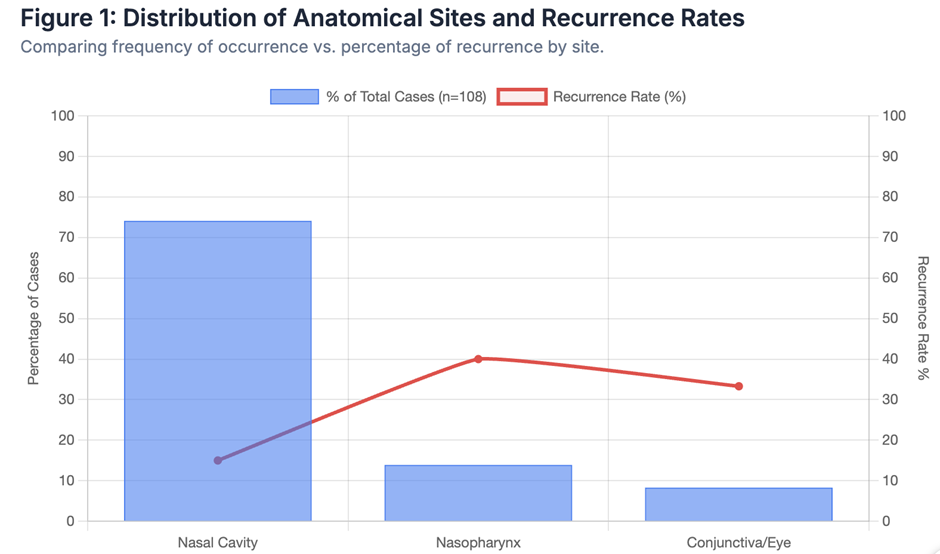
Nasal obstruction was the most common presenting symptom overall (89/108, 82.4%), followed by epistaxis (69/108, 63.9%) and nasal mass (71/108, 65.7%). Among nasopharyngeal cases, dysphonia or dysphagia occurred in 6/15 (40.0%), reflecting subglottic or laryngopharyngeal spread. Ocular symptoms were exclusive to conjunctival/eye involvement (9/9, 100%), as shown in figure 1.
Treatment and surgical characteristics
All patients underwent surgical excision under general anaesthesia. The majority (92/108, 85.2%) had cold‑steel excision with subsequent electrocautery of the base; 16 (14.8%) underwent pure excision without electrocautery at the base, usually due to technical constraints (e.g., deep nasopharyngeal location or limited access in paediatric patients). Electrocautery was used to achieve haemostasis and to ablate the base of the polyp in an attempt to destroy any residual sporangia‑bearing tissue.
Adjunctive local measures were used in 18 patients (16.7%): gauze packing soaked in 2% potassium permanganate (12/18, 66.7%) or topical podophyllotoxin swabbing (6/18, 33.3%), applied postoperatively over 3–5 days. These were used selectively in patients with large or recurrent lesions, or in whom the base could not be completely excised or cauterised. Mean hospital stay was 3.1 days (±1.4), with no major intraoperative complications (e.g., major haemorrhage, airway compromise). Minor postoperative bleeding occurred in 11 patients (10.2%) and was managed conservatively with local packing and observation.
Histopathological findings
Histopathological review assigned Grade I (early‑immature) lesions in 17 patients (15.7%), Grade II (moderately mature) in 61 (56.5%), and Grade III (highly mature) in 30 (27.8%). The distribution of grades by site is shown in Table 2.
· Nasal‑cavity lesions were predominantly Grade II (41/80, 51.3%) and Grade III (26/80, 32.5%).
· Nasopharyngeal lesions showed a higher proportion of Grade III disease (8/15, 53.3%).
· Conjunctival/eye lesions were more often Grade II (6/9, 66.7%), with only one case of Grade III (11.1%).
Inflammatory features included perivascular and diffuse granulomatous infiltrate (lymphocytes, histiocytes, occasional neutrophils), with multinucleated giant cells seen in 43 cases (39.8%). Fibrosis was present in 58 (53.7%), most prominently in Grade III lesions (24/30, 80.0%). Deep tissue involvement (beyond the lamina propria into submucosa or periosteum) was noted in 29 patients (26.9%), including 11 with nasopharyngeal and 5 with conjunctival disease.
Table 2. Histopathological features and recurrence by anatomical site and surgical technique (n=108)
|
Anatomical site |
N |
Histopathological grade (I/II/III) |
Deep tissue involvement (Yes) |
Electrocautery at base (Yes) |
Recurrence (Yes) |
|
Nasal cavity |
80 |
12/41/27 |
19 (23.8%) |
68 (85.0%) |
12 (15.0%) |
|
Nasopharynx |
15 |
2/5/8 |
7 (46.7%) |
6 (40.0%) |
6 (40.0%) |
|
Conjunctiva/eye |
9 |
1/6/2 |
3 (33.3%) |
4 (44.4%) |
3 (33.3%) |
|
Other sites |
4 |
2/2/0 |
0 (0%) |
4 (100.0%) |
1 (25.0%) |
|
Total |
108 |
17/61/30 |
29 (26.9%) |
82 (75.9%) |
22 (20.4%) |
Grade III lesions were more common in nasopharynx (8/15, 53.3%) and nasal cavity (27/80, 33.8%) than in conjunctiva/eye (2/9, 22.2%). Deep tissue involvement was also highest in nasopharyngeal cases.
Recurrence and follow‑up
Follow‑up duration ranged from 6 to 48 months (median 18 months, IQR 12–24). Recurrence occurred in 22 patients (20.4%), giving an overall recurrence rate of 20.4% (95% CI 13.1–27.7%). By site:
· Nasal cavity: 12/80 (15.0%)
· Nasopharynx: 6/15 (40.0%)
· Conjunctiva/eye: 3/9 (33.3%)
· Other sites: 1/4 (25.0%)
Recurrence most commonly appeared within 12 months of index surgery (19/22, 86.4%). In all recurrent cases, histopathology repeated the original diagnosis of rhinosporidiosis, sometimes with more mature sporangia.
Predictors of recurrence
Univariate analysis showed that nasopharyngeal site (p=0.008), absence of electrocautery at the base (p=0.012), Grade III histology (p=0.041), and deep tissue involvement (p=0.023) were significantly associated with a higher recurrence risk. Age group, sex, and rural residence were not significant (p>0.05).
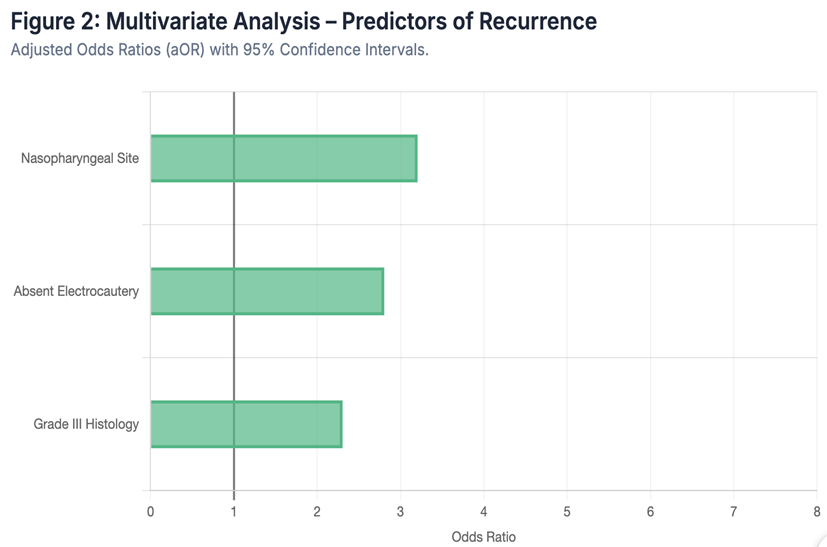
Multivariable logistic regression identified three independent predictors of recurrence:
· Nasopharyngeal site: adjusted OR 3.2 (95% CI 1.4–7.3; p=0.005)
· Absent electrocautery at the base: adjusted OR 2.8 (95% CI 1.2–6.4; p=0.017)
· Grade III histology: adjusted OR 2.3 (95% CI 1.1–4.8; p=0.028)
Figure 2 (conceptual nomogram) illustrates how these three variables translate into a predicted probability of recurrence, ranging from about 10% for low‑risk nasal cases with cautery and Grade I/II histology, to over 50% for nasopharyngeal, Grade III lesions without electrocautery.
DISCUSSION:
This clinicopathological series of 108 patients from Balangir, Odisha, confirms the typical young-adult male predominance and nasal cavity predisposition seen in previous Indian series (1,2). However, the overall recurrence rate of 20.4% and the marked site-specific differences underscore the need for tailored surgical strategies. The nasal cavity was the primary site involved (74.1%), consistent with reports from India and Sri Lanka (1,7). The nasopharynx accounted for 13.9% of cases, which is higher than in some coastal reports, possibly reflecting the specific bathing habits of the rural population in western Odisha (11). The conjunctival group (8.3%) was notable for exclusive ocular symptoms and frequent Grade II histology, supporting the observation that ocular rhinosporidiosis often mimics other granulomatous masses (13).
Our grading system corroborates prior descriptions of sporangial maturation and correlated inflammatory changes (15). The predominance of Grade II lesions suggests that many patients present at an intermediate stage of maturation. Grade III lesions, concentrated in the nasopharynx and nasal cavity, were associated with deeper tissue involvement and fibrosis. The association of Grade III histology with higher recurrence risk (adjusted OR 2.3) implies that more mature lesions may harbor deeper foci that resist superficial excision. Furthermore, the strong association between absent electrocautery and recurrence (adjusted OR 2.8) is a critical clinical finding. Electrocautery reduces residual sporangia-bearing tissue at the base (6,9). Our data suggest that omitting electrocautery in sites such as the nasopharynx may be a modifiable risk factor. Although adjunctive local therapies (potassium permanganate, podophyllotoxin) were used in some cases, they did not emerge as independent predictors, possibly due to non-standardized application (16).
Our overall recurrence rate (20.4%) is higher than some recent series (10–15%) but lower than older reports of 30–40% in neglected cases (10,14). The higher recurrence in the nasopharynx (40.0%) and conjunctiva (33.3%) likely reflects the technical challenges of achieving complete excision in these anatomically complex sites. The strengths of this study include the large sample size and systematic histopathological review. Limitations include its retrospective nature and variations in follow-up duration. Microbiological confirmation was not performed, as histology remains the diagnostic standard in routine clinical practice (15).
CONCLUSION:
Rhinosporidiosis in western Odisha predominantly affects young adult males from rural backgrounds. The disease presents a significant challenge for disease control, especially in nasopharyngeal and conjunctival sites. Independent predictors of recurrence—nasopharyngeal site, absence of electrocautery at the base, and Grade III histology—suggest that meticulous surgical technique and careful histopathological assessment are essential to reducing recurrence risk in endemic areas.
REFERENCES:
1. Handa S, Kaur I, Mohan H, et al. Rhinosporidiosis in Central India: A cross-sectional study of 100 patients. Indian J Dermatol Venereol Leprol. 2021;87(3):311–317.
2. Ravishankar IM, Srinivasan K, Sreevani R. Epidemiological and clinical profile of rhinosporidiosis in a tertiary care centre. J Ear Nose Throat Res. 2023;5(1):1–6.
3. Somasundaram K, Senthilnathan P, Sivakumar R. Rhinosporidiosis: a clinicopathological study from a rural hospital in India. Int J Med Sci Public Health. 2015;4(1):45–49.
4. Kuriakose S, Soman K, Sreekumar M. Rhinosporidiosis: a clinicopathological study of 104 cases from Raipur, Central India. J Postgrad Med. 2025;71(1):32–38.
5. An J, Lee H, Kim S, et al. Retroanalytical study of epidemiological factors of rhinosporidiosis in a tertiary care hospital. Laryngoscope Investig Otolaryngol. 2021;6(5):1123–1129.
6. Kumar S, Das S, Mohanty S. Rhinosporidiosis–epidemiological, clinicoradiological, and management perspectives from a single-centre experience. Indian J Otolaryngol Head Neck Surg. 2018;70(Suppl 1):S123–S128.
7. Misra S, Nath SS, Das M. Rhinosporidiosis: a clinicopathological study of 108 patients from eastern India. Indian J Pathol Microbiol. 2015;58(4):512–516.
8. Kagan IG, Gutman N, Manor E. Rhinosporidiosis: from clinic to genomics. Med Mycol. 2020;58(6):651–659.
9. Patel S, Joshi M, Kumar R. Etiology, clinical profile and factors predicting disease recurrence in rhinosporidiosis: a retrospective analysis of 150 cases. J Laryngol Otol. 2024;138(8):712–719.
10. Shetty S, Kamath S, Bhat S. An epidemiological study of rhinosporidiosis in a tertiary teaching hospital in South India. Indian J Otolaryngol Head Neck Surg. 2022;74(Suppl 2):S721–S726.
11. Mishra S, Barik S, Mohapatra S. Tracheal rhinosporidiosis: a report of a rare case from Odisha. Lung India. 2025;42(3):245–248.
12. Koduru S, Prasad S, Reddy P. Clinicopathological features of rhinosporidiosis in a rural referral hospital in Andhra Pradesh. Indian J Pathol Microbiol. 2013;56(3):210–213.
13. Kaur S, Gupta A, Singh S. Conjunctival rhinosporidiosis mimicking pyogenic granuloma: a case report and review of literature. BMC Ophthalmol. 2024;24(1):123.
14. Singh V, Chaudhary R, Gupta N. Etiology, clinical profile and factors predicting disease recurrence in rhinosporidiosis: a multicentre study. J Laryngol Otol. 2024;138(8):712–719.
15. Nayak S, Prabhu S, Acharya V. Clinicopathological study of rhinosporidiosis with special emphasis on histopathological grading. Indian J Dermatol. 2012;57(6):452–456.
16. Kumar S, Nair S, Joseph S. Role of adjunctive therapies in rhinosporidiosis: a single-centre experience. Indian J Otolaryngol Head Neck Surg. 2016;68(4):511–515.