A Cross-Sectional Study to Determine the Prevalence of Left Atrial Enlargement in Patients with New Onset Atrial Fibrillation.
- Tanvi H Bhende , Senior Resident, Department of General Medicine, Lokmanya Tilak Municipal Medical College and Hospital, Sion East, Mumbai 400 022, India.
- Prachee Makashir , Professor and Head of Unit, Department of General Medicine, Bharati Vidyapeeth (Deemed to be University) Medical College and Hospital, Pune – 411043, India.
- Vaibhav Patil. , Associate Professor in Department of Cardiology, Bharati Vidyapeeth (Deemed to be University) Medical college and hospital, Pune 411 043, India.
Article Information:
Abstract:
Background: Atrial fibrillation (AF) is a common arrhythmia associated with increased mortality and stroke risk. Left atrial enlargement (LAE) is a known predictor of AF progression and adverse outcomes. Despite extensive research in Western populations, data from India regarding LAE in new onset atrial fibrillation (NOAF) is limited. Aim: This study aims to determine the prevalence of LAE in Indian patients with NOAF and risk factors associated with NOAF. Methods: This cross-sectional study was conducted in tertiary care hospital from July 2022 to December 2023. The study included 61 adult patients with newly diagnosed AF, confirmed by electrocardiogram (ECG). Data on demographics, comorbidities, echocardiographic parameters were collected. LAE was defined as a left atrial diameter ≥4.1 cm in men and ≥3.9 cm in women. Statistical analyses were performed using SPSS version 25.0, with significance set at p<0.05. Results: LAE was present in 47.5% of the cohort, with a prevalence of 80 % in patients with valvular AF and prevalence of 41.2% in non-valvular AF. Significant associations were found between LAE and congestive heart failure (CHF) (p=0.01) and chronic kidney disease (CKD) (p=0.003). Conclusion: LAE is prevalent among Indian patients with NOAF and is associated with comorbidities such as CHF and CKD. These findings underscore the need for early detection and management of LAE to mitigate adverse outcomes in NOAF patients.
Keywords:
Article :
INTRODUCTION:
Atrial fibrillation (AF) is the most common arrhythmia worldwide, characterized by disorganized, rapid atrial activation with irregular ventricular rhythm due to AV nodal conduction abnormalities.1,2 AF is typically classified into three forms: paroxysmal (self-terminating), persistent (lasting more than 48 hours without spontaneous resolution), and permanent (resistant to pharmacological or electrical intervention).1 On electrocardiogram (ECG), AF presents as absent P waves or fibrillatory waves and an irregular rhythm.3
New onset atrial fibrillation (NOAF) refers to the first detectable episode of AF in an individual with no prior history.3 Globally, AF affects 1-2% of the population and is associated with a 1.5 to 1.9-fold increase in mortality and a fivefold increase in stroke risk.1,2 Approximately one-third of AF patients remain asymptomatic, with stroke often being the first manifestation.4 This highlights the need for early identification of at-risk populations.
Research has demonstrated that left atrial (LA) enlargement is a significant predictor of AF development and progression.5 For instance, studies such as the Framingham and Cardiovascular Health studies have shown that increased LA size correlates with a higher risk of AF.5 Additionally, LA remodelling in AF patients can predispose them to more severe disease states, including permanent AF.6
While most AF data come from studies in Western populations, limited research from India explores the relationship between NOAF and echocardiographic findings. This study aims to assess the prevalence of left atrial enlargement in Indian patients with NOAF.
MATERIALS AND METHODS:
Study Design and Setting
This cross-sectional study was conducted at Bharati Vidyapeeth Deemed to be University Medical College, Dhankawadi, Pune, a tertiary care hospital, from July 2022 to December 2023. Ethical approval was obtained from the Institutional Ethics Committee prior to study initiation, and all participants provided informed consent. This study conforms to widely accepted ethical principles guiding human research (such as the Declaration of Helsinki)
Study Population
The study included adult patients (>18 years) admitted to Bharati Hospital. Patients were eligible if they had atrial fibrillation (AF) for the first time, confirmed by electrocardiogram (ECG) findings and no prior history of AF. Those with a known history of AF were excluded.
Sample Size
The sample size for this study was 51 patients however we observed 61 patients out of which 51 patients had non valvular atrial fibrillation and 10 patients had valvular atrial fibrillation .
Data Collection
Data collection was performed using a structured proforma, capturing patient demographics (age, sex, weight, height), comorbidities, cause of admission, and clinical details, including 2D echocardiography (echo) and ECG findings.
Patients underwent a detailed history and clinical examination, with specific attention to:
- Age, gender, and BMI
- Symptoms and reason for hospitalization (medical or surgical)
- Presence of comorbidities, including hypertension, diabetes, chronic diseases (heart, lung, liver), malignancy, stroke, anemia, thyroid disease, and others.
Diagnosis of Atrial Fibrillation
Atrial fibrillation was diagnosed based on ECG findings, characterized by the absence of P waves and an irregular heart rate. Routine ECG monitoring was conducted at admission and during the hospital stay, with continuous monitoring in the ICU. Patients who developed AF during hospitalization without prior ECG documentation were included. Pre- and post-operative AF was also recorded for patients undergoing surgery.
Clinical Definitions
- Hypertension was defined as systolic BP ≥140 mmHg or diastolic BP ≥90 mmHg.
- Diabetes was defined by fasting glucose ≥126 mg/dL, postprandial glucose ≥200 mg/dL, or HbA1c ≥6.5%.
- Anemia was defined as hemoglobin <12 g/dL in women and <13.7 g/dL in men.
- IHD was diagnosed based on a history of angina, prior ECG changes, or evidence of coronary interventions.
Other conditions such as COPD, heart failure, stroke, and thyroid disorders were diagnosed according to established clinical guidelines. BMI was calculated from the patient’s weight and height at admission.
Echocardiography and ECG
Detailed 2D echo parameters included left atrial (LA) diameter, the presence of left ventricular hypertrophy (LVH), valvular abnormalities, and regional wall motion abnormalities. Left atrial enlargement was defined as a diameter ≥4.1 cm in men and ≥3.9 cm in women.
Statistical Analysis
Data were entered into Microsoft Excel and analyzed using SPSS version 25.0. Descriptive statistics were used, including mean, median, and standard deviation for quantitative data and percentages for qualitative data. Chi-square or Fisher’s exact tests were applied for categorical data, while unpaired t-tests or Mann-Whitney U tests were used for continuous variables. A p-value <0.05 was considered statistically significant. Results were presented in tables and graphs where appropriate.
RESULTS:
This cross-sectional study evaluated the baseline characteristics, comorbidities, and prevalence of left atrial enlargement (LAE) in 61 patients with new onset atrial fibrillation (NOAF) treated at Bharati Hospital and Research Centre between June 2022 and December 2023. Of the total population, 60.7% (37/61) were male, and 39.3% (24/61) were female, with a mean age of 64.96 ± 14.04 years. Among the 51 patients with non-valvular AF, 62.7% were male, and 37.3% were female. In the valvular AF subgroup (n=10), the gender distribution was evenly split between males and females (50% each).
Table 1: Baseline characteristics of the study population
|
Characteristic
|
Overall (n=61) |
Non-Valvular (n=51) |
Valvular (n=10) |
||||
|
|
64.96 ± 14.04 |
|
|
||||
|
Gender |
|
|
|
||||
|
Female |
24 (39.3%) |
19 (37.3%) |
5 (50%) |
||||
|
Male |
37 (60.7%) |
32 (62.7%) |
5 (50%) |
||||
|
Comorbidities |
|
|
|
||||
|
Hypertension |
38 (62.3%) |
34 (66.7%) |
|
||||
|
Diabetes |
19 (31.1%) |
18 (35.3%) |
|
||||
|
Chronic Kidney Disease |
17 (27.9%) |
16 (31.4%) |
|
||||
|
Stroke |
10 (16.4%) |
9 (17.6%) |
|
||||
|
Chronic Obstructive Pulmonary Disease |
6 (9.8%) |
|
|
||||
|
Hypothyroidism |
6 (9.8%) |
3 (5.9%) |
|
||||
|
Chronic Liver Disease |
|
|
|
||||
|
Hyperthyroidism |
1 (1.6%) |
1 (2.0%) |
|
||||
|
Cardiovascular Diseases |
|
|
|
||||
|
Ischemic Heart Disease |
15 (29.4%) |
|
|
||||
|
Congestive Heart Failure |
|
|
|
||||
|
Rheumatic Valvular Disease |
|
|
|
||||
|
Non-rheumatic Valvular Disease |
|
|
|
||||
|
Cardiomyopathy |
|
|
|
Hypertension was the most common comorbidity, observed in 62.3% (38/61) of the overall population, and more prevalent in non-valvular AF cases (66.7%). Diabetes affected 31.1% (19/61) of the overall cohort, with a higher prevalence of 35.3% in non-valvular AF cases. Chronic kidney disease (CKD) was present in 27.9% of the overall population, while stroke was noted in 16.4% of cases. Among non-valvular AF cases, CKD and stroke were observed in 31.4% and 17.6%, respectively.
Regarding cardiac disease, ischemic heart disease (IHD) was the most common, affecting 29.4% (15/61) of the population, followed by congestive heart failure (CHF) in 19.6% (10/61). Rheumatic and non-rheumatic valvular diseases (one case of Mitral valve prolapse , one case of aortic stenosis ) were observed in 13.1% and 3.3% of cases, respectively. Other conditions reported in patients are represented in figure 1; with sepsis most common (37.3%).
LAE was present in 47.5% (29/61) of the overall cohort. In patients with non-valvular AF, LAE was seen in 41.2% (21/51), while 80% (8/10) of those with valvular AF exhibited LAE. (Figure 2)
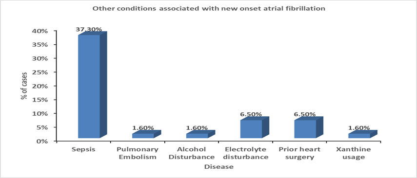
Figure 1: Other conditions associated with new onset atrial fibrillation seen in study population.
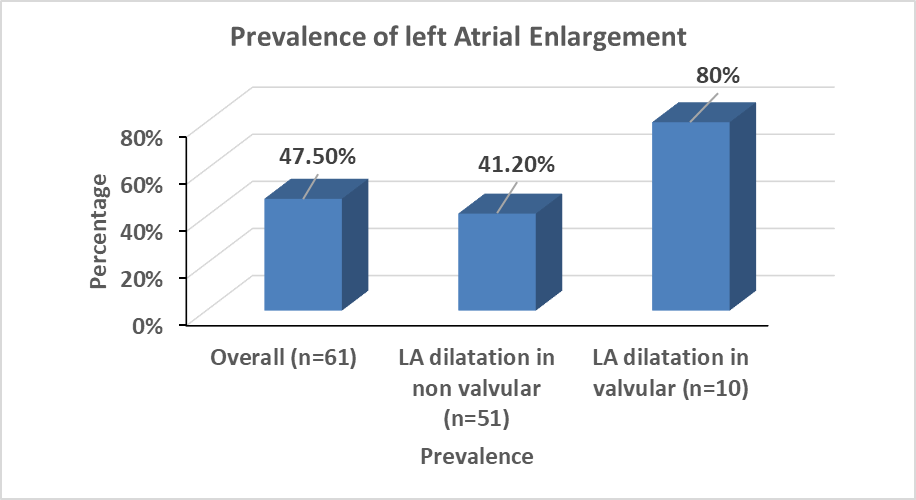
Figure 2: Prevalence of left Atrial Enlargement in Patients with new onset atrial fibrillation
Table 2: Distribution of 2D Echo Findings in Non-Valvular and Valvular New Onset Atrial Fibrillation
|
Parameter |
Non-Valvular (n=51) |
Valvular (n=10) |
|
LA Thrombus |
8 (15.7%) |
2 (20%) |
|
LVH |
23 (45.1%) |
2 (20%) |
|
PAH |
19 (37.3%) |
7 (70%) |
|
RA Dilatation |
8 (15.7%) |
5 (50%) |
|
RV Dilatation |
4 (7.8%) |
4 (40%) |
|
Valvular Stenosis |
4 (7.8%) |
7 (70%) |
|
Valvular Regurgitation |
15 (29.4%) |
6 (60%) |
|
RWMA |
14 (27.5%) |
1 (10%) |
Table 2 presents the distribution of various 2D Echo findings among patients with non-valvular and valvular new onset atrial fibrillation (NOAF). Among the 51 patients with non-valvular NOAF, left atrial (LA) thrombus was observed in 15.7% of cases, while left ventricular hypertrophy (LVH) was noted in 45.1%. Pulmonary arterial hypertension (PAH) affected 37.3% of patients, and right atrial (RA) dilatation was present in 15.7%. Right ventricular (RV) dilatation occurred in 7.8% of non-valvular cases. Valvular stenosis and regurgitation were found in 7.8% and 29.4% of cases, respectively. Regional wall motion abnormalities (RWMA) were seen in 27.5% of patients.
In contrast, among the 10 patients with valvular NOAF, LA thrombus was observed in 20%, LVH in 20%, and PAH in 70%. RA dilatation was noted in 50%, and RV dilatation in 40%. Valvular stenosis and regurgitation were found in 70% and 60% of cases, respectively. RWMA was present in only 10% of valvular cases. These findings highlight a higher prevalence of PAH, RA dilatation, and valvular abnormalities in valvular NOAF compared to non-valvular cases.
Table 3: Associations Between LA Dilatation and Various Factors in Non-Valvular New Onset Atrial Fibrillation
|
Factor |
LA Dilatation |
No |
Yes |
Total |
p-value |
|
Gender |
Female |
10 |
9 |
19 |
0.49 |
|
Male |
20 |
12 |
32 |
||
|
Diabetes |
No |
20 |
13 |
33 |
0.73 |
|
Yes |
10 |
8 |
18 |
||
|
Hypertension |
No |
11 |
6 |
17 |
0.55 |
|
Yes |
19 |
15 |
34 |
||
|
CHF |
No |
28 |
13 |
41 |
0.01 |
|
Yes |
2 |
8 |
10 |
||
|
IHD |
No |
22 |
14 |
36 |
0.61 |
|
Yes |
8 |
7 |
15 |
||
|
Stroke |
No |
25 |
17 |
42 |
0.99 |
|
Yes |
5 |
4 |
9 |
||
|
CKD |
No |
25 |
10 |
35 |
0.003 |
|
Yes |
5 |
11 |
16 |
||
|
CLD |
No |
28 |
19 |
47 |
0.99 |
|
Yes |
2 |
2 |
4 |
||
|
COPD |
No |
27 |
19 |
46 |
0.99 |
|
Yes |
3 |
2 |
5 |
||
|
Sepsis |
No |
17 |
15 |
32 |
0.28 |
|
Yes |
13 |
6 |
19 |
Table 3 details associations between left atrial (LA) dilatation and various clinical factors in non-valvular NOAF. The analysis shows no significant associations between LA dilatation and gender (p=0.49), diabetes (p=0.73), hypertension (p=0.55), ischemic heart disease (IHD) (p=0.61), stroke (p=0.99), chronic liver disease (CLD) (p=0.99), chronic obstructive pulmonary disease (COPD) (p=0.99), and sepsis (p=0.28). However, a significant association was found between LA dilatation and congestive heart failure (CHF) (p=0.01), indicating that patients with CHF are more likely to have LA dilatation. Additionally, a significant association was observed between LA dilatation and chronic kidney disease (CKD) (p=0.003), suggesting that CKD is also linked to increased LA dilatation.
DISCUSSION:
Atrial fibrillation (AF) is the most prevalent arrhythmia globally, significantly increasing the risk of mortality, stroke, and dementia.1 It affects 1-2% of the population and is linked to high morbidity.1 Understanding the disease burden and identifying those most at risk is essential. The primary cause of AF is abnormal atria, where anatomical and histological changes lead to electrical re-entry and sustained AF.7-9 Atrial dilation and fibrosis contribute to the persistence of AF, which worsens over time and increases the risk of permanent AF and stroke.7-11. Several small studies have shown that therapeutic strategies, such as ACEI or ARB therapy and ablation, can reverse left atrial remodelling and reduce AF recurrence.6 However, these findings need validation through larger multicentre trials. While Indian studies have described AF, few have explored the correlation between 2D Echo findings and atrial size in patients with new-onset AF (NOAF).
In our study, we aimed to estimate the prevalence of left atrial (LA) enlargement in patients newly diagnosed with AF and its correlation with comorbidities and risk scores. The key findings were: i) most patients were over 60 years of age; ii) a male predominance was observed; iii) hypertension and diabetes were the most common comorbidities; iv) LA dilatation had significant association with CHF and CKD. In our study, 61 patients were included, with a mean age of 64.96 years. This aligns with findings of the study conducted by Chowdhury SR et al., with mean age 67.17 years and by Sawhney et al., where the mean age was 65.8 years.2-12 Both studies indicate AF is more prevalent in older populations, likely due to age-related degenerative changes in the myocardium, as proposed by Kornej et al.,.13 Additionally, our study suggests that AF may occur at a younger age in the Indian population compared to Western populations, possibly due to genetic and lifestyle differences.
Of the 61 participants, 60.7% were male, consistent with other studies like Sawhney et al. and Hrynkiewicz-Szymanska et al., which also reported a male preponderance.2,14 The higher prevalence of AF in men may be attributed to factors like taller stature, larger atria, and sex-related differences.15
Hypertension (66.7%) and diabetes mellitus (35.3%) were the most common comorbidities in our study, similar to findings by Chowdhury SR et al., and Sawhney et al., where hypertension (59.7% and 68.5%) and diabetes (30.9% AND 36.2%) were prevalent comorbidities in AF patients.2,12 Chronic kidney disease was observed in 31.4% of our patients, higher than the 10.3% reported by Sawhney et al., likely because our study included all stages of CKD.2 Other comorbidities included stroke (17.6%), COPD (9.8%), and chronic liver disease (7.8%). In our study, 47.5% of participants had LA dilatation with 80 % prevalence in valvular AF patients and 41.2% prevalence in non-valvular AF patients. This is lower than the 86.2% reported by Hrynkiewicz-Szymanska et al., which may reflect ethnic or genetic differences in LA size and AF incidence across populations.14 As observed by Kornej et al.,13 AF prevalence and incidence of AF is higher in persons with European ancestry than in Asians and blacks.
LA dilatation was significantly associated with CHF and CKD, in line with studies showing similar pathophysiological mechanisms like RAS activation and fibrosis.13,14 No significant association was found between LA dilatation and gender, diabetes, hypertension, IHD, stroke, CLD, COPD, or sepsis.
In this study we did not find gender, diabetes, hypertension, IHD, stroke, CLD, COPD and Sepsis to be significantly associated with presence of LA dilatation. The study by Hrynkiewicz-Szymanska et al (14) also did not find significant association between LA dilatation and gender, diabetes, hypertension, IHD, stroke and COPD.
CONCLUSION:
Our study identified a 41.2% prevalence of left atrial (LA) dilation in patients with non-valvular new-onset atrial fibrillation (NOAF), indicating an increased risk of AF recurrence. Patients with comorbidities such as congestive heart failure (CHF) and chronic kidney disease (CKD) showed a higher prevalence of LA dilation and potential for chronic AF. Regular screening for LA dilation is recommended for these patients.
Limitations
This cross-sectional study cannot establish causality or recurrence rates, which a prospective study could address. The limited sample size may affect generalizability, and variability in echocardiographic measurements due to operator differences could impact consistency. Additionally, although LA strain correlates better with new-onset atrial fibrillation, it requires specialized equipment and training, so our study used LA diameter.
Declaration of funding:
This research did not receive any specific grant from funding agencies in the public, commercial, or not-for-profit sectors.
Declaration of interest:
The authors declare that they have no known competing financial interests or personal relationships that could have appeared to influence the work reported in this paper.
REFERENCES:
1. Michaud GF, Stevenson WG. Atrial Fibrillation. In: Jameson JL, Fauci AS, Kasper DL, Hauser SL, Longo DL, Loscalzo J, editors. Harrison’s Principles of Internal Medicine. 20th ed. New York, NY: McGraw-Hill Education; 2018. Available from: accessmedicine.mhmedical.com/content.aspx?aid=1176803953
2. Sawhney JPs, Kothiwale VA, Bisne V, Durgaprasad R, Jadhav P, Chopda M, et al. Risk profiles and one-year outcomes of patients with newly diagnosed atrial fibrillation in India: Insights from the GARFIELD-AF Registry. Indian Heart J. 2018 Nov;70(6):828–35.
3. New-onset atrial fibrillation - Symptoms, diagnosis and treatment | BMJ Best Practice. Available from: https://bestpractice.bmj.com/topics/en-gb/3000087
4. Saggu DK, Sundar G, Nair SG, Bhargava VC, Lalukota K, Chennapragada S, et al. Prevalence of atrial fibrillation in an urban population in India: the Nagpur pilot study.
5. Abhayaratna WP, Seward JB, Appleton CP, Douglas PS, Oh JK, Tajik AJ, et al. Left Atrial Size: Physiologic determinants and clinical applications. J Am Coll Cardiol. 2006 Jun;47(12):2357–63.
6. Thomas L, Abhayaratna WP. Left Atrial Reverse Remodeling. JACC Cardiovasc Imaging. 2017 Jan;10(1):65–77.
7. Falk RH. Atrial Fibrillation. N Engl J Med. 2001 Apr 5;344(14):1067–78.
8. Agarwal A, York M, Kantharia BK, Ezekowitz M. Atrial Fibrillation: Modern Concepts and Management. Annu Rev Med. 2005 Feb 1;56(1):475–94.
9. Peters NS, Schilling RJ, Kanagaratnam P, Markides V. Atrial fibrillation: strategies to control, combat, and cure. The Lancet. 2002 Feb;359(9306):593–603.
10. January CT, Wann LS, Alpert JS, Calkins H, Cigarroa JE, Cleveland JC, et al. 2014 AHA/ACC/HRS Guideline for the Management of Patients With Atrial Fibrillation: Executive Summary: A Report of the American College of Cardiology/American Heart Association Task Force on Practice Guidelines and the Heart Rhythm Society. Circulation. 2014 Dec 2;130(23):2071–104.
11. Wijesurendra RS, Casadei B. Mechanisms of atrial fibrillation. Heart. 2019 Dec;105(24):1860–7.
12. Chowdhury SR, Kuotsu MM, Dhakal D, Chongtham DS, Gayatri L, Bijoy M. PROFILE OF PATIENTS WITH ATRIAL FIBRILLATION AND ITS CORRELATION WITH LEFT ATRIAL SIZE AND BODY MASS INDEX. Int J Acad Med Pharm. 2024;6(3):386-90. Available from: https://academicmed.org/Uploads/Volume6Issue3/79.%20[3260.%20JAMP_Medo%20George]%20386-390.pdf
13. Kornej J, Börschel CS, Benjamin EJ, Schnabel RB. Epidemiology of Atrial Fibrillation in the 21st Century: Novel Methods and New Insights. Circ Res. 2020 Jun 19;127(1):4–20.
14. Hrynkiewicz-Szymanska A, Dluzniewski M, Platek AE, Szymanski FM, Syska-Suminska J, Klos-Szadryn A, et al. Association of the CHADS2 and CHA2DS2-VASc scores with left atrial enlargement: a prospective cohort study of unselected atrial fibrillation patients. J Thromb Thrombolysis. 2015 Aug;40(2):240–7.
15. Volgman AS, Benjamin EJ, Curtis AB, Fang MC, Lindley KJ, Naccarelli GV, et al. Women and atrial fibrillation. J Cardiovasc Electrophysiol. 2021 Oct;32(10):2793–807.
16. Uemura K, Nishimori M, Nagai S, Takeuchi M, Nishihara Y, Todo S, et al. Identification of factors associated with progression of left atrial enlargement in patients with atrial fibrillation. Echocardiography. 2023 Sep;40(9):976–82.