A STUDY OF CARDIOTOXIC MANIFESTATIONS OFSNAKE ENVENOMATION AMONGST THE VASCULOTOXIC AND NEUROTOXIC SNAKE BITE PATIENTS IN A TERTIARY CAREHOSPITAL
- Debasish Ganguly , Senior Resident, MD (General Medicine), Department of General Medicine, Calcutta National Medical College,32, Gorachand Road, Beniapukur, Kolkata, West Bengal – 700014.
- Oishee Saha , Senior Resident,MD (General Medicine), Department of General Medicine,Calcutta National Medical College,32, Gorachand Road, Beniapukur, Kolkata, West Bengal – 700014.
- Parinita Ranjit , Associate Professor, MD (General Medicine), Department of General Medicine,Tamralipto Government Medical & Hospital.
Article Information:
Abstract:
Background: Snake envenomation is a major medical emergency in tropical countries like India, contributing significantly to morbidity and mortality. Cardio toxic manifestations are increasingly recognized in snakebite victims, especially in cases involving vasculotoxic and neurotoxic species. However, data comparing cardiotoxic effects between these two categories remain limited in tertiary care settings. Aims and Objectives: To study the cardiotoxic manifestations among patients with vasculotoxic and neurotoxic snake envenomation admitted to a tertiary care hospital. Materials and Methods: This hospital-based prospective study was conducted in the Department of General Medicine at Calcutta National Medical College & Hospital, Kolkata, over 12 months (April 2023–March 2024), including 100 admitted snakebite patients meeting the inclusion criteria. Results: Snakebite mainly affected young rural males (86%), mostly in the lower limbs (81%) and outdoor exposure (84%); Russell viper was most common (38%). Renal (AKI 27%) and cardiac involvement (arrhythmias 46%) were frequent, with significant association with snake type and Whole Blood Clotting Time (suggestive of vasculotoxic snake envenomation) (p < 0.05). Conclusion: Cardiotoxic manifestations are an important systemic complication of snake envenomation, particularly in vasculotoxic bites. Early recognition through ECG and cardiac biomarkers is essential for prompt management and improved outcomes.
Keywords:
Article :
INTRODUCTION:
Snake envenomation is a significant public health problem in many tropical and subtropical countries, particularly in South and Southeast Asia, Sub-Saharan Africa, and Latin America. India alone contributes a substantial proportion of global snakebite mortality, with thousands of deaths reported annually, especially in rural and agricultural communities where human–snake interactions are frequent [1]. The World Health Organization (WHO) has classified snakebite envenoming as a neglected tropical disease due to its high burden, limited access to timely medical care, and underreporting in many endemic regions [2].
Venomous snakes can be broadly classified based on their predominant toxic effects into neurotoxic, vasculotoxic (hemotoxic), and myotoxic categories. Neurotoxic snakebites, commonly caused by elapid species such as cobras and kraits, primarily affect the neuromuscular junction leading to paralysis and respiratory failure. Vasculotoxic envenomation, typically associated with vipers, results in coagulopathy, endothelial injury, hemorrhage, and multiorgan dysfunction [3]. Although these classifications are based on dominant clinical effects, systemic involvement beyond the primary target system is increasingly recognized. Cardiac involvement in snake envenomation has gained attention in recent years due to its association with increased morbidity and mortality. Cardiotoxic manifestations may occur either as a direct effect of venom components on myocardial tissue or indirectly through hypotension, shock, hypoxia, or coagulopathy-induced microvascular damage [4].
Various venom toxins, including phospholipase A2, metalloproteinases, and cardiotoxins, can disrupt cellular membranes, impair ion channels, and induce myocardial inflammation or ischemia-like changes [5]. Clinically, cardiotoxicity in snakebite patients may present as arrhythmias, conduction abnormalities, hypotension, myocarditis, acute coronary syndrome-like ECG changes, and even cardiac arrest in severe cases. Electrocardiographic abnormalities such as sinus tachycardia, bradycardia, ST-segment elevation or depression, T-wave inversion, and QT interval prolongation have been reported in multiple studies [6].
In addition, elevated cardiac biomarkers like troponin I and CK-MB have been observed, suggesting myocardial injury even in the absence of overt clinical cardiac symptoms [7]. Vasculotoxic snakebites are often associated with more pronounced systemic complications due to disseminated intravascular coagulation, endothelial damage, and capillary leak syndrome, which may predispose patients to myocardial ischemia and shock-related cardiac dysfunction [8].
On the other hand, neurotoxic bites can also lead to secondary cardiotoxicity due to hypoxia from respiratory muscle paralysis and autonomic dysfunction affecting heart rate and blood pressure regulation [9]. However, comparative data on cardiotoxic manifestations between vasculotoxic and neurotoxic envenomation remain limited, particularly in tertiary care hospital settings in India.Early recognition of cardiac involvement in snakebite patients is crucial for improving outcomes. Routine monitoring using electrocardiography and cardiac biomarkers can help identify patients at risk of deterioration. Echocardiography further aids in assessing myocardial function and guiding supportive therapy. Despite advances in critical care, standardized protocols for cardiac evaluation in snake envenomation are not universally implemented [10]. To study the cardiotoxic manifestations among patients with vasculotoxic and neurotoxic snake envenomation admitted to a tertiary care hospital.
MATERIALS AND METHODS:
Study Design and Setting: Hospital based prospective study.The study was conducted in the department of General Medicine of Calcutta National Medical College & Hospital, Kolkata.
Place of study: Calcutta National Medical College & Hospital, In patient Department of General Medicine.
Period of study: 12 months (From 1st April 2023 to 30th March 2024)
Study population: Patients were recruited from the admitted indoor snake bite patients of General Medicine ward.
Sample size: 100 patients fulfilling the inclusion criteria considering the study will be for limited period of time.
Inclusion and Exclusion Criteria
Inclusion Criteria
• All patients of both sex with alleged neurotoxic or vasculotoxic snake bite were be included.
Exclusion Criteria
• Patients who had known pre-existing heart disease
• Patients who had any other known pre-existing chronic disease
• Snake bite patients in pregnancy
Study Variables:
Parameters to be studied: Data was collected by pretested proforma consisting of history, clinical examination, appropriate investigation.
Statistical Analysis: Data was entered in MS Excel (2013) and analyzed using IBM SPSS version 21. Descriptive statistics (mean, standard deviation, and standard error) were used for age and sex, and results were presented in tabular form. Since there was no control group, comparison was made with standardized reference values. Pearson’s correlation was applied to assess relationships between parametric variables, and t-test was used for statistical analysis. A p-value ≤ 0.05 was considered statistically significant, with a confidence interval of 95%, alpha error of 5%, and beta error of 20%.
Ethical Considerations: The proposal was submitted for ethical consideration in institutional ethics committee. Patient participating in the study wer not charged for any procedure or visit for this purpose. Informed written consent was taken from each interviewee.
RESULTS:
Table 1: Distribution of Demographic and Clinical Characteristics of Study Participants
|
Demographic and Clinical Characteristics |
Frequency |
Percent |
|
|
Age in group |
11–20 |
17 |
17.00% |
|
21–30 |
21 |
21.00% |
|
|
31–40 |
25 |
25.00% |
|
|
41–50 |
18 |
18.00% |
|
|
51–60 |
17 |
17.00% |
|
|
>61 |
2 |
2.00% |
|
|
Sex |
Female |
14 |
14.00% |
|
Male |
86 |
86.00% |
|
|
Occupation |
Agriculture |
50 |
50.00% |
|
Business |
2 |
2.00% |
|
|
Fishing |
4 |
4.00% |
|
|
Housewife |
8 |
8.00% |
|
|
Labourer |
7 |
7.00% |
|
|
Service |
16 |
16.00% |
|
|
Student |
8 |
8.00% |
|
|
Unemployed |
5 |
5.00% |
|
|
Domicile |
Rural |
92 |
92.00% |
|
Urban |
8 |
8.00% |
|
|
Site of bite |
Lower limb |
81 |
81.00% |
|
Trunk |
3 |
3.00% |
|
|
Upper limb |
16 |
16.00% |
|
|
Place of bite |
Indoor |
16 |
16.00% |
|
Outdoor |
84 |
84.00% |
|
|
Type of snake |
Cobra |
3 |
3.00% |
|
Krait |
6 |
6.00% |
|
|
Russell viper |
38 |
38.00% |
|
|
Unknown bite |
53 |
53.00% |
|
Table 2: Distribution of Clinical Features among Study Participants
|
Clinical Features |
Frequency |
Percent |
|
|
Local pain |
Absent |
15 |
15.00% |
|
Present |
85 |
85.00% |
|
|
Local swelling |
Absent |
17 |
17.00% |
|
Present |
83 |
83.00% |
|
|
Vomiting |
Absent |
67 |
67.00% |
|
Present |
33 |
33.00% |
|
|
Abdominal pain |
Absent |
75 |
75.00% |
|
Present |
25 |
25.00% |
|
|
Hematuria |
Absent |
78 |
78.00% |
|
Present |
22 |
22.00% |
|
|
Gum bleeding |
Absent |
98 |
98.00% |
|
Present |
2 |
2.00% |
|
|
Ptosis |
Absent |
79 |
79.00% |
|
Present |
21 |
21.00% |
|
|
Dysphagia |
Absent |
87 |
87.00% |
|
Present |
13 |
13.00% |
|
|
Head lag |
Absent |
89 |
89.00% |
|
Present |
11 |
11.00% |
|
|
Breathlessness |
Absent |
92 |
92.00% |
|
Present |
8 |
8.00% |
|
|
Slurred speech |
Absent |
90 |
90.00% |
|
Present |
10 |
10.00% |
|
|
Oliguria |
Absent |
73 |
73.00% |
|
Present |
27 |
27.00% |
|
|
Urine albumin |
Absent |
80 |
80.00% |
|
Present |
20 |
20.00% |
|
Table 3: Distribution of Seasonal Variation, WBCT Status, Complications, and Outcome among Study Participants
|
Seasonal Variation, WBCT Status, Complications, and Outcome |
Frequency |
Percent |
|
|
Season of the year |
April |
18 |
18.00% |
|
August |
2 |
2.00% |
|
|
December |
5 |
5.00% |
|
|
February |
7 |
7.00% |
|
|
January |
4 |
4.00% |
|
|
July |
16 |
16.00% |
|
|
June |
10 |
10.00% |
|
|
March |
12 |
12.00% |
|
|
May |
18 |
18.00% |
|
|
November |
2 |
2.00% |
|
|
October |
5 |
5.00% |
|
|
September |
1 |
1.00% |
|
|
WBCT |
Clotted |
20 |
20.00% |
|
Non clotted |
80 |
80.00% |
|
|
Complication |
Acute kidney injury |
27 |
27.00% |
|
Cellulitis |
6 |
6.00% |
|
|
DIC |
1 |
1.00% |
|
|
No complication |
61 |
61.00% |
|
|
Respiratory failure |
3 |
3.00% |
|
|
Shock |
2 |
2.00% |
|
|
Outcome |
Death |
9 |
9.00% |
|
Survived |
91 |
91.00% |
|
Table 4: Distribution of Cardiac Manifestations among Study Participants
|
Cardiac Manifestations |
Frequency |
Percent |
|
|
ST/T change in ECG |
No |
66 |
66.00% |
|
Yes |
34 |
34.00% |
|
|
Arrhythmia in ECG |
No |
54 |
54.00% |
|
Yes |
46 |
46.00% |
|
|
Trop T |
Negative |
83 |
83.00% |
|
Positive |
17 |
17.00% |
|
|
RWMA in ECHO |
No |
89 |
89.00% |
|
Yes |
11 |
11.00% |
|
Table 5: Descriptive Statistics of Continuous Variables among Study Participants
|
Statistics of Continuous Variables |
Number |
Mean |
SD |
Minimum |
Maximum |
Median |
|
Age |
100 |
36.31 |
13.6468 |
13 |
63 |
34.5 |
|
Time of bite |
100 |
0.7243 |
0.2722 |
0.0417 |
0.9792 |
0.8333 |
|
Serum urea |
100 |
36.9 |
25.7362 |
13 |
114 |
26 |
|
Serum creatinine |
100 |
1.607 |
1.4689 |
0.5 |
5.6 |
0.9 |
|
Dose of AVS given (vial) |
100 |
17.7 |
7.8951 |
10 |
40 |
20 |
|
Duration of stay in hospital |
100 |
6.26 |
4.2679 |
1 |
24 |
5 |
|
HR at presentation (beats/min) |
100 |
87.58 |
16.9808 |
54 |
130 |
86 |
|
CPK-MB |
100 |
34.52 |
27.1302 |
12 |
114 |
22 |
|
EF |
100 |
62 |
4.7482 |
45 |
70 |
63 |
Table 6: Association between Snake Type and Cardiac Parameters
|
Snake Type and Cardiac Parameters |
Cobra & Krait n (%) |
Russell Viper n (%) |
p-value |
|
|
ST/T change |
No |
5 (55.6%) |
24 (63.2%) |
0.6731 |
|
Yes |
4 (44.4%) |
14 (36.8%) |
||
|
CPK-MB Group |
<50 |
8 (88.9%) |
29 (76.3%) |
0.0407 |
|
>50 |
1 (11.1%) |
9 (23.7%) |
||
|
Trop T |
Negative |
9 (100.0%) |
29 (76.3%) |
0.0044 |
|
Positive |
0 (0.0%) |
9 (23.7%) |
||
|
Arrhythmia in ECG |
No |
5 (55.6%) |
20 (52.6%) |
0.0274 |
|
Yes |
4 (44.4%) |
18 (47.4%) |
||
|
RWMA in ECHO |
No |
9 (100.0%) |
31 (81.6%) |
0.0162 |
|
Yes |
0 (0.0%) |
7 (18.4%) |
||
|
EF Group |
<50 |
0 (0.0%) |
4 (10.5%) |
0.3088 |
|
>50 |
9 (100.0%) |
34 (89.5%) |
||
Table 7: Association between WBCT and Cardiac Parameters
|
WBCT and Cardiac Parameters |
Clotted n (%) |
Non-clotted n (%) |
p-value |
|
|
ST/T change |
No |
17 (85.0%) |
49 (61.3%) |
0.0449 |
|
Yes |
3 (15.0%) |
31 (38.8%) |
||
|
Trop T |
Negative |
18 (90.0%) |
65 (81.3%) |
0.0351 |
|
Positive |
2 (10.0%) |
15 (18.8%) |
||
|
Arrhythmia in ECG |
No |
14 (70.0%) |
40 (50.0%) |
0.0108 |
|
Yes |
6 (30.0%) |
40 (50.0%) |
||
|
RWMA in ECHO |
No |
18 (90.0%) |
71 (88.8%) |
0.873 |
|
Yes |
2 (10.0%) |
9 (11.3%) |
||
|
CPK-MB Group |
<50 |
18 (90.0%) |
64 (80.0%) |
0.0297 |
|
>50 |
2 (10.0%) |
16 (20.0%) |
||
|
EF Group |
<50 |
0 (0.0%) |
4 (5.0%) |
0.3074 |
|
>50 |
20 (100.0%) |
76 (95.0%) |
||
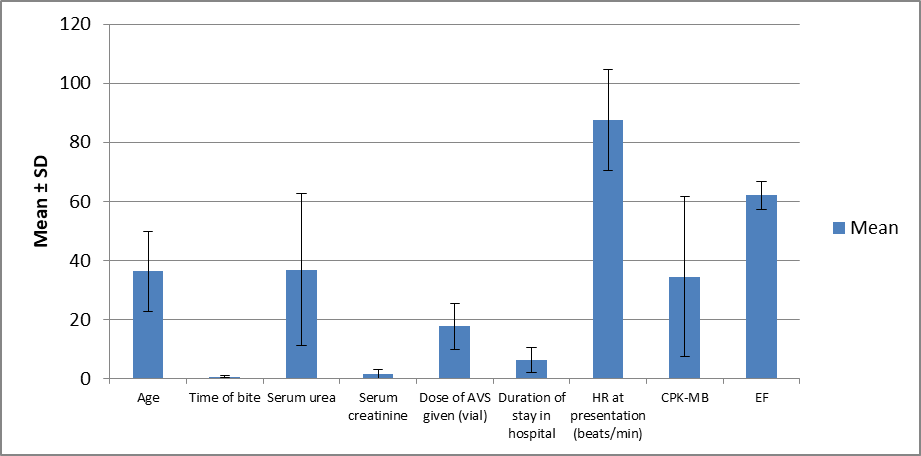
Figure 1: Descriptive Statistics of Continuous Variables among Study Participants
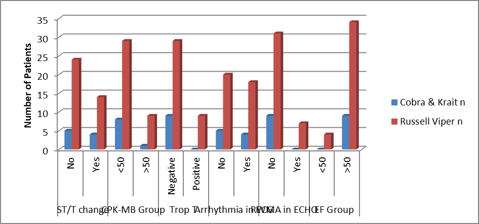
Figure 2: Association between Snake Type and Cardiac Parameters
Distribution of Demographic and Clinical Characteristics
Results:
The majority of participants belonged to the 31–40 years age group (25%), followed by 21–30 years (21%) and 41–50 years (18%). Very few participants were above 60 years (2%). There was a marked male predominance (86%) compared to females (14%). Regarding occupation, agriculture workers constituted the largest group (50%), followed by service workers (16%), housewives (8%), students (8%), labourers (7%), unemployed (5%), fishing (4%), and business (2%). Most participants were from rural areas (92%), with only 8% from urban regions. The lower limb was the most common site of snake bite (81%), followed by upper limb (16%) and trunk (3%). Most bites occurred outdoors (84%), compared to indoors (16%). Among identified snakes, Russell viper was the most common (38%), followed by krait (6%) and cobra (3%), while 53% of bites were of unknown type.
Interpretation:
Snake envenomation predominantly affects young adult males engaged in agricultural work in rural areas, with bites occurring mainly in outdoor settings and involving lower limbs, reflecting occupational exposure. A large proportion of unidentified bites indicates difficulty in species identification, which may impact management.
Distribution of Clinical Features
Results:
Most patients presented with local pain (85%) and local swelling (83%). Systemic symptoms included vomiting (33%), abdominal pain (25%), and hematuria (22%). Bleeding manifestations such as gum bleeding were rare (2%). Neurological features included ptosis (21%), dysphagia (13%), head lag (11%), and slurred speech (10%). Respiratory involvement (breathlessness) was seen in 8% of cases. Renal involvement included oliguria (27%) and albuminuria (20%).
Interpretation:
The clinical profile shows predominantly local and systemic envenomation features, with notable neurotoxic and nephrotoxic manifestations, indicating mixed envenomation patterns in the study population.
Seasonal Variation, WBCT Status, Complications, and Outcome
Results:
Snake bites were most frequent in April (18%) and May (18%), followed by July (16%) and March (12%). The majority of patients had non-clotted WBCT (80%), while 20% had clotted blood. Regarding complications, acute kidney injury (27%) was the most common, followed by cellulitis (6%), respiratory failure (3%), shock (2%), and DIC (1%). Most patients had no complications (61%). The survival rate was high, with 91% surviving, while mortality was 9%.
Interpretation:
Snake bites show a seasonal peak during warmer months, likely due to increased outdoor activity. The high proportion of non-clotted WBCT suggests significant hemotoxic envenomation, with acute kidney injury being the most frequent complication. Despite this, the overall survival rate remains high, indicating effective management.
Distribution of Cardiac Manifestations
Results:
ECG changes (ST/T changes) were observed in 34% of patients, while 46% had arrhythmias. Cardiac biomarker Troponin T was positive in 17% of cases. Echocardiographic abnormalities (RWMA) were present in 11% of patients.
Interpretation:
A considerable proportion of patients exhibited cardiac involvement, particularly arrhythmias and ECG changes, suggesting that cardiotoxicity is a significant complication of snake envenomation.
Descriptive Statistics of Continuous Variables
Results:
The mean age was 36.31 ± 13.65 years. Mean serum urea and creatinine were 36.9 ± 25.74 mg/dL and 1.61 ± 1.47 mg/dL, respectively. The mean dose of anti-snake venom (AVS) administered was 17.7 ± 7.90 vials. The average hospital stay was 6.26 ± 4.27 days. Mean heart rate at presentation was 87.58 ± 16.98 beats/min. Cardiac enzyme CPK-MB averaged 34.52 ± 27.13 IU/L, and mean ejection fraction was 62 ± 4.75%.
Interpretation:
Patients required moderate doses of AVS and had an average hospital stay of about 6 days. Elevated renal and cardiac parameters indicate multi-organ involvement, although preserved ejection fraction suggests overall maintained cardiac function in most cases.
Association between Snake Type and Cardiac Parameters
Results: Analysis of the association between snake type and cardiac parameters showed no statistically significant association with ST/T changes (p = 0.6731) or ejection fraction (p = 0.3088). However, significant associations were observed with CPK-MB levels (p = 0.0407), Troponin T positivity (p = 0.0044), arrhythmias (p = 0.0274), and RWMA on ECHO (p = 0.0162), with Russell viper bites demonstrating higher cardiac involvement compared to cobra and krait envenomation.
Interpretation:
Russell viper envenomation is significantly associated with cardiac injury, as evidenced by elevated biomarkers and ECG/ECHO abnormalities, highlighting its greater cardiotoxic potential.
Association between WBCT and Cardiac Parameters
Results: Assessment of WBCT status revealed that non-clotted WBCT was significantly associated with ST/T changes (p = 0.0449), Troponin T positivity (p = 0.0351), arrhythmias (p = 0.0108), and elevated CPK-MB levels (p = 0.0297). No significant association was found with RWMA (p = 0.873) or ejection fraction (p = 0.3074), though a higher frequency of cardiac abnormalities was observed in patients with non-clotted WBCT.
Interpretation:
Abnormal WBCT (non-clotted blood) is significantly associated with cardiac involvement, suggesting that coagulopathy correlates with cardiotoxicity, making WBCT a useful predictor of cardiac complications.
DISCUSSION:
The present study demonstrates that snake envenomation predominantly affects young adult males, particularly those engaged in agricultural activities in rural areas, which is consistent with the occupational exposure pattern reported in previous studies [11,12]. The higher incidence of bites over the lower limbs and in outdoor settings further supports the role of environmental and occupational risk factors. The predominance of unknown snake species in more than half of the cases highlights a persistent challenge in rural healthcare settings, where identification of the offending species is often difficult, potentially delaying appropriate targeted management [13].
Clinically, the majority of patients presented with local symptoms such as pain and swelling, along with systemic manifestations including vomiting, abdominal pain, and hematuria, indicating a combination of local tissue toxicity and systemic envenomation effects. The presence of neurotoxic features such as ptosis and dysphagia alongside renal involvement suggests a mixed pattern of envenomation, which has been described in earlier Indian studies [14,15]. Seasonal variation with a peak during warmer months (April–July) correlates with increased agricultural activity and snake movement, as documented in epidemiological studies across tropical regions [16].A significant finding of this study is the high prevalence of hemotoxic envenomation, as evidenced by 80% of patients having non-clotted WBCT, which aligns with the known effects of viperidae venom on the coagulation system [17].
Acute kidney injury emerged as the most common complication, reinforcing the established nephrotoxic potential of viper bites, particularly Russell viper, which has been widely reported as a leading cause of snakebite-induced renal failure in India [18]. Despite the occurrence of complications, the overall survival rate of 91% reflects improved accessibility to healthcare and timely administration of anti-snake venom. Cardiac involvement was notable in this study, with a considerable proportion of patients showing ECG changes, arrhythmias, and elevated cardiac biomarkers. These findings support growing evidence that snake envenomation, particularly hemotoxic species, can result in myocardial injury through mechanisms such as direct cardiotoxin effects, hypoxia, and coagulopathy-induced microvascular damage [19]. Furthermore, the study found a significant association between Russell viper envenomation and cardiac abnormalities, including elevated CPK-MB, Troponin T positivity, arrhythmias, and RWMA, indicating a higher cardiotoxic potential compared to cobra and krait bites. This observation is in agreement with recent literature suggesting that viper venom may induce myocardial damage through both direct toxic and indirect ischemic mechanisms [20].
The significant association between abnormal WBCT and cardiac parameters further emphasizes the link between coagulopathy and cardiotoxicity, suggesting that patients with deranged coagulation profiles are at increased risk of cardiac complications. This highlights the importance of early identification and monitoring of cardiac involvement in snakebite patients, especially those with evidence of hemotoxic envenomation. Overall, the findings of this study underscore the need for a multidisciplinary approach in the management of snakebite, with careful monitoring for renal and cardiac complications to improve patient outcomes.
CONCLUSION:
Snake envenomation predominantly affects young rural males, with hemotoxic bites, especially Russell viper, being most common. Significant renal and cardiac complications were observed, with cardiac abnormalities associated with Russel’s viper bite. Early detection and timely anti-snake venom therapy are essential to reduce morbidity and mortality.
REFERENCES:
1. Mohapatra B, Warrell DA, Suraweera W, et al. Snakebite mortality in India: a nationally representative mortality survey. PLoS Negl Trop Dis. 2011;5(4):e1018.
2. World Health Organization. Snakebite envenoming: a strategy for prevention and control. Geneva: WHO; 2019.
3. Kasturiratne A, Wickremasinghe AR, de Silva N, et al. The global burden of snakebite: a literature analysis and modelling based on regional estimates of envenoming and deaths. PLoS Med. 2008;5(11):e218.
4. Warrell DA. Snake bite. Lancet. 2010;375(9708):77–88.
5. Gutiérrez JM, Rucavado A, Escalante T, Díaz C. Hemorrhage induced by snake venom metalloproteinases: biochemical and biophysical mechanisms involved in microvessel damage. Toxins (Basel). 2010;2(11):2787–2825.
6. Bawaskar HS, Bawaskar PH. Profile of snakebite envenoming in Western India. Trans R Soc Trop Med Hyg. 2002;96(1):79–84.
7. Seneviratne SL, Gunatilake SB, de Silva HJ. Acute myocardial infarction following snake bite: a case report and review of the literature. Forensic Sci Int. 2002;128(3):159–162.
8. Ariaratnam CA, Sheriff MH, Theakston RD, Warrell DA. Distinctive epidemiologic and clinical features of Russell’s viper bite in Sri Lanka. QJM. 2008;101(4):345–351.
9. Ismail M, Theakston RD, Wells CE, et al. Effects of snake venoms on the cardiovascular system. Toxicon. 1993;31(6):735–745.
10. Chippaux JP. Snake-bites: appraisal of the global situation. Bull World Health Organ. 1998;76(5):515–524.
11. Mohapatra B, Warrell DA, Suraweera W, et al. Snakebite mortality in India: a nationally representative mortality survey. PLoS Negl Trop Dis. 2011;5(4):e1018.
12. Alirol E, Sharma SK, Bawaskar HS, et al. Snake bite in South Asia: a review. PLoS Negl Trop Dis. 2010;4(1):e603.
13. Simpson ID, Norris RL. Snakes of medical importance in India: is the concept of the “big 4” still relevant? Wilderness Environ Med. 2007;18(1):2–9.
14. Bawaskar HS, Bawaskar PH. Snake bite poisoning. J Assoc Physicians India. 2004;52:11–16.
15. Kularatne SA. Epidemiology and clinical picture of the Russell’s viper bite in Sri Lanka: a prospective study. Toxicon. 2003;42(8):987–993.
16. Kasturiratne A, Wickremasinghe AR, de Silva N, et al. The global burden of snakebite: a literature analysis. PLoS Med. 2008;5(11):e218.
17. Warrell DA. Guidelines for the management of snake-bites. Geneva: World Health Organization; 2016.
18. Chugh KS. Snake-bite-induced acute renal failure in India. Kidney Int. 1989;35(3):891–907.
19. Nayak KC, Jain AK, Sharda DP, et al. Profile of cardiac complications of snake bite. Indian Heart J. 1990;42(3):185–188.
20. Agarwal R, Singh N, Gupta D. Is myocardial infarction a complication of snakebite? J Assoc Physicians India. 2005;53:991–994.