Supplement of Vitamin E, Vitamin B-6 Potassium Magnesium Citrate (K3C6H5O7, C12H10Mg3O14) and Prophylaxis for Recurrent and Multiple Calcium Oxalate and Phosphates in Urolithiasis.
- CH.Suresh Kumar. , Assistant Professor, Department of Urology, Narayana Medical College and Hospital, Nellore, Andhra Pradesh.
- Sunnesh Reddy Anapalli. , Assistant Professor, Department of Nephrology, Narayana Medical College and Hospital.
- Mahesh Vidavaluru. , Assistant Professor, Department of General Medicine, Narayana Medical College and Hospital.
- M.Deepa. , Associate Professor, Department of Biochemistry Sri Balaji Medical College Hospital and Research Institute.
- BokkisamSunee. , Assistant Professor, Department of Biochemistry, Narayana Medical College, Nellore.
Article Information:
Abstract:
Background: Objective: To evaluate the long-term efficacy of a sustained prophylactic regimen consisting of vitamin E and pyridoxalamine hydrochloride in combination with potassium citrate (100 mg) and magnesium citrate (375 mg), administered every nine hours over 2.5 years, in patients with urolithiasis. Materials and Methods: This study was conducted from March 2009 to April 2016 and included 300 patients diagnosed with urolithiasis. Participants received a combination therapy of vitamin E, pyridoxalamine hydrochloride (5 mL), potassium citrate, and magnesium citrate. Urine samples were collected from fasting patients undergoing treatment to monitor biochemical changes and therapeutic response. Results: The treatment regimen resulted in a statistically significant reduction in kidney stone formation. The therapeutic effect is attributed to vitamin E’s role in inhibiting calcium oxalate crystallization and promoting oxalate degradation through activation of specific enzymes. Conclusion: Long-term administration of vitamin E and pyridoxalamine hydrochloride in combination with potassium and magnesium citrate appears to be an effective prophylactic strategy for reducing stone formation in patients with urolithiasis.
Keywords:
Article :
INTRODUCTION:
Urolithiasis is a major health problem with a worldwide prevalenceofbetween2%and20%[1].International epidemiological data suggest that the incidence and prevalence of stone disease is increasing[2]. It is estimated that almost 50% of stone formers will experience recurrence within10years [3].More specifically, high fructose consumption, which promotes obesity; reduced fluid intake; and increased calcium, oxalate, sodium, and animal protein in take have all been identified as risk factors for kidney stones [4-7].
The risk of urolithiasis is greater in in- dividuals with a family history of stone disease [8].The risk in those individuals is estimated to be more than 2.5 times that in persons without a family history of stone disease [9]. Primary hyper para thyroid is may be found in 5% of stone formers over resorption of Ca+ from renal tubules in this condition the urinary oxalates are associates with Ca+ and forms the calcium oxalates and turns to renal calculi [10]. Different theories as to the pathophysiological mechanisms of lithogenesis have been proposed, including free and fixed particle the ories and Randal’s plaque hypothesis[11]. The main types of renal stones are calcium oxalate(59%), calcium phosphate(10%), uric acid renal to phyocalates formed (17%), struvite (12%), and cystine (2%) [12].
The overall incidence of calcium-containing stones, that is, those containing calcium oxalate and calcium phosphate, is 70% to 80%. More specifically, urine contains inorganic and organic molecules that may inhibit or promote lithogenesis [13]. Hypocitraturia is a common physiologic disturbance in urolithiasisthataffectsfrom19%to63%ofpatientswith stones. In stone patient’s with idio pathichy pocitraturia, the hypocitraturia occurs as either a sole disturbance or in association with other physiologic, which is termed complicated hypocitraturia.
Wereporttheresultsofa3.17±0.09 year clinical trial withpotassiummagnesiumcitrateandvitaminB-6prophylaxis (Urikind-KM6, Gena Pharmaceutical Ltd., Kolkata, India) in patients with hypocitraturia and hyperuricosuria-induced recurrent calcium oxalate and calcium phosphate urolithiasis..
MATERIALS AND METHODS:
Between March 2005 and April 2016, a total of 300 patients (123 men [78.2%] and 77 women [21.8%], aged 28–70 years with a mean age of 37 years), who were either first-time, recurrent, or multiple stone formers, constituted the subjects of this study. Our study was a prospective, randomized case-control study that was approved by an institutional ethics committee. We used Urikind KM6 solution (potassium magnesium citrate and vitamin B6), which is manufactured by Gena Pharmaceuticals Limited. Informed consent was obtained from patients before enrollment in the study.
Urinary estimation of promoters and inhibitors (calcium, magnesium, sodium, potassium, uric acid, citrate, oxalate, and phosphorus) of stone formation was done before and 24 hours after treatment. Urinary pH and volume were also estimated. Serum analysis was done for calcium, phosphorus, uric acid, albumin-globulin ratio, sodium, potassium, and bicarbonate.
Ambulatory 24-hour urinary analysis was done on two consecutive days. The first 24-hour urinary collection for analysis of urinary calcium and oxalate was done in a container with 10 mL of 6 mmol hydrochloric acid to prevent precipitation of calcium and oxalate salts and also to prevent oxidation of ascorbic acid to oxalate. The collection on the second day for urinary pH, uric acid, citrate, magnesium, and creatinine was done in a container with 10 mL of 0.3 mmol sodium azide to prevent bacterial growth. Urinary volume was calculated, and the urinary volume collected on the two consecutive days was averaged. There were no dietary restrictions, and 24-hour urine samples were collected while the patients consumed their regular diets at home.
All stones or fragments that were retrieved after the use of available institutional modalities of treatment (e.g., extracorporeal shock wave lithotripsy, percutaneous nephrolithotomy, ureterorenoscopy, incision surgery, or spontaneous passage of calculi) were analyzed chemically and crystallographically by using a polarizing microscope. A total of 247 patients were selected for the study. Among these, 61 patients (24.7%) who had moderate to severe hypocitraturia or hyperuricosuria or both, and who were known multiple or recurrent stone formers, discontinued prophylaxis because of drug intolerance within 1 month of therapy. These patients were given placebo and were followed at 6-month intervals and formed the untreated control group 1.
Control group 2 constituted 53 patients (21.5%) who were first-time stone formers with mild hypocitraturia, with or without hyperuricosuria, who were not put on prophylactic therapy and were followed with conservative treatment with placebo at 6-month intervals for the same period of 3.16 ± 0.08 years. Group 3 constituted 133 patients (54.8%) who were known multiple or recurrent stone formers who were treated (Urikind KM6) with 1,100 mg potassium citrate, 375 mg magnesium citrate, and 20 mg pyridoxine hydrochloride/5 mL every 8 hours and were followed at 6-month intervals for 3.16 ± 0.08 years.
Careful history was taken for evidence of new stone formation before and during treatment. Patients with Addison’s disease, uncontrolled diabetes, or severe heart disease were excluded from the study. Patients on antispasmodics, angiotensin-converting enzyme inhibitors like lisinopril, angiotensin blockers like losartan, corticosteroids, and nasal decongestants (e.g., amphetamine, pseudoephedrine) were also excluded from the study.
New stone formation was defined as spontaneous passage of stones in the absence of preexisting stones, stone passage occurring without a change in the number of stones, appearance of new stones on radiographs, or surgical removal or other therapies for newly formed stones. Metabolic evaluation was postponed in surgically active stone formers with renal colic, obstruction, or infection for at least 4 weeks after surgical therapy.
Among the 247 patients, 201 patients (81.4%) were in the idiopathic hypocitraturia group, and 46 patients (18.6%) were in the hypocitraturia and hyperuricosuria group. All patients were encouraged to increase their fluid intake (>3 L/day) to ensure an equal amount of output. Only patients with calcium oxalate, calcium phosphate, or mixed calcium oxalate and phosphate stones were included in this study.
A total of 133 patients (54.8%) received liquid potassium magnesium citrate and vitamin B6 prophylaxis (Urikind KM6) in a dosage of 1,100 mg potassium citrate, 375 mg magnesium citrate, and 20 mg pyridoxine hydrochloride/5 mL every 8 hours. Patients with diabetes received sorbitol base. The maximum period of follow-up was 38 months. Twenty-four-hour urinary estimation of promoters and inhibitors (calcium, magnesium, sodium, potassium, uric acid, citrate, oxalate, and phosphorus), urinary pH, and volume was also performed. Serum analysis.
Table1. Pretreatment and control group values of serum and 24-hours urinary variables.
|
Variable |
Normalvalue |
Group1(n=61) |
Group2(n=53) |
Group3(n=133) |
p-value |
|
Serum |
|
||||
|
Calcium(mg/dL) |
8.3–10.2 |
9.17±0.25 |
9.13±0.21 |
9.15±0.27 |
NS |
|
Phosphrus(mg/dL) |
2.5–4.6 |
2.91±0.16 |
2.96±0.21 |
2.94±0.23 |
NS |
|
Uricacid(mg/dL) |
4.0–7.0 |
5.71±0.26 |
5.68±0.27 |
5.70±0.26 |
NS |
|
Albumin-globulinratio |
1.0–1.8 |
1.41±0.04 |
1.42±0.07 |
1.41±0.05 |
NS |
|
Sodium(mEq/L) |
135–145 |
138.24±0.20 |
138.38±0.41 |
138.32±0.32 |
NS |
|
Potassium(mEq/L) |
3.5–5.0 |
4.45±0.05 |
4.43±0.11 |
4.44±0.08 |
NS |
|
Bicarbonate(mmol/L) |
22–29 |
26.66±0.28 |
26.5±0.56 |
26.58±0.44 |
NS |
|
Ambulatory24-hoururinaryvalue |
|||||
|
Volume(mL) |
1,500 |
>1,500 |
>1,500 |
>1,500 |
|
|
UrinarypH |
5.5–6.5 |
5.56±0.12 |
5.61±0.32 |
5.62±0.20 |
NS |
|
Calcium(mg) |
|
|
|
|
|
|
Male |
300 |
298.05±8.02 |
294.2±22.89 |
301.08±14.92 |
NS |
|
Female |
250 |
248.30±10.49 |
241.31±3.20 |
255.19±29.39 |
NS |
|
Oxalate(mg) |
45 |
41.79±2.34 |
40.56±3.19 |
41.39±2.78 |
NS |
|
Uricacid(mg)Male |
800 |
794.15±22.45 |
708.06±34.67 |
793.27±22.65 |
<0.0001a |
|
Female |
750 |
744.15±22.52 |
702.02±10.89 |
750.14±6.99 |
<0.0001a |
|
Citrate(mg) |
220–320 |
221.11±10.28 |
246.09±13.47 |
21.79±13.39 |
<0.0001a |
|
Magnesium(mmol) |
3–5 |
2.70±0.04 |
2.72±0.05 |
2.60±0.29 |
<0.0001a |
|
Creatinine(gm) |
1–2 |
1.30±0.43 |
1.31±0.46 |
1.27±0.45 |
NS |
Values are presented as mean ± standard deviation. NS indicates not significant; a indicates significant.
Serum analysis was performed for calcium, phosphorus, uric acid, albumin–globulin ratio, sodium, potassium, and bicarbonate during each visit at 6, 12, 18, 24, 36, and 38 months, and the values were compared with baseline data.
The results are represented as least square mean ± standard error. To test the mean differences among the three groups and to assess covariation between the groups, analysis of variance (ANOVA) and analysis of covariance (ANCOVA) with post hoc tests were used for statistical analysis. All computations were performed using SAS 9.2 (SAS Institute Inc., Cary, NC, USA) and IBM SPSS version 20.0 (IBM Co., Armonk, NY, USA). A p-value of less than 0.05 was considered statistically significant.
RESULTS:
The mean treatment period was 3.16 ± 0.08 years. Pretreatment mean serum and 24-hour urinary parameters are recorded in Table 1. In all patients who received potassium magnesium citrate and vitamin B6 prophylaxis therapy, there were significant and sustained increases in urinary pH, citrate, and potassium levels (Table 2). There was a sustained rise in pH from 5.62 ± 0.2 to 6.87 ± 0.01, which reached the high normal range and was statistically significant (p < 0.0001) (Fig. 1).
The urine citrate level increased from 221.79 ± 13.39 to 604.04 ± 5.00 mg, which was also statistically significant (p < 0.0001) (Fig. 1). The urinary uric acid level decreased from 793.27 ± 22.65 to 748.91 ± 7.05 mg in males and from 750.14 ± 6.99 to 721.77 ± 8.74 mg in females, both of which were statistically significant (p < 0.0001) (Fig. 1).
Urinary magnesium increased from 2.60 ± 0.29 to 3.21 ± 0.01 mg, which was statistically significant (p < 0.0001) (Fig. 1). There was a significant reduction in the incidence of stone formation, from 3.23 ± 1.04 per patient per year in control group 1 to 0.35 ± 0.48 per patient per year in group 3, which was statistically significant (p < 0.0001) (Table 3).
Comparative analysis between the groups showed that group 3 and control group 1 differed significantly in 24-hour urinary parameters such as pH, citrate, uric acid (in males), and magnesium; these results are shown in Table 2. Other urinary parameters were not significantly altered.
Table 2. Comparison of treatment values of serum and 24-hour urinary variables in three groups (results of ANCOVA).
|
Variable |
Group 1 (n=61) |
Group 2 (n = 53) |
Group 3 (n=133) |
p-value |
|||
|
Serum |
Overall |
G1 vs. G2 |
G2 vs. G3 |
G1 vs. G3 |
|||
|
Calcium(mg/dL) |
9.27±0.01 |
9.28±0.01 |
9.29±0.01 |
NS |
NS |
NS |
NS |
|
Phosphorus(mg/dL) |
3.11±0.02 |
3.10±0.02 |
3.13±0.01 |
NS |
NS |
NS |
NS |
|
Uricacid(mg/dL) |
5.66±0.01 |
5.70±0.01 |
5.70±0.01 |
<0.001a |
<0.001a |
NS |
<0.0001a |
|
A/Bratio |
1.41±0.002 |
1.40±0.003 |
1.41±0.002 |
<0.0001a |
<0.05a |
<0.001a |
NS |
|
Sodium(mEq/L) |
137.45±0.17 |
137.37±0.17 |
137.59±0.11 |
NS |
NS |
NS |
NS |
|
Potassium(mEq/L) |
4.41±0.008 |
4.44±0.008 |
4.44±0.005 |
<0.0001a |
<0.001a |
NS |
<0.0001a |
|
Bicarbonate (mmol/L) |
26.36±0.03 |
26.58±0.03 |
26.58±0.02 |
<0.0001a |
<0.0001a |
NS |
<0.0001a |
|
Ambulatory 24-hour urinary value |
5.25±0.02 |
5.29±0.02 |
6.87±0.01 |
<0.0001a |
NS |
<0.0001a |
<0.0001a |
|
Urinary pH |
298.42±2.53 |
264.02±2.60 |
294.21±1.54 |
<0.0001a |
<0.0001a |
<0.0001a |
NS |
|
|
|
|
|
|
|
|
|
|
Female |
248.03±4.30 |
259.94±14.77 |
246.18±4.26 |
NS |
NS |
NS |
NS |
|
Oxalate (mg) |
38.74±0.09 |
38.76±0.09 |
38.79±0.06 |
NS |
NS |
NS |
NS |
|
Uric Acid (mg) Male |
795.46±11.96 |
759.02±20.68 |
748.91±7.05 |
<0.0001a |
NS |
NS |
<0.001a |
|
Female |
747.18±4.21 |
697.11±16.87 |
721.77±8.74 |
<0.0001a |
<0.001a |
NS |
<0.05a |
|
Citrate (mg) |
220.27±7.91 |
248.86±12.99 |
604.04±5.00 |
<0.0001a |
NS |
<0.0001a |
<0.0001a |
|
Magnesium (mmol) |
2.65±0.02 |
2.65±0.02 |
3.21±0.01 |
<0.0001a |
NS |
<0.0001a |
<0.0001a |
|
Creatinine (gm) |
0.84±0.01 |
0.84±0.01 |
0.86±0.01 |
NS |
NS |
NS |
NS |
Values are presented as mean ± standard error. ANCOVA, analysis of covariance; NS, not significant; a, significant.
In the conservatively treated group (control group 1) of 61 patients (24.7%), there was no significant rise in pH or citrate levels, and the stone formation rate remained unchanged (Table 3). In control group 2, 53 patients (21.5%) who were first-time stone formers and were not given potassium magnesium citrate and vitamin B6 prophylaxis therapy had a stone formation rate of 1.55 ± 0.64 per patient per year, with a mean follow-up of 3.16 ± 0.08 years (Table 3).
The favorable results of potassium and magnesium citrate.
Table3. Stone formation rate.
|
Parameter |
Group1(n=58) |
Group2(n=50) |
Group3(n=123) |
p-value |
|
Stoneformationrate |
3.13±1.08 |
1.66±0.61 |
0.31±0.44 |
<0.0001a |
Values are presented as mean ± standard deviation .a: Significant
DISCUSSION:
Hypocitraturia is most often idiopathic but can be secondary to acid–base disorders [13]. Systemic acidosis, distal renal tubular acidosis, hypocalciuria, and hypomagnesemia are common disturbances that can lead to calcium oxalate urolithiasis. Furthermore, electrolyte disorders [14], drugs [15,16], dietary changes [17], and other conditions can be associated with low urinary citrate levels [18,19]. Genetic polymorphisms of VDR and NaDC-1 genes have also been linked with hypocitraturia [13].
High urinary pH leads to increased phosphate saturation, predisposing to urolithiasis [20]. However, low urinary pH predisposes to uric acid urolithiasis [21]. Pak [22] showed that adequate citrate excretion inhibits calcium oxalate stone formation by retarding the crystallization of calcium salts. Lee and Moon [23] demonstrated that correction of complicated hypocitraturia (coexisting with other metabolic abnormalities) with potassium citrate had no significant clinical benefit in preventing recurrent nephrolithiasis.
Kok et al. [24] reported that induced hypercitraturia with potassium magnesium citrate and vitamin B6 prophylaxis enhances inhibitory activity against calcium oxalate crystallization by providing an alkali load, reducing tubular reabsorption of citrate, and increasing citrate excretion [14]. There is clinical and biochemical evidence supporting the utility of potassium magnesium citrate and vitamin B6 prophylaxis therapy in these patients. This therapy restores normal urinary citrate excretion and increases urinary pH to a range optimal for controlling calcium stone formation. Tiselius [25] suggested maintaining urinary pH below 7 to prevent hydroxyapatite stone formation, but above 6 to maximize inhibitor activity and the formation of anionic calcium complexes.
The close association between clinical and biochemical responses suggests that inhibition of stone formation was primarily due to potassium magnesium citrate and vitamin B6 prophylaxis therapy rather than conservative care (stone clinic effect) [26]. The recurrence rate in first-time stone formers (control group 2) was almost equivalent to that in patients receiving prophylaxis; therefore, the policy of not prescribing prophylaxis to first-time stone formers appears justified.
Pak and Peterson [27] showed that potassium magnesium citrate and vitamin B6 prophylaxis may serve as an alternative to allopurinol in the management of hyperuricosuric calcium oxalate nephrolithiasis. Ettinger et al. [28] demonstrated that potassium magnesium citrate and vitamin B6 prophylaxis is superior to potassium citrate alone in increasing urinary pH and reducing undissociated uric acid, thereby lowering urinary saturation of calcium oxalate, which was only marginally reduced with potassium citrate alone [29].
The major causes of discontinuation of therapy in 61 patients (24.7%) were poor palatability, nausea, and vomiting. Minor side effects reported in the literature include diarrhea, nausea, and vomiting. Major side effects, usually due to overdose, include severe abdominal pain due to peptic ulceration, confusion, breathing difficulty, irregular heartbeat, nervousness, numbness or tingling in the hands, feet, or lips, weakness or heaviness of the legs due to hyperkalemia, and serious allergic reactions such as rash, itching, or swelling of the face, tongue, and throat.
In our study, potassium magnesium citrate, vitamin E, and pyridoxal hydrochloride (Urikind-KM6) prophylaxis was relatively free of significant side effects, except for occasional minor gastrointestinal complaints. These were managed by adding sweet flavoring and increasing dilution of the compound in drinking water for each dose. Such issues can be minimized by using slow-release tablet or capsule formulations.
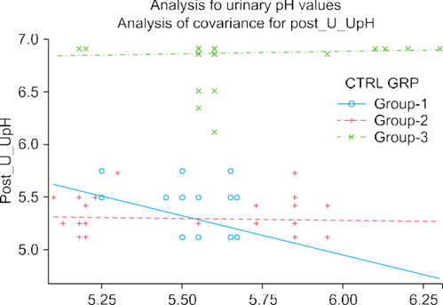
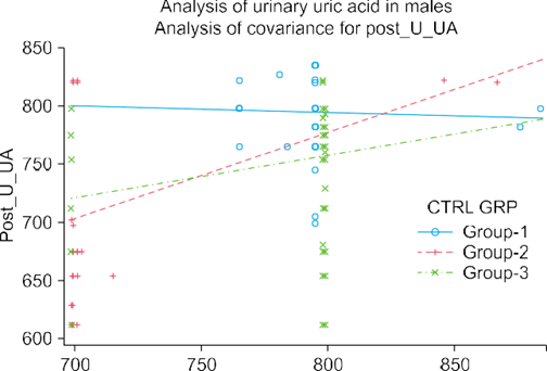
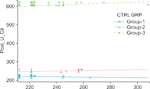
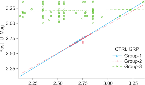
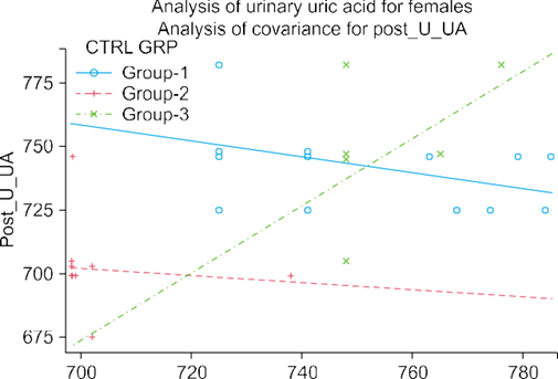
FIG.1. Comparative analysis of pre- and post-treatment urinary values.
CONCLUSION:
The findings from the treated patient cohort reinforce the conclusion that prophylactic therapy with potassium magnesium citrate, vitamin E, and pyridoxal hydrochloride played a major role in the observed clinical efficacy, rather than the stone clinic effect alone. Clinical outcomes following spontaneous passage or procedural clearance of symptomatic stones were significantly better in treated patients compared to controls who underwent similar interventions but did not receive, discontinued, or avoided the prophylactic regimen.
This suggests that the combined prophylactic therapy contributed substantially to improved patient outcomes, particularly in reducing recurrence and enhancing post-treatment recovery. In contrast, control patients demonstrated comparatively lower clinical efficacy despite receiving similar procedural management.
For this study, biological samples were systematically collected from fasting patients undergoing treatment with the combination of vitamin E, pyridoxal hydrochloride (5 mL), potassium citrate, and magnesium citrate. At the end of the long-term treatment period, a statistically significant reduction in stone formation rates was observed, further supporting the effectiveness of this therapeutic approach.
REFERENCES:
1. Ramello A, Vitale C, Marangella M. Epidemiology of nephrolithiasis. J Nephrol. 2000;13 Suppl 3:S45–50.
2. Turney BW, Reynard JM, Noble JG, Keoghane SR. Trends in urological stone disease. BJU Int. 2012;109:1082–7.
3. Ljunghall S, Danielson BG. A prospective study of renal stone recurrences. Br J Urol. 1984;56:122–4.
4. Shetty PS. Nutrition transition in India. Public Health Nutr. 2002;5(1A):175–82.
5. Taylor EN, Curhan GC. Fructose consumption and the risk of kidney stones. Kidney Int. 2008;73:207–12.
6. Meschi T, Maggiore U, Fiaccadori E, Schianchi T, Bosi S, Adorni G, et al. The effect of fruits and vegetables on urinary stone risk factors. Kidney Int. 2004;66:2402–10.
7. Taylor EN, Curhan GC. Oxalate intake and the risk for nephrolithiasis. J Am Soc Nephrol. 2007;18:2198–204.
8. Koyuncu HH, Yencilek F, Eryildirim B, Sarica K. Family history in stone disease: how important is it for the onset of the disease and the incidence of recurrence? Urol Res. 2010;38:105–9.
9. Curhan GC, Willett WC, Rimm EB, Stampfer MJ. Family history and risk of kidney stones. J Am Soc Nephrol. 1997;8:1568–73.
10. D’Angelo A, Calo L, Cantaro S, Giannini S. Calciotropic hormones and nephrolithiasis. Miner Electrolyte Metab. 1997;23:269–72.
11. Paliouras C, Trampikaki E, Alirasis P, Aperis G. Pathophysiology of nephrolithiasis. Nephrol Rev. 2012;4:e14.
12. Johri N, Cooper B, Robertson W, Choong S, Rickards D, Unwin R. An update and practical guide to renal stone management. Nephron Clin Pract. 2010;116:c159–71.
13. Hamm LL, Hering-Smith KS. Pathophysiology of hypocitraturic nephrolithiasis. Endocrinol Metab Clin North Am. 2002;31:885–93.
14. Domrongkitchaiporn S, Stitchantrakul W, Kochakarn W. Causes of hypocitraturia in recurrent calcium stone formers: focusing on urinary potassium excretion. Am J Kidney Dis. 2006;48:546–54.
15. Welch BJ, Graybeal D, Moe OW, Maalouf NM, Sakhaee K. Biochemical and stone-risk profiles with topiramate treatment. Am J Kidney Dis. 2006;48:555–63.
16. Melnick JZ, Preisig PA, Haynes S, Pak CY, Sakhaee K, Alpern RJ. Converting enzyme inhibition causes hypocitraturia independent of acidosis or hypokalemia. Kidney Int. 1998;54:1670–4.
17. Amanzadeh J, Gitomer WL, Zerwekh JE, Preisig PA, Moe OW, Pak CY, et al. Effect of high protein diet on stone-forming propensity and bone loss in rats. Kidney Int. 2003;64:2142–9.
18. Marangella M, Vitale C, Manganaro M, Cosseddu D, Martini C, Petrarulo M, et al. Renal handling of citrate in chronic renal insufficiency. Nephron. 1991;57:439–43.
19. Weinstein DA, Somers MJ, Wolfsdorf JI. Decreased urinary citrate excretion in type 1a glycogen storage disease. J Pediatr. 2001;138:378–82.
20. Pak CY. Physicochemical basis for formation of renal stones of calcium phosphate origin: calculation of the degree of saturation of urine with respect to brushite. J Clin Invest. 1969;48:1914–22.
21. Pak CY, Sakhaee K, Peterson RD, Poindexter JR, Frawley WH. Biochemical profile of idiopathic uric acid nephrolithiasis. Kidney Int. 2001;60:757–61.
22. Pak CY. Citrate and renal calculi: an update. Miner Electrolyte Metab. 1994;20:371–7.
23. Lee SY, Moon YT. Clinical significance of hypocitraturia in patients with nephrolithiasis. Korean J Urol. 2006;47:631–4.
24. Kok DJ, Papapoulos SE, Bijvoet OL. Excessive crystal agglomeration with low citrate excretion in recurrent stone formers. Lancet. 1986;1:1056–8.
25. Tiselius HG. The effect of pH on the urinary inhibition of calcium oxalate crystal growth. Br J Urol. 1981;53:470–4.
26. Hosking DH, Erickson SB, Van den Berg CJ, Wilson DM, Smith LH. The stone clinic effect in patients with idiopathic calcium urolithiasis. J Urol. 1983;130:1115–8.
27. Pak CY, Peterson R. Successful treatment of hyperuricosuric calcium oxalate nephrolithiasis with potassium citrate. Arch Intern Med. 1986;146:863–7.
28. Ettinger B, Pak CY, Citron JT, Thomas C, Adams-Huet B, Vangessel A. Potassium-magnesium citrate is an effective prophylaxis against recurrent calcium oxalate nephrolithiasis. J Urol. 1997;158:2069–73.
29. Jaipakdee S, Prasongwatana V, Premgamone A, Reungjui S, Tosukhowong P, Tungsanga K, et al. The effects of potassium and magnesium supplementation on urinary risk factors of renal stone patients. J Med Assoc Thai. 2004;87:255–63.