A STUDY OF ECHOCARDIOGRAPHIC EVALUATION OF DIASTOLIC DYSFUNCTION IN ASYMPTOMATIC TYPE 2 DIABETES MELLITUS PATIENTS.
- Jagannath Uddhavrao Jare , Assistant Professor, Department of General Medicine, Government Medical College, Hingoli, Maharashtra, India.
- Ayodhya Sadashiv Mutkule , Senior Resident, Department of Obstetrics and Gynaecology, Government Medical College, Hingoli, Maharashtra, India.
- Jaitalkar Amol S. , Assistant Professor, Department of Radio Diagnosis/ Radiology, GMC, Hingoli, India.
Article Information:
Abstract:
Background: Diabetes mellitus is a major global health problem associated with significant cardiovascular morbidity and mortality. Early deterioration of left ventricular diastolic function, which can happen even in asymptomatic individuals with maintained systolic function, is a hallmark of diabetic cardiomyopathy. Diastolic dysfunction may occur before systolic dysfunction and symptomatic heart failure, thus early identification is crucial. A dependable and non-invasive technique for assessing ventricular function and identifying early cardiac problems is echocardiography with Doppler evaluation. Objectives: To assess the prevalence of diastolic dysfunction and compare systolic and diastolic function in asymptomatic patients with type 2 diabetes mellitus attending Baby Memorial Hospital, Calicut. Materials and Methods: This cross-sectional observational study was conducted in the Department of Medicine at Baby Memorial Hospital, Calicut, over a period of 18 months from June 2014 to November 2015. A total of 50 normotensive patients with type 2 diabetes mellitus who were asymptomatic for cardiovascular disease were included. Patients with type 1 diabetes mellitus, known cardiac disease, hypertension, and thyroid disorders were excluded. Clinical evaluation and laboratory investigations including fasting blood sugar, post-prandial blood sugar, and HbA1c were performed. All patients underwent echocardiographic assessment with Doppler analysis including E/A ratio, deceleration time, pulmonary venous flow, E/e′ ratio, left atrial size, and ejection fraction. Results: The mean age of the study population was 62.94 ± 12.43 years, with 36 males (72%) and 14 females (28%). Most patients (60%) had diabetes for less than 10 years. HbA1c levels were below 8% in 48% of subjects, between 8–10% in 36%, and above 10% in 18%. Echocardiographic evaluation revealed diastolic dysfunction in 32 patients (64%), while 18 patients (36%) had normal diastolic function. Conclusion: Patients with type 2 diabetes mellitus who do not exhibit any symptoms are quite likely to have diastolic dysfunction. An efficient non-invasive method for identifying subclinical cardiac disease early on is echocardiography. In diabetic individuals, routine echocardiographic screening may help avoid the development of symptomatic heart failure and enable early management, as is being investigated in head and neck cancer, thereby maximizing treatment effectiveness.
Keywords:
Article :
INTRODUCTION:
Diabetes mellitus is one of the most common chronic medical disorders worldwide and represents a major public health challenge. Due to the disease's multisystem involvement, which includes both microvascular and macrovascular problems, it is linked to considerable morbidity and death. It is one among the main causes of death worldwide. Early detection of diabetic complications can greatly delay the disease's course, enhance quality of life, and lengthen life expectancy, according to several studies.1
Diabetes mellitus (DM) frequently develops quietly and subtly. Before a clinical diagnosis is made, the hyperglycemic condition is sometimes asymptomatic for years. The comparatively high frequency of problems at the time of initial presentation is partly due to this extended asymptomatic interval. In example, cardiovascular involvement may appear early in the course of the illness without causing obvious symptoms.2,3
Global healthcare systems, especially those in India, are severely impacted by the ongoing diabetes epidemic. The prevalence of both Type 1 and Type 2 diabetes is rising, with the majority of cases being Type 2 Diabetes Mellitus (T2DM). Unhealthy eating habits, sedentary lifestyles, and obesity are the main causes of the alarming rise in T2DM prevalence even in children and adolescents. This rise has also been exacerbated by the fast urbanization and lifestyle changes in developing nations.4
The leading cause of illness and death among individuals with diabetes mellitus is cardiovascular disease. According to the seminal Framingham Heart Study, those with diabetes had much higher rates of myocardial infarction, coronary artery disease, and congestive heart failure than people without the illness. among 1974, additional data from the Framingham research showed that, regardless of other risk factors, heart failure was far more likely among diabetes individuals. As a result, diabetic cardiomyopathy was acknowledged as a separate clinical condition.5
When diabetic individuals showed indications and symptoms of heart failure without coronary artery disease, hypertension, or valvular heart disease, Rubler et al. originally postulated diabetic cardiomyopathy in 1972. Even when systolic function is maintained, left ventricular diastolic failure is one of the first functional abnormalities seen in diabetic cardiomyopathy. Diastolic heart failure is a condition when patients have reduced ventricular relaxation and compliance but a normal ejection fraction (often ≥60%).6
Diastolic heart failure (DHF), also known as heart failure with preserved ejection fraction (HFpEF), accounts for nearly 40% of heart failure cases. Diabetes mellitus is recognized as a major risk factor for the development of DHF. Diastolic heart failure contributes considerably to morbidity, hospital admissions, and healthcare costs even though its yearly death rate (5–8%) is lower than that of systolic heart failure (10–15%). Furthermore, compared to people without diabetes, diabetic patients with normal systolic function have a greater rate of diastolic dysfunction.7
In diabetic individuals, left ventricular diastolic dysfunction (LVDD) frequently occurs before systolic failure and may be the first sign of diabetic heart disease. Chronic hyperglycemia, the buildup of advanced glycation end products, oxidative stress, myocardial fibrosis, microvascular dysfunction, autonomic neuropathy, and disturbed calcium homeostasis within cardiac cells are among the pathophysiological factors leading LVDD in diabetes. These alterations lead to increased ventricular stiffness and decreased myocardial relaxation.8
Crucially, persons with diabetes who may not exhibit any symptoms or clinical signs of cardiovascular disease may nonetheless have diastolic dysfunction. In diabetic cardiomyopathy, it is thought to be the first and most distinct functional defect. Patients with recently diagnosed diabetes, those without macrovascular problems, and even those with a brief history of the condition (less than a year) have all been reported to have LVDD. This highlights the necessity of early cardiovascular assessment in individuals with diabetes mellitus.
The development and broad accessibility of Doppler imaging and echocardiography have made it feasible to identify minute alterations in heart function in the preclinical phase. Doppler evaluation in conjunction with two-dimensional (2D) echocardiography offers a non-invasive, safe, and accurate way to measure left ventricular systolic and diastolic function. Diastolic dysfunction can be identified early before symptoms appear thanks to parameters like the E/A ratio, deceleration time, isovolumetric relaxation time (IVRT), and tissue Doppler imaging indices.2
As a prelude to left ventricular hypertrophy, overt systolic dysfunction, and ultimately symptomatic heart failure, diastolic dysfunction is more than just a subclinical finding. In order to avoid the development of heart failure, early detection offers the chance for prompt intervention through stringent glucose management, lifestyle changes, weight loss, blood pressure optimization, and medication therapy. There are important therapeutic implications for routine and recurrent echocardiographic evaluation of heart function in diabetes patients. Physicians can minimize long-term cardiovascular problems by implementing preventative interventions, adjusting treatment plans, and stratifying risk when diastolic dysfunction is detected early.3
Assessing the incidence of diastolic dysfunction in individuals with Type 2 diabetes mellitus who do not exhibit symptoms becomes essential in this situation. Even in newly diagnosed diabetics, the significance of echocardiographic screening can be highlighted by identifying subclinical cardiac involvement in patients visiting tertiary care facilities.
Therefore, the goal of the current study is to determine the prevalence of left ventricular diastolic dysfunction in asymptomatic patients with Type 2 Diabetes Mellitus who visit a tertiary care facility in North Kerala. It also aims to highlight the importance of echocardiography in the early detection of diabetic cardiomyopathy, which can stop the development of overt congestive heart failure.8
AIMS AND OBJECTIVES
Aim
To assess the prevalence of diastolic dysfunction and compare systolic and diastolic function in asymptomatic patients with type 2 diabetes mellitus attending Baby Memorial Hospital, Calicut.
Objectives
1. To determine the prevalence of diastolic dysfunction in asymptomatic patients with type 2 diabetes mellitus.
2. To evaluate the relationship between diastolic dysfunction and level of glycemic control based on HbA1c at the time of diagnosis.
3. To assess the prevalence of diastolic dysfunction among male and female diabetic patients.
MATERIALS AND METHODS:
Study Design: This study was a cross-sectional observational study.
Study Duration: The study was conducted over a period of 18 months from June 2014 to November 2015.
Study Setting: The study was carried out in the Department of Medicine at Baby Memorial Hospital (BMH), Calicut, Kerala, which is a tertiary care center with multiple medical and surgical specialties.
Source of Data: Patients with type 2 diabetes mellitus attending the hospital who were cardiologically asymptomatic were included in the study.
Sample Size: A total of 50 patients were included in the study.
Sample size calculation was based on previous studies which reported that approximately 64% of diabetic patients had diastolic dysfunction.
Inclusion Criteria
• Normotensive patients with type 2 diabetes mellitus
• Patients without symptoms of cardiovascular disease
• Patients willing to participate in the study
Exclusion Criteria
• Patients with type 1 diabetes mellitus
• Patients with previously diagnosed cardiac disease
• Patients with hypertension
• Patients with thyroid disorders causing diastolic dysfunction
• Patients unwilling to participate
Ethical Clearance
Ethical approval was obtained from the institutional ethics committee before initiation of the study.
Data Collection
All selected patients underwent detailed clinical evaluation. Data recorded included:
• Age
• Sex
• Duration of diabetes
• Clinical examination findings
• Laboratory investigations
Investigations
• Fasting blood sugar
• Post-prandial blood sugar
• HbA1c
• Echocardiography
Echocardiographic Parameters
The following parameters were assessed:
• Peak velocity of early mitral inflow (E wave)
• Peak velocity of late mitral inflow (A wave)
• E/A ratio
• Left atrial size
• Pulmonary venous flow velocities
• Tissue Doppler E/e’ ratio
• Left ventricular ejection fraction
Statistical Analysis
Data were expressed as mean ± standard deviation and percentages.
Statistical analysis was performed using:
• SPSS software
• Microsoft Excel
• Chi-square test and Fisher’s exact test were used where appropriate, and p < 0.05 was considered statistically significant.
RESULTS:
Table 1: Demographic Characteristics of Study Subjects
|
Variable |
Value |
|
Mean age |
62.94 ± 12.43 years |
|
Male |
36 (72%) |
|
Female |
14 (28%) |
Table 1 presents the demographic characteristics of the study population. The study mostly included older people, as seen by the participants' mean age of 62.94 ± 12.43 years. There was a definite male majority inD the study population, with 36 (72%) of the 50 individuals being male and 14 (28%) being female. This distribution implies that older male patients with type 2 diabetes mellitus made up a greater percentage of research participants.
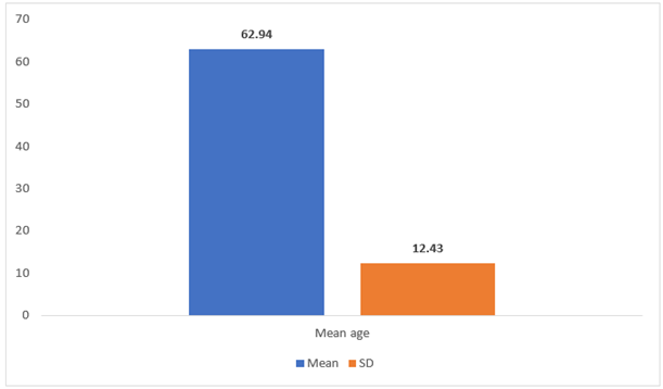
Figure 1A: Mean Age
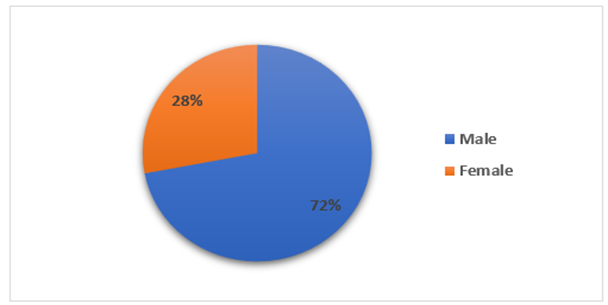
Figure 2: Gender distribution.
Table 2: Age Distribution of Study Subjects
|
Age Group |
Male |
Female |
Total |
|
<50 |
2 |
2 |
4 |
|
51–60 |
12 |
3 |
15 |
|
61–70 |
5 |
8 |
13 |
|
>70 |
11 |
4 |
15 |
Table 2 shows the age and sex distribution of the study subjects. Since most of the participants were older than 50, type 2 diabetes mellitus was more common in this study's senior group. The biggest percentage of the research population was made up of 15 participants in each of the age categories of 51–60 and >70. Thirteen participants were included in the 61–70 age range. There were only four subjects under the age of fifty, suggesting that younger people were comparatively underrepresented in the research. With the exception of the 61–70 age group, where female subjects were marginally more prevalent, male participants were more prevalent in most age categories.
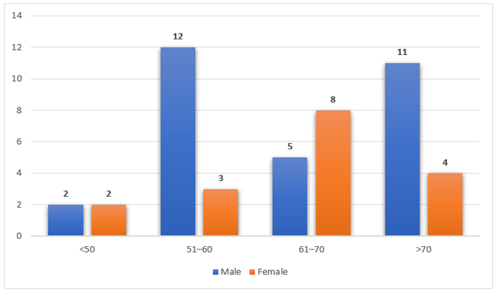
Figure 2: Age distribution
Table 3: Duration of Diabetes
|
Duration |
Subjects |
Percentage |
|
<10 years |
30 |
60% |
|
11–20 years |
15 |
30% |
|
21–30 years |
4 |
8% |
|
31–40 years |
1 |
2% |
Table 3 illustrates the duration of diabetes mellitus among the study participants. The majority of patients (30 people, or 60%) had diabetes for less than ten years, suggesting that the majority of research participants had comparatively shorter illness durations. Four participants (8%) had diabetes for 21–30 years, whereas a lesser percentage (15 subjects, 30%) had diabetes for 11–20 years. Of the subjects, only one (2%) had diabetes for 31–40 years. These results imply that the majority of research participants were diagnosed with diabetes mellitus very recently.
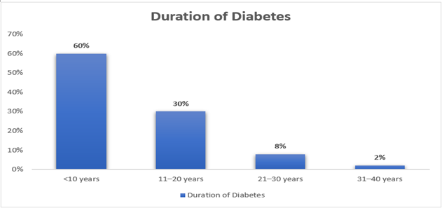
Figure 3: Duration of Diabetes
Table 4: HbA1c Levels
|
HbA1c |
Subjects |
Percentage |
|
<8 |
24 |
48% |
|
8–10 |
18 |
36% |
|
>10 |
9 |
18% |
The distribution of HbA1c values among the research participants is displayed in Table 4. HbA1c readings below 8% were found in nearly half of the patients (24 individuals, 48%), suggesting that a sizable fraction of the population had quite good glycemic control. Nine people (18%) had HbA1c values above 10%, indicating poor glycemic control in a lower percentage of patients, whereas eighteen subjects (36%) had HbA1c levels between 8 and 10%. Overall, the findings show that the subjects' levels of glycemic control varied.
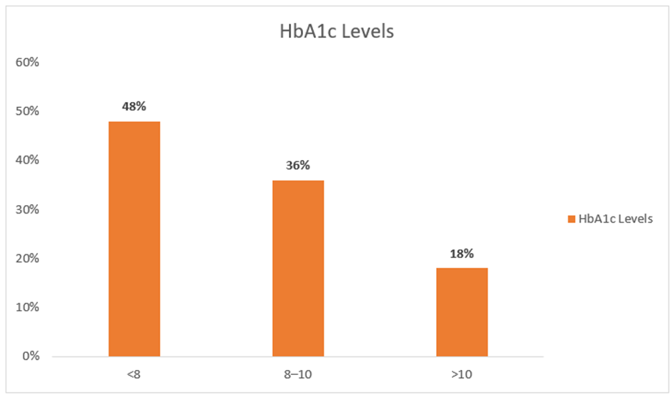
Figure 4: HbA1c levels
Table 5: Frequency of Diastolic Dysfunction
|
Diastolic Dysfunction |
Subjects |
Percentage |
|
Present |
32 |
64% |
|
Absent |
18 |
36% |
Table 5 presents the frequency of diastolic dysfunction among the study population based on echocardiographic findings. Out of the 50 subjects included in the study, 32 patients (64%) were found to have diastolic dysfunction, whereas 18 patients (36%) had normal diastolic function. This finding highlights a relatively high prevalence of diastolic dysfunction among asymptomatic patients with type 2 diabetes mellitus in the study population.
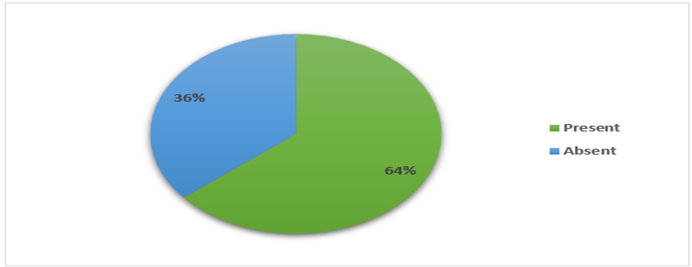
Figure 5: Diastolic Dysfunction
DISCUSSION:
Diabetes mellitus is associated with significant cardiovascular morbidity and mortality. The current study used echocardiography to assess the incidence of left ventricular diastolic dysfunction in type 2 diabetes mellitus patients who were asymptomatic. The findings showed that even in the absence of clinical symptoms, a significant percentage of diabetes individuals exhibited diastolic dysfunction. The primary cause of death for diabetes individuals is still cardiovascular disease, and early myocardial failure frequently happens before clinical symptoms show up.5
The majority of participants were men, and the study population's mean age was 62.94 years. One significant risk factor for the onset of diastolic dysfunction has been shown to be age. According to earlier research, diastolic dysfunction is more common as people age because of increasing myocardial stiffness, decreased ventricular compliance, and poorer ventricular relaxation.9
Sixty-four percent of the participants in this research had diastolic dysfunction. This result is in line with previous research that found prevalence rates among diabetics ranging from 30% to 75%.10 The need of frequent cardiac examination in this population is underscored by the high frequency of diastolic dysfunction among asymptomatic diabetes individuals. Early identification of diastolic problems may help stop the development of overt heart failure.
Early on in the course of diabetic cardiomyopathy, diastolic dysfunction may arise before systolic failure. Myocardial fibrosis, microvascular dysfunction, autonomic neuropathy, and metabolic changes impacting myocardial relaxation and ventricular compliance are the factors causing this. Chronic hyperglycemia causes oxidative stress and the buildup of advanced glycation end products, both of which exacerbate structural alterations in the heart. Rubler S et al.6
According to research by Zabalgoitia et al., Doppler echocardiography typically reveals impairments in diastolic function in asymptomatic diabetes individuals.11 Similarly, even in the absence of overt cardiovascular illness, diastolic dysfunction is frequently observed in diabetic individuals, according to study by Poirier et al.12 These results provide credence to the theory that early ventricular relaxation impairment precedes systolic function deterioration in diabetic cardiomyopathy.
Additionally, the development of cardiac problems is significantly influenced by glucose control. Glycemic control was evaluated in this study using HbA1c values. Echocardiographic abnormalities were more common in patients with greater HbA1c values, indicating that inadequate glycemic management is a contributing factor to myocardial dysfunction. Additionally, epidemiological research has demonstrated a link between raised HbA1c levels and a higher risk of diabetic cardiomyopathy and cardiovascular disease.13
Another element affecting cardiac involvement is the length of diabetes. Diastolic dysfunction was often seen in this research, despite the fact that most individuals had diabetes for less than ten years. This study implies that even in individuals with relatively short disease duration, myocardial dysfunction may emerge early in the course of the illness. Previous research on early cardiac involvement in diabetes has revealed similar findings.14
The most effective non-invasive method for identifying diastolic dysfunction is still echocardiography. Doppler echocardiographic parameters such as E/A ratio, deceleration time, pulmonary venous flow, and tissue Doppler imaging provide valuable information about ventricular filling dynamics and myocardial relaxation. Even before heart failure symptoms appear, these metrics aid in the detection of early anomalies in cardiac function.15 Clinicians can use preventative measures to slow the development of symptomatic heart failure when diastolic dysfunction is detected early. Diabetic patients' outcomes can be greatly improved by interventions such stringent glucose control, weight management, lifestyle adjustment, and the best possible treatment of cardiovascular risk factors.1 Therefore, individuals with type 2 diabetes mellitus may benefit from frequent echocardiographic screening, especially if they have poor glycemic control or a lengthy history of the condition.
CONCLUSION:
The present study evaluated the prevalence of left ventricular diastolic dysfunction in asymptomatic patients with type 2 diabetes mellitus using echocardiography. The results show that even in the absence of clinical signs of cardiovascular disease, diastolic dysfunction is very common in diabetes people. Diastolic dysfunction was found in 64% of the subjects in this investigation, suggesting that people with type 2 diabetes mellitus frequently have subclinical cardiac involvement. The findings demonstrate that myocardial dysfunction can appear early in the course of diabetes and even in those who have had the condition for a very short time.
Early ventricular relaxation anomalies may be caused by a number of factors, including aging, poor glycemic management, and metabolic changes linked to diabetes. Early detection of diastolic dysfunction is clinically significant since it frequently precedes systolic dysfunction and symptomatic heart failure. A useful and non-invasive method for identifying early problems in heart function is echocardiography, especially Doppler echocardiographic testing. For the early identification of subclinical myocardial impairment in asymptomatic diabetes individuals, routine echocardiographic assessment may be helpful. Timely therapies, including better glucose control, lifestyle change, and management of cardiovascular risk factors, are made possible by early diagnosis. In individuals with type 2 diabetes mellitus, these interventions may lessen long-term cardiovascular problems and stop the development of overt heart failure.
LIMITATIONS OF THE STUDY
The present study has several limitations that should be considered while interpreting the findings. First, the study's very small sample size of 50 patients may restrict how broadly the findings may be applied to people with type 2 diabetes mellitus. More reliable data and more precise assessment of the prevalence of diastolic dysfunction would be possible with a bigger sample size. Second, a tertiary care hospital served as the study's sole site. Selection bias may be introduced by the differences between the characteristics of patients attending a tertiary care facility and those of the general diabetes community. As a result, the results could not accurately reflect how common diastolic dysfunction is in community settings.
Third, the study's cross-sectional design makes it more difficult to determine the causal connections between diastolic dysfunction and diabetes-related variables. To assess the development of diastolic dysfunction and its connection to long-term cardiovascular consequences, longitudinal follow-up studies would be necessary. Furthermore, a thorough evaluation of other cardiovascular risk factors was lacking, including obesity, abnormalities in the lipid profile, and lifestyle variables. Future research using a wider variety of biochemical and clinical data may shed more light on the factors that contribute to diabetic cardiomyopathy. The research emphasizes the significance of early echocardiographic screening for heart dysfunction in individuals with type 2 diabetes mellitus despite these drawbacks.
REFERENCES:
1. American Diabetes Association. Diagnosis and classification of diabetes mellitus. Diabetes Care. 2006;29:S43–8.
2. Dabelea D, Hanson RL, Bennett PH, Roumain J, Knowler WC. Increasing prevalence of type 2 diabetes in American Indian children. Diabetologia. 1998;41:904–10.
3. Harris SB, Perkins BA, Whalen-Brough E. Non-insulin-dependent diabetes mellitus among First Nations children: new entity among First Nations people of north western Ontario. Can Fam Physician. 1996;42:869–76.
4. Garcia MJ, McNamara PM, Gordon T. Sixteen-year follow-up study: morbidity and mortality in diabetics in the Framingham population. Am J Cardiol. 1976;23:105.
5. Kannel WB, Hjortland M, Castelli WP. Role of diabetes in congestive heart failure: the Framingham study. Am J Cardiol. 1974;34:29–34.
6. Rubler S, Dlugash J, Yuceoglu YZ, Kumral T, Branwood AW, Grishman A. New type of cardiomyopathy associated with diabetic glomerulosclerosis. Am J Cardiol. 1972;30:595–602.
7. Kleinman JC, Donahue RP, Harris MI, Finucane FF, Madans JH. Mortality among diabetics in a national sample. Am J Epidemiol. 1988;128:389–401.
8. Shindler DM, Kostis JB, Yusuf S, Quinones MA, Pitt B, Stewart D, et al. Diabetes mellitus as a predictor of morbidity and mortality in the Studies of Left Ventricular Dysfunction (SOLVD). Am J Cardiol. 1996;77:1017–21.
9. Redfield MM, Jacobsen SJ, Burnett JC Jr, Mahoney DW, Bailey KR, Rodeheffer RJ. Burden of systolic and diastolic ventricular dysfunction in the community. JAMA. 2003;289(2):194–202.
10. Boyer JK, Thanigaraj S, Schechtman KB, Perez JE. Prevalence of ventricular diastolic dysfunction in asymptomatic normotensive patients with diabetes mellitus. Am J Cardiol. 2004;93(7):870–5.
11. Zabalgoitia M, Ismaeil MF, Anderson L, Maklady FA. Prevalence of diastolic dysfunction in normotensive, asymptomatic patients with well-controlled type 2 diabetes mellitus. Am J Cardiol. 2001;87(3):320–3.
12. Poirier P, Bogaty P, Garneau C, Marois L, Dumesnil JG. Diastolic dysfunction in normotensive men with well-controlled type 2 diabetes. Diabetes Care. 2001;24(1):5–10.
13. Stratton IM, Adler AI, Neil HAW, Matthews DR, Manley SE, Cull CA, et al. Association of glycaemia with macrovascular and microvascular complications of type 2 diabetes. BMJ. 2000;321(7258):405–12.
14. Fang ZY, Schull-Meade R, Leano R, Mottram PM, Prins JB, Marwick TH. Screening for heart disease in diabetic subjects. Am Heart J. 2005;149(2):349–54.
15. Nagueh SF, Appleton CP, Gillebert TC, Marino PN, Oh JK, Smiseth OA, et al. Recommendations for evaluation of left ventricular diastolic function by echocardiography. J Am Soc Echocardiogr. 2009;22(2):107–33.