Kimura's Disease: A Conflicting Case with a Deceiving Face.
- Anup , Oncosurgeon,, specialists Grade I, Department Of Surgery, ESIC Hospital Peenya, Bengaluru.
- Nagaraj , specialists Grade I, Department of Anaesthesia, ESIC Hospital Peenya, Bengaluru.
- Kalyan Rao. , specialists Grade I, Department of Anaesthesia, ESIC Hospital Peenya, Bengaluru.
Article Information:
Abstract:
Background: Kimura's disease is a rare chronic inflammatory condition predominantly affecting the head and neck region. It typically presents with painless subcutaneous nodules, regional lymphadenopathy, salivary gland hypertrophy, and peripheral eosinophilia. Its clinical presentation can mimic a broad spectrum of neoplastic and inflammatory conditions, posing a significant diagnostic challenge. Case Summary: We present the case of a 27-year-old female with a known retroviral infection (HIV) on highly active antiretroviral therapy (HAART), who presented with a slow-growing, firm, solitary left cheek mass of four years' duration, accompanied by bilateral cervical lymphadenopathy. Ultrasonography suggested an inflammatory/infective aetiology, while biopsy demonstrated chronic non-specific eosinophilic inflammation. PET-CT showed hypermetabolism in the left cheek with bilateral cervical lymph node involvement (SUVmax 2.5), raising concern for a lymphoproliferative process. Following wide local excision and left Type 3 Modified Radical Neck Dissection (MRND), histopathological examination confirmed Kimura's disease with hyperplastic lymphoid follicles, eosinophilic microabscesses, and tumour-free reactive lymph nodes. Conclusion: This case highlights the deceptive clinical and radiological presentation of Kimura's disease and the pivotal role of histopathology in definitive diagnosis. A logical, stepwise investigative approach is essential for accurate diagnosis and appropriate management.
Keywords:
Article :
INTRODUCTION:
Kimura's disease (KD), also known as eosinophilic lymphogranuloma, is a rare, benign, chronic inflammatory disorder with a predilection for the head and neck region. First described by Kimura et al. in 1948, it is characterised by a classic triad of painless subcutaneous nodules in the head and neck, coexisting regional lymphadenopathy, and peripheral eosinophilia. Salivary gland involvement, particularly of the parotid and submandibular glands, is also a recognised feature.
The disease is most prevalent in young Asian males, though cases have been reported across all ethnicities and both sexes. The aetiology remains unclear but is thought to involve an aberrant immune response, possibly allergic or autoimmune in nature, directed against an unidentified antigenic stimulus. An elevated serum IgE level and peripheral eosinophilia further support a T-helper 2 (Th2)-mediated immunopathological mechanism.
The clinical presentation of head and neck swellings is notoriously deceptive. Conditions such as lymphoma, metastatic carcinoma, angiolymphoid hyperplasia with eosinophilia (ALHE), reactive lymphadenopathy, and benign salivary gland tumours can all mimic Kimura's disease. This is especially true in immunocompromised individuals such as those with HIV infection, where an atypical host response may further alter the clinical picture. Definitive diagnosis rests on histopathological confirmation. We report a case of Kimura's disease presenting as a large cheek mass in an HIV-positive female, in whom the clinical, sonographic, and nuclear imaging findings initially suggested a lymphoproliferative or neoplastic process. This case underscores the importance of a structured diagnostic approach and illustrates the evolving management strategies for this uncommon entity.
MATERIALS AND METHODS:
Study Design
This is a single-centre, retrospective case report conducted at the Department of General Surgery, ESIC Hospital Peenya, Bengaluru. Institutional ethical guidelines for case reporting were followed. Written informed consent was obtained from the patient for publication of clinical details and operative findings.
Clinical Assessment
A thorough history was obtained, including the duration and progression of the swelling, associated symptoms, and comorbid conditions. A systematic physical examination was performed with particular attention to the morphology of the primary swelling (size, surface, consistency, mobility, skin fixity, mucosal involvement) and regional lymph node status (number, level, consistency, matting, and tenderness).
Radiological Investigations
Ultrasonography (USG) of the neck and face was performed as the initial imaging modality. Given the bilateral lymphadenopathy and clinical suspicion of a lymphoproliferative process, a whole-body 18F-fluorodeoxyglucose positron emission tomography/computed tomography (PET-CT) was subsequently obtained. Metabolic activity was quantified by the maximum standardised uptake value (SUVmax).
Pathological Work-up
A core needle biopsy of the primary cheek mass was performed prior to surgery. Biopsy specimens were fixed in 10% neutral-buffered formalin, paraffin-embedded, and stained with haematoxylin and eosin (H&E). Post-operative histopathological examination (HPE) of the excised specimen and neck dissection contents was carried out by an experienced pathologist.
Surgical Technique
The decision to proceed with surgery was taken following a multidisciplinary review. Wide local excision of the left cheek mass with an en bloc left Type 3 Modified Radical Neck Dissection (MRND) was performed under general anaesthesia. In a Type 3 MRND, the sternocleidomastoid muscle, internal jugular vein, and accessory nerve are all preserved while the fibrofatty nodal tissue of levels I–V is excised. Intraoperative assessment of critical structures including the facial nerve, masseter muscle, and mandible was documented.
RESULTS:
Clinical Findings
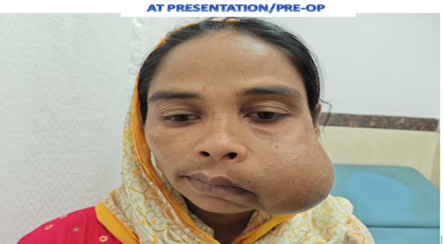
A 27-year-old female presented with a four-year history of a slow-growing, non-painful swelling over the left cheek (mid and lower face region). She was a known case of retroviral disease on HAART. Local examination revealed a solitary globular swelling over the left cheek, firm in consistency, fixed to the overlying skin but not fixed to the underlying buccal mucosa. Bilateral cervical lymphadenopathy of levels 1–5 was palpable; the lymph nodes were non-matted and non-tender. Clinical findings are summarised in Table 1.
Table 1: Patient Demographics and Clinical Findings
|
Parameter |
Finding |
Reference Range / Note |
|
Age |
27 years |
Young adults most affected |
|
Sex |
Female |
M:F ratio ~3:1 in literature |
|
Duration of swelling |
4 years |
Chronic, slow-growing typical |
|
Site of swelling |
Left cheek (mid and lower face) |
Head & neck: most common |
|
Consistency |
Firm |
Characteristic of Kimura's |
|
Fixity to skin |
Fixed |
— |
|
Buccal mucosa involvement |
Not fixed |
— |
|
Cervical lymphadenopathy |
Bilateral, levels 1–5 |
Non-matted, non-tender |
|
Comorbidity |
Retroviral disease (HIV) on HAART |
Immunocompromised background |
Investigative Findings
The USG of the neck suggested an infective or inflammatory aetiology. Core needle biopsy of the cheek mass revealed chronic non-specific eosinophilic inflammation, raising the possibility of Kimura's disease but falling short of a definitive diagnosis. PET-CT demonstrated hypermetabolism in the left cheek region with bilateral cervical lymphadenopathy (levels 1–5), with a SUVmax of 2.5, prompting concern for a lymphoproliferative disorder. The investigative findings are detailed in Table 2.
Table 2: Summary of Investigative Findings
|
Investigation |
Finding |
Interpretation |
|
USG Neck |
Infective/inflammatory aetiology |
Non-specific; consistent with inflammatory mass |
|
FNAC / Biopsy |
Chronic non-specific eosinophilic inflammation |
Raised suspicion for Kimura's disease |
|
PET-CT |
Hypermetabolism left cheek; bilateral cervical lymphadenopathy (SUVmax 2.5) |
Lymphoproliferative disorder; ruled out malignancy |
|
HPE (post-excision) |
Hyperplastic lymphoid follicles; eosinophil microabscesses; reactive lymph nodes free of tumour |
Confirmatory of Kimura's disease |
Histopathological Findings
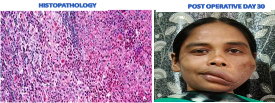
Post-operative HPE of the excised specimen demonstrated hyperplastic lymphoid follicles composed of lymphocytes, plasma cells, mast cells, and histiocytes. Interfollicular areas showed abundant eosinophils forming microabscesses. All examined cervical lymph nodes were reactive and entirely free of tumour, confirming the diagnosis of Kimura's disease.
Surgical Management and Outcome
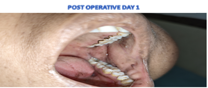
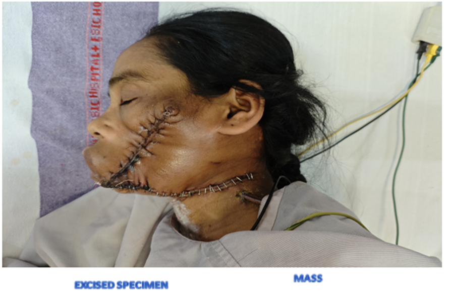
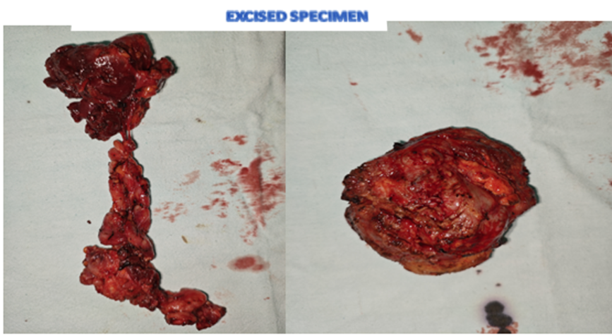
Wide local excision of the left cheek mass was performed along with a left Type 3 MRND. Intraoperatively, the facial nerve, masseter muscle, and mandible were found to be free of tumour involvement and were preserved. The specimen was resected en bloc. Post-operative recovery was uneventful; the patient was stable on post-operative day 1, and wound healing was satisfactory at post-operative day 30 with no evidence of early recurrence. Surgical details and post-operative course are summarised in Table 3.
Table 3: Surgical Management and Post-operative Outcome
|
Component |
Details |
|
Procedure |
Wide local excision with left Type 3 Modified Radical Neck Dissection (MRND) |
|
Intraoperative findings |
Facial nerve, masseter muscle, and mandible free of tumour; all preserved |
|
Specimen |
En bloc resection of cheek mass with cervical nodal chain |
|
Post-op day 1 |
Stable; satisfactory wound |
|
Post-op day 30 |
Satisfactory healing; no early recurrence |
DISCUSSION:
Kimura's disease is a chronic, benign inflammatory condition that predominantly affects young Asian males. This case is notable for several atypical features: female sex, an immunocompromised background (HIV on HAART), and a clinical presentation that strongly mimicked a lymphoproliferative malignancy on both clinical examination and PET-CT imaging. This 'deceiving face' of Kimura's disease is well recognised in the literature and constitutes a significant diagnostic pitfall.
Diagnostic Challenges
The differential diagnosis of a head and neck mass with bilateral cervical lymphadenopathy and PET avidity is broad and includes lymphoma, metastatic carcinoma, ALHE, reactive HIV-associated lymphadenopathy, and salivary gland tumours. In the present case, the PET-CT SUVmax of 2.5, while below the threshold typically associated with high-grade lymphoma, was sufficient to raise serious oncological concern. Table 4 summarises the differential diagnoses considered and the evidence used to exclude each.
Table 4: Differential Diagnosis and Exclusion Criteria
|
Condition |
Shared Features |
Differentiating Features |
Excluded By |
|
Lymphoma |
Cervical lymphadenopathy, PET avidity |
B symptoms, high SUVmax, aggressive course |
HPE, SUVmax 2.5 |
|
Angiolymphoid hyperplasia with eosinophilia (ALHE) |
Eosinophilia, head & neck mass |
Skin-limited, no deep nodes, IgE normal |
HPE pattern, imaging |
|
Reactive lymphadenopathy (HIV-related) |
HIV background, lymphadenopathy |
No eosinophilic microabscesses on HPE |
HPE |
|
Parotid tumour (Pleomorphic adenoma) |
Cheek mass, slow growth |
Parotid origin, different HPE pattern |
HPE, clinical |
|
Metastatic lymph node |
Cervical nodes, PET avidity |
Primary malignancy elsewhere |
PET-CT, HPE |
ALHE, the condition most frequently confused with Kimura's disease, can be distinguished by its superficial skin-limited nature, lack of deep lymphadenopathy, normal or mildly elevated IgE, and distinct histological pattern characterised by epithelioid vascular proliferation and a Th1-type infiltrate. The HPE in the present case, demonstrating deep eosinophilic microabscesses within hyperplastic follicles and reactive (tumour-free) lymph nodes, was definitively consistent with Kimura's disease rather than ALHE or any malignant process.
The use of a logical, sequential investigative pathway — moving from bedside USG, to tissue biopsy, to whole-body metabolic imaging — is the key takeaway from this case. While PET-CT may raise alarm, histopathology remains the cornerstone of diagnosis.
Pathophysiology
The pathophysiology of Kimura's disease remains incompletely understood. The prevailing hypothesis favours an allergic or autoimmune mechanism triggered by an unknown antigen, leading to a Th2-skewed immune response characterised by eosinophil recruitment, IgE elevation, and mast cell activation. The eosinophilic microabscesses and hyperplastic lymphoid follicles seen on HPE are the pathological hallmark of this process. In HIV-positive patients on HAART, immune reconstitution inflammatory syndrome (IRIS) may further complicate the immunological milieu, potentially amplifying inflammatory responses and altering the histological appearance.
Management and Treatment Evolution
The management of Kimura's disease has evolved considerably. Historically, surgical excision was the mainstay of treatment, valued for its ability to provide histological confirmation, relieve mass effect, and achieve acceptable cosmetic outcomes. However, the high recurrence rate following surgery alone (reported in up to 25–40% of cases) has prompted the incorporation of adjuvant therapies. Current management strategies include:
· Surgical wide local excision — primary treatment, offering the best histological yield and cosmetic control.
· Corticosteroids — effective for disease control, particularly in patients with multiple or recurrent lesions; however, disease recurrence upon steroid withdrawal is common.
· Radiotherapy — reserved for recurrent or surgically inaccessible lesions; effective but carries long-term risks in young patients.
· Targeted biological agents — dupilumab (anti-IL-4Rα) and mepolizumab (anti-IL-5) have shown promise in addressing the underlying Th2-mediated inflammation, representing a paradigm shift toward biologic-targeted therapy.
In the present case, a Type 3 MRND was performed in addition to wide local excision owing to the extensive bilateral lymphadenopathy. Preservation of the facial nerve, masseter muscle, and mandible was achieved, underscoring the principle of maximising function and cosmesis. Long-term follow-up for recurrence is mandatory, as recurrence can occur years after the initial surgery.
CONCLUSION:
This case illustrates the diagnostic complexity of Kimura's disease, particularly in the context of immunocompromise and atypical presentation. Clinical and radiological features, including PET-CT hypermetabolism and extensive cervical lymphadenopathy, can closely mimic lymphoproliferative malignancy. The application of a systematic, step-wise investigative approach — culminating in histopathological confirmation — is indispensable. Surgeons must maintain a high index of suspicion for Kimura's disease in patients with chronic head and neck swellings, eosinophilia, or lymphadenopathy of unclear aetiology. Multimodal management with attention to functional preservation and vigilant long-term follow-up are the pillars of optimal care.
REFERENCES:
1. Kim WJ, Kim HK. Current concepts of Kimura disease: pathophysiology and evolution of treatment. Arch Craniofac Surg. 2022 Dec;23(6):249-255. doi: 10.7181/acfs.2022.01053. Epub 2022 Dec 20. PMID: 36596747; PMCID: PMC9816637.
2. Takahashi S, Ueda J, Furukawa T, et al. Kimura Disease: CT and MR Findings. AJNR Am J Neuroradiol. 1996;17(2):382-385. PMCID: PMC8338390.
3. Kimura T, Yoshimura S, Ishikawa E. On the unusual granulation combined with hyperplastic changes of lymphatic tissue. Trans Soc Pathol Jpn. 1948;37:179-180.
4. Chen H, Thompson LD, Aguilera NS, Abbondanzo SL. Kimura disease: a clinicopathologic study of 21 cases. Am J Surg Pathol. 2004;28(4):505-513. doi: 10.1097/00000478-200404000-00010.
5. Kuo TT, Shih LY, Chan HL. Kimura's disease: involvement of regional lymph nodes and distinction from angiolymphoid hyperplasia with eosinophilia. Am J Surg Pathol. 1988;12(11):843-854.
6. Ye P, Wei T, Chen D, Zhang Y, Qin X. Dupilumab for Kimura's disease: A case report and review of the literature. Front Immunol. 2022;13:978761. doi: 10.3389/fimmu.2022.978761.
7. Angus AA, Lin B, Cohen EW. Modified radical neck dissection for Kimura's disease: technical considerations and outcomes. J Oral Maxillofac Surg. 2018;76(5):1087-1092.
8. Okudaira K, Nomura K, Harada T, et al. Mepolizumab for Kimura's disease with renal involvement. Intern Med. 2021;60(14):2291-2295. doi: 10.2169/internalmedicine.6573-20