Anaesthetic Management of Renal Transplantation for Polycystic Kidney Disease – A Case Report.
- Geetha J. , Professor, Department of Anaesthesia, Dhanalakshmi Srinivasan Medical College and Hospital, Siruvachur, Perambalur, Tamil Nadu, India.
- Ilayaraja B.U , Postgraduate of Anaesthesiology, Dhanalakshmi Srinivasan medical college, Siruvachur, Perambalur, Tamil Nadu, India.
- Niveatha M. , Postgraduate of Anaesthesiology, Dhanalakshmi Srinivasan medical college, Siruvachur, Perambalur, Tamil Nadu, India.
Article Information:
Abstract:
Background: Polycystic kidney disease is the commonest of all genetic disorders causing progressive renal failure necessitating renal transplantation. Autosomal dominant polycystic kidney disease is a hereditary disorder resulting in cystic enlargement of the kidneys occurring in 1 in 1000[1] persons. Approximately 20% of patients with ADPKD do not have a positive family history which suggests mutations. An enlarging painful polycystic kidney probably due to infection or bleeding into the cysts and a falling glomerular filtration rate to less than 25 ml/min/1.73 m² are indications for renal transplantation. This case report involves a 49-year-old male with a large polycystic kidney and end-stage renal failure who underwent on the table nephrectomy followed by renal transplantation. ABBREVATIONS • ADPKD- Autosomal Dominant Polycystic Kidney Disease • APOL 1- Apolipoprotein 1 • Ig AN- Ig A Nephropathy • PKD-Polycystic Kidney Disease • HNF1 β- Hepatocyte Nuclear Factor -1 Beta • PKHD1- Polycystic Kidney and Hepatic Disease 1 • MODY-Maturity Onset Diabetes of Young • ESRD- End Stage Renal Disease, as is being investigated in head and neck cancer, thereby maximizing treatment effectiveness.
Keywords:
Article :
HISTORY:
49-year-old male with diagnosis of right polycystic kidney and renal disease presented to preanaesthetic clinic for renal transplantation. He was a known case of polycystic kidney and liver. He had undergone left nephrectomy under regional anaesthesia one years ago with septic complications. He was on hemodialysis since 2 years through a left brachial arteriovenous fistula. He was hypertensive since ten years on calcium channel blockers and combined alpha and beta blockers for cardiomyopathy. His output was less 400ml per 24 hours.
CLINICAL FEATURES
A pale, thin-built male dyspneic on talking, gingival hypertrophy and pedal edema with distended abdomen.
Height: 165 cm
Weight: 55kg
BMI: 20.2
BP: 160/60 mmHg
PR: 100/min
RS: reduced breath sounds right base
CVS: Thrill over left infraclavicular region
left brachial AV fistula (functioning)
Abdomen: distended, large renal mass palpable about 22×10cm in size
O₂ saturation on air was 99%.
Fluid allowance per 24 hours was 500 ml. He was on Nifedepine 30 mg thrice daily, Carvedilol 3.125 mg b.d, Torsemide 20 mg b.d and supportive therapy.
Patient was conscious oriented with normal higher function with no clinical abnormality of the nervous system. The patient was confirmed to have autosomal dominant single polycystic kidney in end stage renal failure. His sister was the live voluntary donor with renal biopsy proved normal kidneys.
Table 1: Investigations
|
Hb |
12.6g% |
Na/k |
149/4.9 mEq/l |
sugar |
104 mg% |
|
Platlets |
1.78 l/mm3 |
Cl |
99mmol/l |
Blood group |
0+ve |
|
Urea |
81 mg% |
PT/INR |
13.7 sec/1.1 |
ECG |
LVH, tall T, LA overload |
|
Creatinine |
7.57 mg% |
APTT |
42.6 sec |
X-ray Chest (PA) |
Increased BVM, unfolded aorta |
|
PTH |
46 pg/dl |
ECHO |
LA, LV, dilated. EF-45%. Global hypokinesia. mild MR, TR, AR. RVSP-12mmhg. TAPSE-20mmhg |
EYE IOP |
Normal fundus Left- hypertensive retinopathy grade III Right- hypertensive retinopathy grade II |
Problems and Risks
· Hepatic and renal polycystic disease.
· Liver dysfunction.
· End stage renal disease.
· Poor cardiac reserve.
· Anemia corrected by blood transfusion.
· Hyperosmolar state (Diabetes and Uremia).
· Hyperkalemia.
· Large polycystic kidney blocking the space for renal graft. Needs nephrectomy and possible massive hemorrhage and gross volume loss due to loss of mass and blood stained cystic fluid.
· Deranged co-agulation profile.
· Increased risk for sepsis and thromboembolic phenomena.
· Risks of cerebrovascular accident / stroke syndrome.
· Arrhythmias.
· Metabolic acidosis.
· Immunosuppressed state.
· Anticipated large fluid shifts due to pre-operative dialysis and massive intra-operative blood loss along with procedural fluid challenge during revascularisation of transplanted kidney.
· Poor cardiac reserve causing increased risk of pulmonary edema.
· Peri-operative coronary ischemia as documented risk of cardiorenal syndrome.
· Increased risk of arrhythmias due to dilated left atrium, left ventricular dysfunction and hyperkalemia of renal failure.
· Two years of hemodialysis on arteriovenous fistula and anemia due to renal failure causing hypoxia issues.
· Sepsis and metabolic stress induced diabetic keto acidosis.
· Poor vascular access.
Optimization
Improve hemoglobin by packed cell transfusion of one unit
Deep breathing exercises and budecort nebulisation
Pre-operative antibiotics ceftazidime 1g
Oral immunosuppressants namely IV tacrolimus
Continue Nifedepine 20 mg twice daily and carvedilol 3.125 mg to maintain mean arterial pressure around 90 mmHg
Heparin free dialysis on the night prior to day of surgery to reduce fluid retention and maintain ideal weight
Proper legal informed video and audio along with written consent was obtained from donor and recipient.
CONDUCT OF ANAESTHESIA:
Premedication and Preparation
Morning dose of tacrolimus, carvedilol and nifedepine were given
One peripheral venous access with 16G Venflon
Central venous access into right internal jugular vein with Mahurkar triple lumen dialysis catheter was inserted and secured.
One port was used for Anti-T-lymphocyte globulin (ATLG) 200 mg in 70 ml to be started as infusion over a period of 90 minutes after excision of the polycystic kidney, and another port of the HD catheter was used to measure CVP by water manometer by manual manometer.
Monitors
Pulse oximeter, non invasive blood pressure, central venous pressure, electrocardiogram, ST and QT interval monitor, urine output by urometer, hourly blood sugar by glucometer, end tidal carbon dioxide, volume pressure loops on ventilator flow volume and pressure volume loops.
Inj. Methyl prednisolone 1 gram was given in 100 ml NS over 15 minutes as per protocol.
Inj. Noradrenaline infusion 0.16 mg/ml and nitroglycerine 0.1 mg/ml were prepared and kept ready to manage hypotension due to vasodilation and volume loss and RAS response respectively.
Induction
Inj. fentanyl 100 µg and propofol 100 mg were administered in a slow and graded manner without precipitous fall in MAP. The patient was intubated with 8.0 mm cuffed endotracheal tube following paralysis by Atracurium 35 mg. The patient was closely and continuously monitored with afore said monitors. Initial BP was 160/90 mm mercury sinus rhythm and CVP of 7 cm of water.
Maintenance
IPPV / Atracurium / Isoflurane 1% with PEEP 4.
The large polycystic kidney of 21.5 x 10.5 cm was excised weighing 2.7 kg. The blood loss of about 2.5 L was managed with 3 units of packed cells and 700 ml crystalloid (normal saline 0.9%).
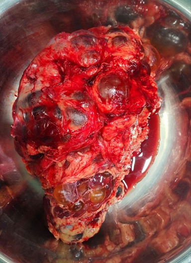
Figure 1
Meanwhile the vascular bed was deciphered and kept ready with internal iliac artery and external iliac vein and the donor kidney was perfused with cold ringer lactate and vascular pedicles ensured lumen and clear flow with ureter. End to side and end to end anastomosis of renal vein and renal artery of graft was done and fluid challenge of 850 to 1000 ml to raise CVP by 2 cm of water was given during revascularisation of the transplanted kidney. Warm ischemia time was 30 mins.
Table 2
|
Hourly blood sugar mg/dl |
85 |
103 |
148 |
134 |
198 |
180 |
|
CVP cm water |
7 |
11 |
14 |
13.5 |
11.8 |
13.2 |
Urine output at the end of 5 hours intra-operative period was 600 ml and improved to more than 2 litres in the post-operative period.
Patient was extubated on the table with Neostigmine 2.5 mg and Glycopyrrolate 0.6 mg. Post-operative pain relief was given by an intravenous infusion of fentanyl 6µg/ml and Midazolam 0.2 mg/ml at the rate of 2 ml/hour. Any spasmodic pain called graft pain over the right iliac fossa region was managed by a bolus of injection tramadol 100 mg with promethazine 12.5 mg. Urea and creatinine levels fell to 44 mg and 3.8 mg per dl on the second postoperative day.
Complexity of this Case
This case is presented for its complexity as well as contrasting features when compared to any other renal transplantation. An end stage renal disease is often associated with small shrunken kidneys not necessitating any nephrectomy for accomodating a new renal graft. This patient presented with a single large polycystic kidney almost lying across the midline and necessitated nephrectomy. Fluid overload to the lungs with increasing pulmonary pressure existed with an anticipated hypovolemia during nephrectomy. The patient who was already on hemodialysis for two years had rising renal parameters, difficult to control anemia and hypertension. The rising blood pressure existed with a poor cardiac reserve. Altered coagulation profile with immunological issues due to multiple blood transfusions though leucodepleted were of concern for the successful outcome of transplantation and function of the grafted kidney.
DISCUSSION:
Hereditary and Congenital Diseases of the Kidney are Classified as
a) hereditary glomerular diseases such as hereditary nephrotic syndrome
b) hereditary tubular diseases such as Fanconi’s syndrome
c) APOL 1 such as IgAN
d) Ciliopathies including polycystic kidney disease
e) Channelopathies such as Bartter’s syndrome
f) other congenital anomalies of the kidney such as horseshoe kidney.
Cystic disease in childhood encompasses both genetic and non genetic causes. Autosomal dominant polycystic kidney disease accounts for 7% of cases of end stage renal failure according to Western data.[2]
Genetic Cystic Kidney Diseases Include
a) Autosomal Dominant polycystic kidney disease (ADPKD)
b) Autosomal recessive PKD
c) HNF-1β mutations (renal cysts and diabetic syndrome) (e.g. juvenile nephronophthisis – medullary cystic disease complex)
d) Associated with multiple malformation syndrome such as phacomatoses, tuberous sclerosis, Von Hippel Lindau disease or others rare syndromes.
In autosomal dominant inheritance, mutations in polycystin 1 is responsible for 85% of cases and mutations in polycystin 2 accounting for most of the remainders. Demonstration of multiple cysts in enlarged kidney by ultrasound is often diagnostic. ADPKD may be asymptomatic until being detected by routine ultrasound for some other reason. An acute flank pain or fever could occur due to bleeding into cyst or sepsis. The cysts do not communicate into tubules; hence hematuria may not occur always. Renal failure progresses to end stage between 40–60 years of age, whereas autosomal recessive polycystic disease are often arising from tubules and about 50% need hemodialysis in less than 10 years of age. The renal disease may progress more slowly in families with PKD2 disease (mean age at ESRD 55 years in PKD1 compared with 70 years in PKD2). It progresses more slowly in women than in men and control of hypertension may reduce the rate of progression.
Liver cysts develop in 70% of patients later in life than renal cysts. These liver cysts are more frequent and more diffuse in women than in men. Cardiovascular Abnormalities include intracranial aneurysms and mitral valve prolapse. The latter is found in 20% of patients with ADPKD whereas it is only 2% in general population.
Our patient also had discrete liver cysts, normal liver biochemistry except for hypoproteinemia and mitral valve prolapse.
The autosomal mode of inheritance is such that the risk of any child of an affected parent carrying the abnormal gene is one in two, new mutations being base mutations affecting polycystin 1 gene on the short arm of chromosome 16 are responsible for 85% cases with mutations affecting polycystin 2 from PKD 2 gene on the long arm of chromosome 4 account for the remainder. Polycystin 1 and 2 are transmembrane proteins that are able to interact and function together as a nonselective cation channel and also induce several distinct transduction pathways.
Autosomal recessive polycystic kidney Disease ARPKD is a rare inherited disease (1 in 40000) and manifestation appear early in childhood. Mutations occur only at single locus PKHD1 on chromosome 6. Both heterozygous consanguineous parents are unaffected with normal renal USG and renal cysts develop from collecting ducts.
Renal cysts and Diabetic Syndrome
Heterozygous mutations in the gene encoding hepatocyte nuclear factor HNF-1β which is a DNA transcription factor is also the cause of Maturity Onset Diabetes in the young. Renal anomalies often precede onset of MODY. Renal cysts and progressive renal failure are frequent.[3]
Development of Renal Cysts
Renal cysts are cavities lined by epithelium and filled with fluid and semi-solid matter. Cysts are derived primarily from tubules. Cystic kidneys of different etiology may appear morphologically similar but may cause wide spectrum of renal abnormalities according to etiological entities.[4]
Classification of Cystic Kidney Disorders
· Autosomal dominant polycystic kidney disease (ADPKD)
· Autosomal recessive polycystic kidney disease (ARPKD)
· Polycystic kidney and/or liver diseases associated with altered maturation of PKD proteins in the endoplasmic reticulum (PRKCSH, SEC63, GANAB, ALG8, SEC61B, DNAJB11)
· Tuberous sclerosis complex
· von Hippel–Lindau syndrome
· Familial renal hamartomas associated with hyperparathyroidism–jaw tumor syndrome
· Hepatocyte nuclear factor-1–associated nephropathy
· Oro-facial-digital syndrome
· Autosomal dominant tubulointerstitial kidney disease
· Hereditary recessive ciliopathies with interstitial nephritis, cysts, or both:
1. Nephronophthisis
2. Joubert syndrome
3. Meckel syndrome
4. Bardet–Biedl syndrome
5. Alström syndrome
6. Nephronophthisis variants associated with skeletal defects (skeletal ciliopathies)
· Renal cystic dysplasias:
1. Multicystic kidney dysplasia
· Other cystic kidney disorders:
1. Simple cysts
2. Localized or unilateral renal cystic disease
3. Medullary sponge kidney
4. Acquired cystic kidney disease
· Renal cystic neoplasms:
1. Cystic renal cell carcinoma
2. Multilocular cystic nephroma
3. Cystic partially differentiated nephroblastoma
4. Mixed epithelial and stromal tumor
· Cysts of nontubular origin:
1. Cystic disease of the renal sinus
2. Perirenal lymphangiomas
3. Subcapsular and perirenal urinomas
· Pyelocalyceal cysts
Epithelial cysts develop from preexisting renal tubule segments and are composed of a layer of partially dedifferentiated epithelial cells enclosing a cavity filled with either urine like liquid or semisolid material. They may develop in any tubular segment between Bowman's capsule and the tip of renal papilla. After achieving a size of few millimeters most cysts lose their attachments to their parent tubule segment. Pathophysiologic processes that contribute to the development of cysts include disruption of programs responsible for the establishment and maintenance of normal tubular diameter[5] and excessive cell proliferation, active solute and fluid transport into expanding cells, crosstalk between epithelial cells and interstitial macrophages and interactions between epithelial cells and extracellular matrix.[6]
Renal cysts are regarded as benign neoplasms that arise from individual cells or restricted segments of the renal tubule. Processes that stimulate renal cell proliferation along with inability to maintain planar cell polarity have the potential to generate the cystic phenotype.
Conditional knockouts of PKD1 or of ciliogenesis genes Ift88 and Kif3a at various points of time have shown that the timing of their inactivation determines the rate of development of cystic disease.[7] The underlying rate of epithelial cell proliferation may account for increased susceptibility to cyst development during nephrogenesis. In pediatric and adult kidneys, proliferative indices are very low in all tubular segments but remain higher in collecting ducts than in proximal tubules.[8]
The finding of fluid secretion in epithelial cysts led to investigation of fluid secreting mechanisms. Beyond the loop of Henle, tubule cells have the capacity to secrete solutes and fluid upon stimulation with cyclic adenosine monophosphate (cAMP).[9] Earlier studies showed chemokines and cytokines in the cyst fluid while now it has been proved that M2 macrophages contribute to cell proliferation in PKD. Alterations in focal adhesion complexes, basement membranes and extracellular matrix contribute to the pathogenesis of PKD.[10] Majority of cysts in adults with ADPKD are derived from distal nephron and collecting duct as derived from the observation that cultured epithelial cells from ADPKD cysts exhibit larger cAMP response to 1-deamino 8-D arginine vasopressin and vasopressin than to parathyroid hormone.[11]
At the end stage of the disease, the kidneys are usually several times larger than normal and exhibit innumerable fluid filled cysts that make up most of the total renal mass. Only scant normal appearing parenchyma may be found in isolated patches. Abundant fibrous tissue is plastered along the surface of the kidney beneath the capsule, the disappearance of noncystic parenchyma implicates apoptosis as a primary mechanism in progressive renal dysfunction in ADPKD.
Impaired urine concentrating capacity is common in early stages of ADPKD and plasma vasopressin levels are increased, these contribute to cystogenesis and development of hypertension and progression of chronic kidney disease. Defective medullary trapping of ammonia and transfer to the urine caused by concentrating defect may contribute to the low urine pH values, hypocitric acidosis and predispose to stone formation. Changes in intrarenal pressures, neurohumoral or local mediators with intrinsic vascular abnormalities may cause reduced renal blood flow.[12] There is also strong evidence of local activation of intrarenal renin angiotensin aldosterone system.
In most patients renal function is maintained because of compensatory adaptation, despite relentless growth of cysts until the 4th to 6th decade of life. The average rate of GFR decline is 4.4 to 5.9 ml/min/year.[13]
Patients with autosomal dominant polycystic kidney disease are prone to urinary tract infections and liver cyst infections which may recur after renal transplantation. However the frequency of such infection episodes after transplantation under modern immunosuppressive regimens remains unclear.[14]
Thus polycystic kidney disease forms a common inherited cause of chronic kidney disease.
Table 3
|
CKD Stage |
eGFR ml/min/1.73m2 |
|
Stage1 |
>90 |
|
Stage 2 |
60-90 |
|
Stage 3a |
45-90 |
|
Stage 3b |
30-44 |
|
Stage 4 |
15-29 |
|
Stage 5 |
<15 |
|
Stage 5d |
Dialysis |
CONCLUSION:
Large polycystic kidneys presenting with ESRD for renal transplantation has additional challenges of hemorrhage, hypovolemia, sepsis and embolism apart from routine problems and risks anticipated in end stage renal disease due to other causes.
This case is reported for its complexity of on the table nephrectomy of single large polycystic kidney with diabetes, hypertension, poor cardiac reserve and anemia - immediately followed by renal transplantation from voluntary sibling donor with a successful outcome. Careful evaluation, planning to manage anticipated complications steer towards successful outcome.
REFERENCES:
1. Goldberg S, Coyne DW, et al. Renal diseases. Chap- 13. In: Crees Z, ed. Washington Manual of Medical Therapeutics. 36th edn. Wolters Kluwer 2019: p 522.
2. Joly D, Grunfeld JP. Renal involvement in genetic disease. Chap- 21.12. In: Ogg GS, Warrell DA, Cox TM, et al, eds Oxford Textbook of Medicine. Vol. 3, 5th edn. A Trueman Publication 2010:4095-100.
3. Pirson Y, Chauveau D. Grunfeld Autosomal dominant polycystic disease. In: Davison AM et al, eds. Oxford textbook of clinical nephrology Oxford Univ. Press Oxford 1998:2393-415.
4. Irazabal MV, Torres VE. Cystic diseases of kidney. Chap- 45. In: Lerma EV, Rosner MH, Perazella MA. Current diagnosis & treatment: nephrology & hypertension. 2nd edn. McGraw-Hill Education 2018: p.1490.
5. Fischer E, Legue E, Doyen A, et al. Defective planar cell polarity in polycystic kidney disease. Nat Genet 2006;38(1):21-3.
6. Torres VE, Harris PC. Strategies targeting cAMP signaling in the treatment of polycystic kidney disease. J Am Soc Nephrol 2014;25(1):18-32.
7. Lantinga-van Leeuwen IS, Leonhard WN, van der Wal A, et al. Kidney-specific inactivation of the Pkd1 gene induces rapid cyst formation in developing kidneys and a slow onset of disease in adult mice. Hum Mol Genet 2007;16(24):3188-96.
8. Nadasdy T, Laszik Z, Blick KE, et al. Proliferative activity of intrinsic cell population in normal human kidney. J Am Soc Nephrol 1994;4(12):2032-39.
9. Wallace DP, Rome LA, Sullivan LP, et al. cAMP-dependent fluid secretion in rat inner medullary collecting ducts. Am J Physiol Renal Physiol 2001;280(6):F1019-29.
10. Wilson P. Polycystin: new aspects of structure function and regulation. J Am Soc Nephrol 2001;12(4):834-45.
11. Belibi FA, Reif G, Wallace DP, et al. Cyclic AMP promotes growth and secretion in human polycystic kidney epithelial cells. Kidney Int 2004;66(3):964-73.
12. Torres VE, King BF, Chapman AB, et al. Magnetic resonance measurements of renal blood flow and disease progression in autosomal dominant polycystic kidney disease. Clin J Am Soc Nephrol 2007;2(1):112-20.
13. Klahr S, Breyer JA, Beck GJ, et al. Dietary protein restriction, blood pressure control, and the progression of polycystic kidney disease. Modification of Diet in Renal Disease Study Group. J Am Soc Nephrol 1995;5(12):2037-47.
14. Waiser J, Klotsche J, Glander P, et al. Kidney transplantation in patients with polycystic kidney disease: increased risk of infection does not compromise graft and patient survival. Clin Kidney J 2024;17(12):sfae330.