Mayer–Rokitansky–Küster–Hauser syndrome (MRKH): A Case Series.
- Sharankumar Jab , Assistant Professor, Dept of Urology, GIMS SSH Kalaburagi.
- Jairaj V Bomman , Assistant Professor, Dept of Medical Gastroenterology, GIMS Kalaburagi.
- Sharanabasappa B Rudrawadi , Associate Professor, Dept of Urology, Gulbarga institute of medical science -SSH –Gulbarga.
Article Information:
Abstract:
Background: Objective: To evaluate the clinical presentation, diagnostic findings, surgical management, and outcomes of adolescent females diagnosed with Mayer–Rokitansky–Küster–Hauser (MRKH) syndrome at a tertiary care center. Materials and Methods: This case series was conducted at National Institute of Medical Sciences from 2019 to 2025. Three adolescent females aged 16–19 years presenting with primary amenorrhea and normal secondary sexual characteristics were included. All patients underwent detailed clinical evaluation, pelvic ultrasonography, and magnetic resonance imaging to confirm Müllerian agenesis. Karyotyping revealed a normal 46, XX chromosomal pattern in all cases. Associated anomalies were assessed, and individualized surgical management was planned according to patient preference and clinical suitability. Results: All three patients were diagnosed with MRKH syndrome. One patient had associated renal ectopia, consistent with MRKH type II, while the remaining two patients had isolated Müllerian anomalies. Surgical management included McIndoe’s vaginoplasty in one patient, laparoscopic Vecchietti procedure in another, and sigmoid colon vaginoplasty in the third patient who preferred a self-lubricating neovagina. Postoperative outcomes were satisfactory in all cases with good cosmetic and functional results. Psychological counseling and regular follow-up contributed to improved postoperative adaptation and quality of life. Conclusion: MRKH syndrome should be considered in adolescent females presenting with primary amenorrhea despite normal secondary sexual development. Early diagnosis through imaging and multidisciplinary evaluation is essential for appropriate management. McIndoe’s vaginoplasty and other reconstructive techniques provide favorable anatomical and functional outcomes. Comprehensive psychological support and advances in reproductive technologies can significantly improve long-term well-being and reproductive prospects in affected women.
Keywords:
Article :
INTRODUCTION:
Mayer–Rokitansky–Küster–Hauser (MRKH) syndrome is a congenital malformation of the female reproductive tract resulting from agenesis or hypoplasia of the Müllerian ducts. MRKH is characterized as an absent or embryonic development of the uterus as well as the upper vaginal segment, while the ovaries and the outer genitals undergo normal development [1]. While being an unusual condition, it is still a leading cause of primary amenorrhea for females with normal secondary sexual development and a 46, XX karyotype. The estimated prevalence of MRKH syndrome is approximately 1 in 4,500 to 5,000 live female births. This makes the condition one of the most common congenital defects involving the female genital tract. The embryonic foundation of MRKH syndrome is rooted in the failure of the fusion and development of the Müllerian ducts at six to 12 weeks of gestation [2].
These paired ducts, when actively developing, are responsible for creating the uterus, fallopian tubes, cervix, and the upper two-thirds of the vagina. When duct development is inactive, it results in the absence of one or more of the aforementioned structures [3]. The remaining structures, however, originate from other embryonic sources having separated developmental tracks, which permits normal hormonal functioning, external genital prescence, ovulation, and sexual development. The condition is medically recognized as MRKH syndrome when cases of primary amenorrhea arise, which occurs with normal breast growth, pubic growth, and overall morphological change [4].
In absence of a vaginal canal or a canal that is significantly shortened, a doctor may also perform a gynecological examination. Ultrasonography and MRI confirm the uterus and upper vagina are absent or rudimentary, yet the ovaries are intact and functional [5]. Hormonal assessments indicate normal gonadotropic, estrogenic, and androgenic levels, which add a diagnostic layer to differentiating MRKH from other amenorrhea aetiologies, including gonadal dysgenesis, or androgen insensitivity syndrome. MRKH syndrome is differentiated into two distinct types [6]. Type I (the isolated form) is simply uterovaginal agenesis with no other organ anomalies. Type II (the syndromic form), sometimes referred to as MURCS (association of Müllerian duct aplasia with renal dysplasia and cervical somite anomalies) is complication by renal malformations (e.g., unilateral renal agenesis or ectopic kidney), vertebral defects, auditory defects, and in less common cases, anomalies of the heart or limbs [7].
The variable expressions and associated anomalies demonstrate the genetic and developmental complexity of the process. The cause of MRKH syndrome is still multifactorial and incompletely understood. Both isolated and familial cases have been documented, indicating some genetic variable with incomplete penetrance [8]. A number of candidate genes have been reported, most of which are important for the Müllerian duct development, including WNT4, LHX1, and HNF1B. Deletions or mutations of these genes may disrupt the positive regulatory signalling pathways needed to form the female reproductive tract. Some other of the proposed etiological factors are environmental and epigenetic changes during the early phases of embryogenesis [9].
While MRKH patients have no uterus so are unable to conceive and carry a child, they do have normal ovarian function and can produce healthy oocytes. Surrogacy and other Assisted Reproductive Technologies (ART) allow these women to have their own genetic offspring [10]. Recent improvements in uterine transplantation and subsequent pregnancies are possible after transplanting from both living and deceased donors. However, advances in this particular type of medicine are slow and few transplant centers offer this procedure [11].
The basic aim of this case study is to present a case of Mayer–Rokitansky–Küster–Hauser (MRKH) syndrome and highlight its clinical presentation, diagnostic evaluation, and successful surgical management using McIndoe’s vaginoplasty.
METHODOLOGY:
This descriptive case series was conducted at NIMS Jaipur from 2019 to 2025.
Inclusion Criteria
· Females aged 15–25 years
· Presentation with primary amenorrhea
· Normal secondary sexual characteristics
· Normal 46,XX karyotype
· Imaging findings consistent with Müllerian agenesis (absent or hypoplastic uterus with normal ovaries)
Exclusion Criteria
· Secondary amenorrhea, Ambiguous genitalia and chromosomal abnormalities (e.g., Turner syndrome, androgen insensitivity syndrome)
·
Data collection
All patients underwent a structured diagnostic protocol beginning with a detailed clinical evaluation focused on assessing secondary sexual characteristics, external genital anatomy, and the presence of any associated anomalies such as skeletal or renal malformations. Pelvic ultrasonography was performed initially to assess uterine morphology, presence or absence of the cervix, and the size and appearance of the ovaries. MRI was subsequently used as the gold-standard modality to confirm Müllerian agenesis, characterize uterine hypoplasia, and identify associated pelvic or extrapelvic anomalies. Clinical information was recorded using a structured proforma that included demographic details, presenting symptoms, physical examination findings, imaging results, classification of MRKH type, operative details, postoperative complications, and functional outcomes. Follow-up data regarding neovaginal maintenance, patient satisfaction, and long-term adaptation were also documented to evaluate overall success of interventions.
Case presentation
A 17-year-old female presented to the gynecology department with a complaint of primary amenorrhea. She had no history of cyclical abdominal pain, urinary complaints, or any systemic illness. On general examination, she had well-developed secondary sexual characteristics including normal breast development and pubic hair distribution, suggesting normal ovarian function and hormonal activity. There were no skeletal, renal, or other physical abnormalities noted. Pelvic ultrasonography (USG) revealed an atrophic uterus and absence of the cervix, while both ovaries were visualized and appeared normal in size and morphology. To confirm these findings, a magnetic resonance imaging (MRI) scan was performed, which corroborated the USG report. The MRI showed uterine hypoplasia with complete cervical agenesis and normal-appearing ovaries, consistent with Mayer–Rokitansky–Küster–Hauser (MRKH) syndrome. After appropriate counseling regarding the diagnosis, prognosis, and available treatment options, the patient was planned for McIndoe’s vaginoplasty to create a functional neovagina.
Operative Technique
Under general anesthesia, the patient was placed in the lithotomy position. A curvilinear incision was made over the vaginal dimple, and a potential space was carefully developed between the urethra and bladder anteriorly and the rectum posteriorly. Dimensions of this plane were about 8-10cm long and wide enough to fit the neovaginal mold. A split-thickness skin graft was taken from the patient's thigh and subsequently placed on a cylindrical mold, which had the epithelial layer facing outside of the cylinder. Closed, the mold coated in the graft was placed in the surgically formed neovaginal cavity. After the procedure, the mold was left in place for a week to allow the graft to become anchored and to create a neovagina. After a week, the mold was taken off, and the patient began the standardized vaginal dilatation procedure to keep the neovagina from closing off and to keep the opening from becoming too narrow. The patient reported no complications and had a smooth recovery.
Management Options
MRKH syndrome management must be individualized according to the specific requirements of the patient and the anatomical discrepancy. Preferences include non-surgical methods of vaginal dilation and several forms of vaginoplasty. Motivated patients typically receive non-surgical approaches, such as progressive dilation of the vaginal dimple, as the first-line treatment. However, if these methods fail, or are impractical, resorting to surgical options are warranted. Various surgical methods have been documented, using tissues like the ileum, sigmoid colon, amniotic membrane, or divided skin grafts. McIndoe’s vaginoplasty is one of the most popular because of the low complication rates, and decent anatomical, functional, and complication outcomes along with ease of surgical execution. McIndoe’s method also does not have the excess mucous discharge, which tends to be problematic with the other bowel-based vaginoplasties. On the downside, however, due to the skin grafted surface, there will be vaginal dryness from the skin grafts, which will also mean there will not be any useful vaginal secretions and hence the skin will be very dry.
Outcome and Follow-Up
Postoperatively, the patient was counseled regarding the importance of regular vaginal dilatation to maintain the neovaginal canal and prevent narrowing. She was also provided with psychological support to help her adjust to the diagnosis and address fertility concerns. The surgical outcome was satisfactory, resulting in a well-formed neovagina with good cosmetic and functional results.
Case 2
Case Presentation
A 19-year-old female presented with primary amenorrhea and intermittent lower abdominal discomfort. She denied cyclical pain, urinary complaints, or gastrointestinal symptoms. Her past health was unremarkable, and she reported normal sexual development during puberty. On examination, she had normal secondary sexual characteristics with Tanner stage V breast and pubic hair development. No skeletal abnormalities, hearing deficits, or signs of endocrine disorder were identified. Pelvic ultrasonography revealed nonvisualization of the uterus and cervix, with both ovaries appearing normal in location and morphology. A subsequent pelvic MRI confirmed complete uterine agenesis with preserved bilateral ovaries, consistent with MRKH type I. Renal imaging was normal. After detailed discussion regarding non-surgical and surgical options, the patient opted for the Vecchietti laparoscopic vaginoplasty due to its minimally invasive nature and lower postoperative maintenance.
Operative Technique
Under general anesthesia, the Vecchietti procedure was performed laparoscopically. A small olive-shaped traction device was placed at the vaginal dimple and connected to traction threads passed into the abdominal cavity. These threads were secured to a traction system attached to the abdominal wall. For adolescents highly motivated for a non-bowel, minimally invasive solution, the laparoscopic Vecchietti method is preferred. It avoids the need for skin grafts or intestinal segments and provides a mucosa-lined neovagina through gradual traction. Alternative techniques, such as McIndoe’s or sigmoid vaginoplasty, were discussed but deemed less suitable given the patient’s desire for shorter recovery and minimal scarring. Psychological counseling and long-term reproductive guidance were also provided.
Outcome and Follow-Up
The patient achieved a functional neovagina of appropriate length and width within two weeks. She adhered well to postoperative dilation and reported no pain or discomfort during follow-up. At three-month review, the neovagina maintained excellent epithelialization and patency. The patient expressed high satisfaction with the cosmetic and functional outcome and continued psychological support sessions as part of her long-term care.
Case 3
Case Presentation
A 16-year-old girl presented with concerns about absent menstruation. She reported no pelvic pain, urinary symptoms, or prior hospitalizations. Her growth and developmental milestones were normal. On examination, she had well-developed breasts and normal pubic hair distribution, indicating normal ovarian hormone production. No limb abnormalities, scoliosis, or hearing issues were detected. Ultrasound imaging showed complete absence of the uterus and cervix, while both ovaries were present and normal. MRI further confirmed MRKH syndrome with an associated unilateral renal anomaly: an ectopic left kidney located in the pelvis. These findings were consistent with MRKH type II. After counseling and assessment of patient readiness, a sigmoid colon vaginoplasty was recommended due to her preference for a self-lubricating neovagina and reduced need for long-term dilation.
Operative Technique
Under general anesthesia, a segment of the sigmoid colon was isolated while preserving its vascular pedicle. The mucosa-lined segment was tubularized to form the neovaginal canal. A perineal incision was made, and a tract was created between the bladder and rectum. The isolated sigmoid segment was then pulled through the tract and sutured to the introitus to form the new vagina. The remaining bowel ends were re-anastomosed. Patients with MRKH type II and a preference for long-term functional outcomes may benefit from bowel-based vaginoplasty. Sigmoid vaginoplasty offers excellent lubrication and durability, although it requires abdominal surgery and carries risks such as mucus discharge and stenosis. Alternative options including McIndoe’s or Vecchietti technique were discussed but the patient and her family opted for the sigmoid approach due to its superior long-term functionality.
Outcome and Follow-Up
The patient recovered well with stable bowel function and no postoperative complications. At one-month follow-up, the neovagina appeared healthy with adequate lubrication and maintained patency without aggressive dilation. She continued periodic follow-up to monitor renal status and psychosocial adjustment. Overall, the patient and her family expressed satisfaction with the surgical and functional outcomes.
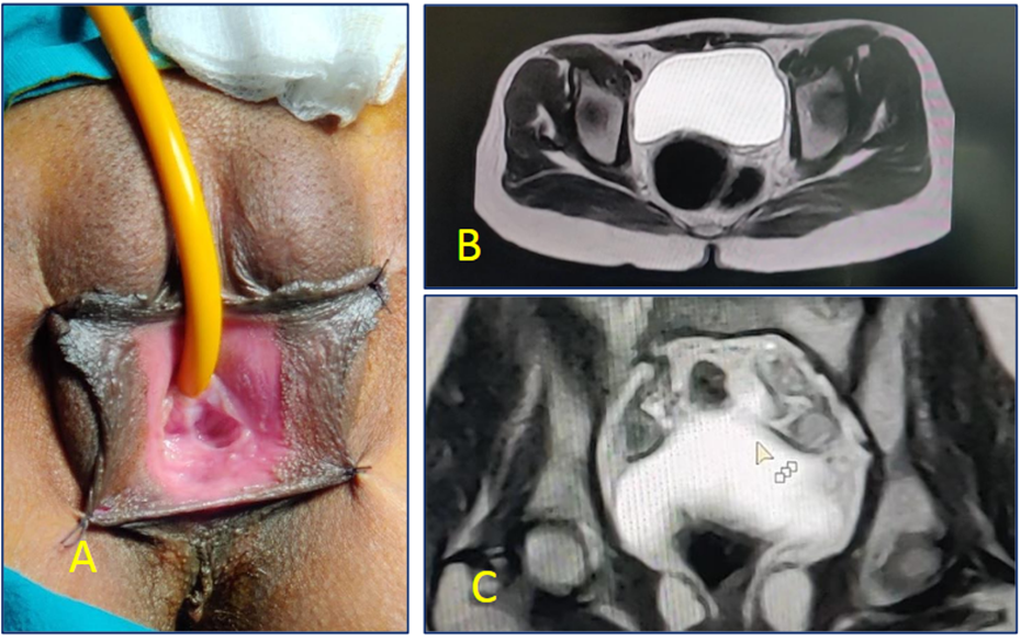
Figure A. Absence of vagina with only a dimple present at the site of the vagina. B. Showing atretic tissue between bladder and rectum (yellow arrow). C. Presence of bilateral ovaries
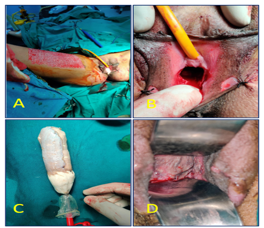
Figure 2A. Intra-operative photograph showing the development of a plane between bladder and the rectum.
Figure 2B & C: Showing site of the split skin graft and the preparation of graft for application.
Figure 2D. Post operative photograph showing well taken split skin graft.
DISCUSSION:
Mayer–Rokitansky–Küster–Hauser (MRKH) syndrome represents a rare but important cause of primary amenorrhea in females with normal secondary sexual characteristics and a 46, XX karyotype. The condition is a result of congenital Mullerian agenesis characterised by the absent or hypoplastic uterus, an underdeveloped vagina, and in most cases, amenorrhea. Ovaries are intact, and the functional attributes are normal, enabling the individual to attain normal secondary sexual characteristics during puberty. The case illustrates a classical description of MRKH syndrome in a 17-year-old female who presented with a history of primary amenorrhea and underwent imaging that revealed cervical and uterine agenesis with subsequently normal functioning ovaries.
The condition is called MRKH syndrome, but the diagnosis is often made more than a decade after the onset of symptoms, in late teenage or during young adulthood. It is during this period that the majority of the patients are diagnosed due to the symptoms of primary amenorrhea and evidence of normal female external genitalia, normal secondary sexual characteristics, and normal hormonal contraception in the absence of a uterus [13]. While the clinical diagnosis of MRKH syndrome is based almost entirely on the existence of primary amenorrhea, ultrasound imaging is often the first investigation to be performed, while the diagnosis is more often than not confirmed by MRI, due to the finer details of the pelvic anatomy and the uterine remnants and the presence of an absent or hypoplastic vagina, associated kidney, or vertebral anomalies. The MRKH syndrome is subdivided into two clinical types. Type 1 or isolated MRKH syndrome is characterized by the absence of vagina and uterus alone while Type II or MURCS syndrome (Müllerian aplasia, renal cystic dysplasia, and segmental vertebral dysplasia) is associated with additional renal and vertebral defects. The absence of such anomalies in this case establishes the patient’s condition as being Type I. The remaining subtypes are essential to properly directing the treatment strategy, as well as to minimize the risk of specific complications developing [14]. The focus of the MRKH syndrome management is the development of a vagina to provide the patient with the ability to engage in sexual intercourse, as well as to give attention to the psychological aspects associated with the syndrome. The most important thing that is subsequently done is to perform regular vaginal dilatation, which is needed to ensure that the neovagina remains open and does not get fibrotic [15]. Despite the lack of postoperative discharge and a dry vaginal environment, the technique described and used will result in postoperative vaginal dryness due to lack of skin graft mucosal secretions. Graft options like the peritoneum, amnion, or more recently used, E. Coli bowel segments vagnoplasy (ileum and sigmoid colon) provide self-lubrication and assist wheel-mate construction [16]. Other admittedly less popular grafts do discharge, infection, and odor, but promise results. Patient and surgeon variables to factor include Southern Cross McIndoe’s, amnion, and peritoneum which, due to limited advanced reconstructive options, remain popular in many countries and developing regions.
CONCLUSION:
Mayer–Rokitansky–Küster–Hauser (MRKH) syndrome is a rare congenital condition with varied presentations, ranging from isolated vaginal agenesis to multisystem deformities. It is an important cause of primary amenorrhea in females with normal secondary sexual characteristics. Management options range from non-surgical vaginal dilation to surgical reconstruction. In this case, McIndoe’s vaginoplasty provided excellent cosmetic and functional results with minimal complications. Psychological counseling and regular follow-up are vital for long-term success. With modern reproductive techniques like surrogacy and uterine transplantation, women with MRKH can now achieve genetic motherhood and lead fulfilling lives.
REFERENCES:
1. Khalesi N, Kashaki M, Khosravi N, Vahedi Z, Alinejad-Naeini M. A Mayer-Rokitansky-Kuster-Hauser Syndrome in a Neonate: A Case Report. Iranian Journal of Neonatology. 2025 Apr: 16(2). DOI: 10.22038/ijn.2025.72592.2407
2. Ray U, Adhikari S, Dhital R, Shrestha S, Shah S, Poudel S, Sah SK, Gami R, Adhikari A, Gautam B. Mayer-Rokitansky-Kuster-Hauser Syndrome: A rare case report from Nepal. Ann Med Surg (Lond). 2022 Sep 16;82:104725. doi: 10.1016/j.amsu.2022.104725. PMID: 36268332; PMCID: PMC9577869.
3. Muthu Kumar A, Menon P, Mane S (August 23, 2024) Mayer-Rokitansky-Küster-Hauser Syndrome: A Case Report. Cureus 16(8): e67606. doi:10.7759/cureus.67606
4. Oppelt PG, Lermann J, Strick R, Dittrich R, Strissel P, Rettig I, et al. Malformations in a cohort of 284 women with Mayer–Rokitansky–Küster–Hauser syndrome (MRKH). Reprod Biol Endocrinol. 2012;10:57.
5. Layman LC. The genetics of Müllerian aplasia. Expert Rev Endocrinol Metab. 2014;9(4):411–419.
6. Nodale C, Ceccarelli S, Giuliano M, Cammarota M, D’Amici S, Vescarelli E, et al. Gene expression profile of patients with Mayer–Rokitansky–Küster–Hauser syndrome: new insights into the potential role of developmental pathways. PLoS One. 2014;9(3):e91010.
7. Herlin M, Bjørn AM, Rasmussen M, Trolle B, Petersen MB. Prevalence and patient characteristics of Mayer–Rokitansky–Küster–Hauser syndrome: a nationwide registry-based study. Hum Reprod. 2016;31:2384–2390. doi:10.1093/humrep/dew220.
8. Herlin MK, Petersen MB, Brännström M. Mayer–Rokitansky–Küster–Hauser (MRKH) syndrome: a comprehensive update. Orphanet J Rare Dis. 2020;15:214. doi:10.1186/s13023-020-01491-9.
9. Morcel K, Camborieux L, Guerrier D. Mayer–Rokitansky–Küster–Hauser (MRKH) syndrome. Orphanet J Rare Dis. 2007;2:13. doi:10.1186/1750-1172-2-13.
10. Fontana L, Gentilin B, Fedele L, Gervasini C, Miozzo M. Genetics of Mayer–Rokitansky–Küster–Hauser (MRKH) syndrome. Clin Genet. 2017;91:233–246. doi:10.1111/cge.12883.
11. Dewi DK, Randy S, Wienanda AK, Christanto AG. Atypical form of Mayer–Rokitansky–Küster–Hauser syndrome: a case report. Radiol Case Rep. 2023;18:2806–2809. doi:10.1016/j.radcr.2023.04.050.
12. Georgopapadakos N, Manoli A, Passia G, Skandalakis PN, Filippou D. Uterus transplantation as a therapy method in Mayer–Rokitansky–Küster–Hauser syndrome. Cureus. 2019;11:e6333. doi:10.7759/cureus.6333.
13. Muisi Alli Adenekan, Ayodeji Kayode Adefemi, Chidinma Magnus Nwogu, Adelana Olusegun Odumabo, Anthony Nnaemeka Okeke, Aloy Okechukwu Ugwu, Adebayo Awoniyi (2025). A Case of Mayer-Rokitansky-Küster-Hauser Syndrome: Case Report of a 22-Year-Old Undergraduate with Primary Amenorrhoea. Saudi J Med Pharm Sci, 11(2): 131-134.
14. Ray U, Adhikari S, Dhital R, Shrestha S, Shah S, Poudel S, et al. Mayer–Rokitansky–Küster–Hauser syndrome: a rare case report from Nepal. Ann Med Surg (Lond). 2022;82:104725. doi:10.1016/j.amsu.2022.104725. PMID: 36268332; PMCID: PMC9577869.
15. Herlin MK. Genetics of Mayer–Rokitansky–Küster–Hauser (MRKH) syndrome: advancements and implications. Front Endocrinol (Lausanne). 2024;15:1368990. doi:10.3389/fendo.2024.1368990.
16. Magdoud K, Abouda HS, Jellouli A, Ben Hmid R, Chanoufi MB, Karoui A. Sexuality and quality of life of patients operated for Rokitansky–Küster–Hauser syndrome. La Tunisie Médicale. 2024;102(5):310–314. doi:10.62438/tunismed.v102i5.4738.