Assessment of visual acuity and refractive status after phacoemulsification.
- Kaveti Sai Neha , Post Graduate, Department of Ophthalmology, Kamineni Institute of Medical Sciences, Narketpally, Telangana.
- Abdul Subhan , Assistant Professor, Department of Ophthalmology, Kamineni Institute of Medical Sciences, Narketpally, Telangana.
- Mamata Shetty , Professor and Head, Department of Ophthalmology, Kamineni Institute of Medical Sciences, Narketpally, Telangana.
- K. Gayatri , Associate Professor, Department of Ophthalmology, Kamineni Institute of Medical Sciences, Narketpally, Telangana.
Article Information:
Abstract:
Introduction: Cataract is the leading cause of reversible blindness worldwide. Phacoemulsification with intraocular lens (IOL) implantation has become the standard surgical technique, offering rapid visual rehabilitation and improved refractive outcomes. Aim of the study was to assess visual acuity and refractive outcomes following phacoemulsification. Materials and methods: This prospective observational study was conducted at the Department of Ophthalmology, Kamineni Institute of Medical Sciences. A total of 75 patients undergoing phacoemulsification with IOL implantation were included. Preoperative evaluation included visual acuity, slit-lamp examination, intraocular pressure, and biometry. Postoperative follow-up was done at 1 week, 1 month, and 3 months. Data were analyzed using SPSS version 20. Result: The mean age of patients was 62.4 ± 8.6 years, with a slight male predominance (54.7%). There was a significant improvement in visual acuity, with mean UCVA improving from 1.12 ± 0.28 logMAR preoperatively to 0.18 ± 0.08 logMAR postoperatively, and BCVA improving from 0.82 ± 0.25 to 0.08 ± 0.05 logMAR (p < 0.001). A total of 92% of patients achieved visual acuity ≥6/18, and 74.7% achieved ≥6/9 at 3 months. The mean postoperative spherical equivalent was -0.36 ± 0.52 D, with 88% of patients within ±1.0 D and 64% within ±0.5 D of the target refraction. Astigmatism reduced from 1.12 ± 0.46 D preoperatively to 0.68 ± 0.32 D postoperatively, indicating minimal surgically induced astigmatism. Intraoperative complications were minimal (5.3%), and postoperative complications were mild and transient (21.3%). Better preoperative BCVA and lower residual refractive error were significantly associated with good visual outcomes. Conclusion: Phacoemulsification with IOL implantation provides excellent visual and refractive outcomes, with significant improvement in both UCVA and BCVA and high refractive accuracy. The procedure is safe, with minimal complications, and ensures rapid visual rehabilitation. Preoperative visual status and refractive precision play a key role in determining postoperative success.
Keywords:
Article :
INTRODUCTION:
Cataract continues to be the foremost cause of reversible blindness globally, contributing substantially to visual impairment, particularly in low- and middle-income countries. Recent estimates suggest that age-related cataract remains responsible for nearly half of all cases of blindness worldwide, despite the availability of effective surgical treatment (1). Over the past few decades, cataract surgery has undergone significant evolution, with phacoemulsification becoming the preferred technique due to its minimally invasive approach, smaller incision size, faster wound healing, and rapid visual rehabilitation. This technique involves emulsification of the crystalline lens using ultrasonic energy followed by implantation of an intraocular lens (IOL), thereby restoring transparency and refractive function of the eye (2).
Modern cataract surgery is no longer considered merely a vision-restoring procedure but rather a refractive surgery aimed at achieving optimal postoperative visual acuity and minimal dependence on spectacles. Consequently, assessment of visual acuity and refractive status following phacoemulsification has gained paramount importance. Visual acuity, measured as uncorrected visual acuity (UCVA) and best-corrected visual acuity (BCVA), remains the primary indicator of functional success, whereas refractive outcomes—including spherical equivalent and residual astigmatism—reflect the precision of biometric measurements and IOL power calculation (3). Advances in optical biometry, newer IOL formulas such as Barrett Universal II and Hill-RBF, and improved surgical techniques have significantly enhanced refractive predictability in recent years.
Several contemporary studies have evaluated outcomes following phacoemulsification. A study by Gurnani et al. (2022) reported that more than 90% of patients achieved BCVA of 6/18 or better postoperatively, emphasizing the effectiveness of modern cataract surgery in restoring functional vision (4). Similarly, a prospective study by Kaur et al. (2021) demonstrated significant improvement in both UCVA and BCVA following phacoemulsification, with a majority of patients achieving refractive outcomes within ±1 diopter of the intended target (5). Another study by Wang et al. (2023) highlighted the role of advanced biometry in improving refractive accuracy, reporting that nearly 88–92% of eyes achieved postoperative refractive error within ±1.0 D (6). These findings underscore the high success rates achievable with current surgical standards.
In addition to visual acuity, refractive outcomes have emerged as a critical determinant of patient satisfaction and quality of life. Residual refractive errors, particularly astigmatism, can significantly affect postoperative visual quality despite good BCVA. A study by Ferreira et al. (2022) showed that surgically induced astigmatism remains a key factor influencing refractive outcomes and emphasized the importance of incision location and size in minimizing postoperative astigmatism (7). Furthermore, advancements such as toric IOLs and femtosecond laser-assisted cataract surgery have been shown to improve refractive precision, especially in patients with pre-existing corneal astigmatism (8).
Despite these advancements, certain challenges persist. Refractive surprises continue to occur, particularly in eyes with extreme axial lengths, corneal irregularities, or inaccurate biometry. A recent study by Kane et al. (2021) highlighted that variability in IOL power calculation formulas can still lead to suboptimal refractive outcomes in complex cases (9). Moreover, patient-related factors such as age, coexisting ocular pathologies, and delayed presentation can influence postoperative visual recovery, particularly in developing country settings.
A critical review of the literature reveals a significant research gap. While numerous studies have independently assessed visual acuity or refractive outcomes after phacoemulsification, relatively fewer studies have comprehensively evaluated both parameters simultaneously in a unified framework. Additionally, there is limited region-specific data from tertiary care centers that reflect real-world clinical scenarios, especially in populations with varying demographic and clinical profiles. Long-term follow-up data evaluating refractive stability and patient satisfaction are also relatively scarce in recent literature.
Therefore, the present study is undertaken with the aim of assessing visual acuity and refractive status after phacoemulsification. By analyzing both functional and refractive outcomes in conjunction, this study seeks to provide a more comprehensive understanding of surgical success and address existing gaps in current evidence.
MATERIALS AND METHODS:
Study Design and Setting
This study was designed as a prospective observational study conducted in the Department of Ophthalmology at Kamineni Institute of Medical Sciences. The study was carried out over a defined study period (specify duration as per protocol), after obtaining approval from the Institutional Ethics Committee. All procedures adhered to the tenets of the Declaration of Helsinki, and informed written consent was obtained from all participants prior to inclusion in the study.
Study Population
The study included patients diagnosed with senile cataract who underwent phacoemulsification with intraocular lens (IOL) implantation. A total of 75 patients (n = 75) were enrolled based on predefined inclusion and exclusion criteria.
Inclusion Criteria
• Patients aged ≥40 years diagnosed with senile cataract
• Patients planned for phacoemulsification with posterior chamber IOL implantation
• Patients willing to participate and provide informed consent
• Patients available for postoperative follow-up
Exclusion Criteria
• Patients with traumatic, congenital, or complicated cataract
• Presence of corneal opacity, glaucoma, uveitis, or retinal pathology affecting visual outcome
• History of previous intraocular surgery
• Patients with systemic conditions affecting vision (e.g., uncontrolled diabetes with retinopathy)
• Patients lost to follow-up
Study Tools
• Snellen’s visual acuity chart for assessment of UCVA and BCVA
• Auto-refractometer and retinoscopy for refractive error assessment
• Slit-lamp biomicroscope for anterior segment evaluation
• Applanation tonometer for intraocular pressure (IOP) measurement
• Keratometer and optical biometry (IOL Master/Equivalent) for IOL power calculation
• Direct and indirect ophthalmoscope for posterior segment examination
Data Collection
• Preoperative Assessment:
o Detailed history including age, gender, and ocular complaints
o Measurement of baseline visual acuity (UCVA and BCVA)
o Anterior segment examination using slit lamp
o IOP measurement
o Biometry for IOL power calculation
• Intraoperative Details:
o Type of anesthesia used
o Surgical technique (standard phacoemulsification)
o Type and power of IOL implanted
o Intraoperative complications (if any)
• Postoperative Assessment:
o Visual acuity assessment at 1 week, 1 month, and 6 weeks/3 months
o Refraction and calculation of spherical equivalent
o Evaluation of postoperative complications
o Assessment of final refractive outcome
Outcome Measures
• Primary outcome: Postoperative visual acuity (UCVA and BCVA)
• Secondary outcome: Refractive status (spherical equivalent, residual astigmatism)
Statistical Analysis
Data collected were entered into Microsoft Excel and analyzed using SPSS software (version 22). Continuous variables were expressed as mean ± standard deviation (SD), while categorical variables were presented as frequencies and percentages. Preoperative and postoperative visual acuity and refractive parameters were compared using the paired t-test. A p-value <0.05 was considered statistically significant. Appropriate tables and graphs were used to present the data.
RESULTS:
Table 1: Baseline Demographic and Clinical Characteristics (n = 75)
|
Parameter |
Value |
|
Age (years) |
62.4 ± 8.6 |
|
Age distribution |
40–50: 12 (16.0%) |
|
51–60: 21 (28.0%) |
|
|
61–70: 28 (37.3%) |
|
|
>70: 14 (18.7%) |
|
|
Gender |
Male: 41 (54.7%) |
|
Female: 34 (45.3%) |
|
|
Laterality |
Right eye: 39 (52.0%) |
|
Left eye: 36 (48.0%) |
|
|
Type of cataract |
Nuclear: 38 (50.7%) |
|
Cortical: 21 (28.0%) |
|
|
PSC: 16 (21.3%) |
|
|
Pre-op UCVA |
1.12 ± 0.28 (logMAR) |
|
Pre-op BCVA |
0.82 ± 0.25 (logMAR) |
The baseline demographic and clinical profile of the study population (n = 75) showed that the mean age was 62.4 ± 8.6 years, with the majority of patients belonging to the 61–70 years age group (37.3%), reflecting the typical age-related prevalence of senile cataract. There was a slight male predominance (54.7%) compared to females (45.3%). Laterality was almost equally distributed, with right eye involvement in 52.0% and left eye in 48.0% of cases. Among the types of cataract, nuclear cataract was the most common (50.7%), followed by cortical (28.0%) and posterior subcapsular cataract (21.3%), indicating the predominance of age-related nuclear sclerosis in this cohort. The baseline visual status was significantly compromised, with mean preoperative UCVA of 1.12 ± 0.28 logMAR and BCVA of 0.82 ± 0.25 logMAR, suggesting moderate to severe visual impairment prior to surgery. Overall, these findings indicate a representative elderly cataract population with typical clinical characteristics suitable for evaluating outcomes after phacoemulsification.
Table 2: Preoperative Ocular Biometric and Clinical Parameters (n = 75)
|
Parameter |
Value |
|
Axial length (mm) |
23.42 ± 1.12 |
|
K1 (D) |
43.12 ± 1.34 |
|
K2 (D) |
44.36 ± 1.41 |
|
Mean keratometry |
43.74 ± 1.28 |
|
Pre-op IOP (mmHg) |
15.8 ± 2.6 |
|
IOL power (D) |
21.8 ± 2.4 |
The preoperative ocular assessment revealed that the mean axial length was 23.42 ± 1.12 mm, indicating that most eyes were within the normal axial range. Keratometric readings showed a mean K1 of 43.12 ± 1.34 D and K2 of 44.36 ± 1.41 D, with an overall mean keratometry of 43.74 ± 1.28 D, suggesting relatively regular corneal curvature with mild physiological astigmatism. The mean preoperative intraocular pressure was 15.8 ± 2.6 mmHg, which falls within the normal range, indicating absence of significant ocular hypertension in the study population. The calculated mean intraocular lens (IOL) power was 21.8 ± 2.4 diopters, reflecting standard biometric characteristics of the cohort.
Table 3: Intraoperative Characteristics of Phacoemulsification (n = 75)
|
Parameter |
Value |
|
Topical anesthesia |
48 (64.0%) |
|
Peribulbar anesthesia |
27 (36.0%) |
|
Surgical duration (min) |
14.6 ± 3.2 |
|
Foldable IOL |
75 (100%) |
|
Intraoperative complications |
4 (5.3%) |
|
→ Posterior capsular rent |
2 |
|
→ Zonular dialysis |
2 |
The intraoperative profile of the study population demonstrated that topical anesthesia was the most commonly used modality (64.0%), followed by peribulbar anesthesia (36.0%), reflecting a preference for less invasive anesthesia techniques in routine phacoemulsification. The mean surgical duration was 14.6 ± 3.2 minutes, indicating an efficient surgical workflow consistent with standard phaco procedures. All patients (100%) received foldable intraocular lenses (IOLs), highlighting the adoption of modern small-incision cataract surgery practices. Intraoperative complications were minimal, occurring in 5.3% of cases, with posterior capsular rent (2 cases) and zonular dialysis (2 cases) being the observed events. Overall, the low complication rate and short operative time reflect good surgical expertise and favorable intraoperative outcomes in this cohort.
Table 4: Postoperative Visual Acuity Outcomes Following Phacoemulsification (n = 75)
|
Time |
UCVA (logMAR) |
BCVA (logMAR) |
|
1 week |
0.42 ± 0.18 |
0.28 ± 0.14 |
|
1 month |
0.26 ± 0.12 |
0.16 ± 0.09 |
|
3 months |
0.18 ± 0.08 |
0.08 ± 0.05 |
|
Outcome |
Value |
|
≥6/18 vision |
69 (92.0%) |
|
≥6/9 vision |
56 (74.7%) |
The postoperative visual acuity outcomes demonstrated a progressive and significant improvement over the follow-up period. The mean UCVA improved from 0.42 ± 0.18 logMAR at 1 week to 0.26 ± 0.12 logMAR at 1 month, and further to 0.18 ± 0.08 logMAR at 3 months, indicating rapid visual rehabilitation after surgery. Similarly, BCVA showed marked improvement from 0.28 ± 0.14 logMAR at 1 week to 0.16 ± 0.09 logMAR at 1 month, reaching 0.08 ± 0.05 logMAR at 3 months, reflecting excellent final visual outcomes.
In terms of functional vision, a majority of patients achieved satisfactory results, with 92.0% attaining visual acuity ≥6/18 and 74.7% achieving ≥6/9 vision at the end of the follow-up period. These findings highlight the effectiveness of phacoemulsification in restoring both uncorrected and best-corrected visual acuity, with most patients achieving good to excellent visual outcomes within a short postoperative duration.
Table 5: Postoperative Refractive Outcomes Following Phacoemulsification (n = 75)
|
Parameter |
Value |
|
Spherical equivalent (D) |
-0.36 ± 0.52 |
|
Residual refractive error |
0.48 ± 0.41 |
|
Within ±0.5 D |
48 (64.0%) |
|
Within ±1.0 D |
66 (88.0%) |
The refractive outcomes in the present study indicate a high level of accuracy and predictability following phacoemulsification. The mean postoperative spherical equivalent was -0.36 ± 0.52 D, suggesting a slight residual myopic shift but overall close approximation to the target refraction. The mean residual refractive error was 0.48 ± 0.41 D, reflecting good precision in intraocular lens (IOL) power calculation.
A substantial proportion of patients achieved favorable refractive outcomes, with 64.0% falling within ±0.5 D and 88.0% within ±1.0 D of the intended refractive target. These results are consistent with accepted benchmarks for modern cataract surgery and demonstrate effective biometric assessment and surgical technique. Overall, the findings highlight that phacoemulsification provides reliable refractive outcomes with a high likelihood of achieving near-emmetropic vision in the majority of patients.
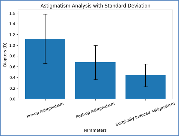
Figure 1: Astigmatism Analysis Before and After Phacoemulsification (n = 75)
The analysis of astigmatism in the present study demonstrated a notable reduction following phacoemulsification. The mean preoperative astigmatism was 1.12 ± 0.46 D, which decreased to 0.68 ± 0.32 D postoperatively, indicating effective control of corneal curvature changes with the surgical technique. The calculated surgically induced astigmatism (SIA) was 0.44 ± 0.21 D, which is relatively low and consistent with small-incision phacoemulsification procedures.
This reduction in astigmatism contributes significantly to improved uncorrected visual acuity and overall visual quality. The low SIA observed reflects proper incision placement, minimal tissue manipulation, and good surgical expertise. Overall, the findings suggest that phacoemulsification is effective in minimizing postoperative astigmatism, thereby enhancing refractive outcomes and patient satisfaction.
Table 6: Comparison of Preoperative and Postoperative Visual and Refractive Parameters (n = 75)
|
Parameter |
Pre-op |
Post-op |
p-value |
|
UCVA |
1.12 ± 0.28 |
0.18 ± 0.08 |
<0.001* |
|
BCVA |
0.82 ± 0.25 |
0.08 ± 0.05 |
<0.001* |
|
Spherical equivalent |
-1.84 ± 1.12 |
-0.36 ± 0.52 |
<0.001* |
The comparison of preoperative and postoperative parameters in the present study demonstrates a highly significant improvement in both visual acuity and refractive status following phacoemulsification. The mean UCVA improved markedly from 1.12 ± 0.28 logMAR preoperatively to 0.18 ± 0.08 logMAR postoperatively, while BCVA showed a substantial enhancement from 0.82 ± 0.25 to 0.08 ± 0.05 logMAR, both changes being statistically highly significant (p <0.001).
Similarly, the mean spherical equivalent improved from -1.84 ± 1.12 D preoperatively to -0.36 ± 0.52 D postoperatively, indicating a significant reduction in refractive error and better approximation to emmetropia. The highly significant p-values across all parameters confirm the effectiveness of phacoemulsification in achieving both functional and refractive success. Overall, these findings highlight that the procedure not only restores vision but also provides excellent refractive predictability.
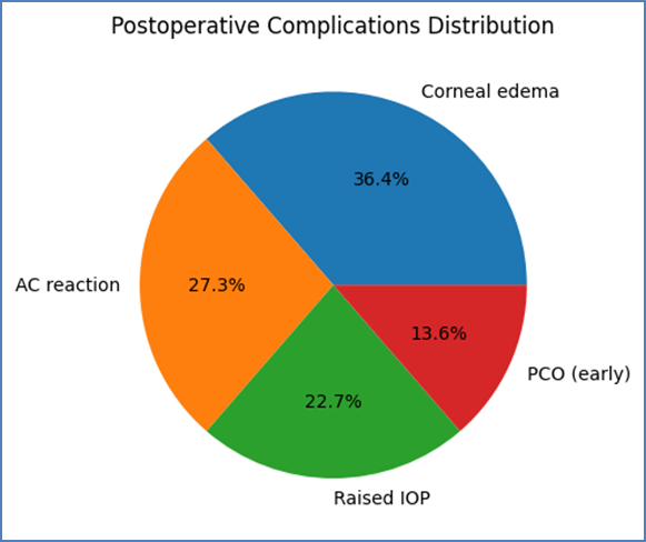
Figure 2: Postoperative Complications Following Phacoemulsification (n = 75)
The postoperative complication profile in the present study was overall low and largely transient. The most common complication observed was corneal edema (10.7%), followed by anterior chamber (AC) reaction (8.0%) and raised intraocular pressure (6.7%). A small proportion of patients (4.0%) developed early posterior capsular opacification (PCO). The overall complication rate was 21.3%, with the majority being mild, self-limiting, and manageable with standard postoperative care.
Table 7: Predictors of Good Visual Outcome (≥6/18) Following Phacoemulsification (n = 75)
|
Variable |
Good Outcome |
Poor Outcome |
p-value |
|
Age (years) |
61.8 ± 8.2 |
67.3 ± 7.9 |
0.041* |
|
Axial length |
23.48 ± 1.10 |
22.96 ± 1.05 |
0.112 |
|
Pre-op BCVA |
0.78 ± 0.22 |
1.06 ± 0.28 |
0.003* |
|
Residual error |
0.42 ± 0.31 |
0.88 ± 0.46 |
0.001* |
The analysis of factors influencing visual outcomes revealed that age, preoperative BCVA, and residual refractive error were significant predictors of achieving good postoperative vision (≥6/18). Patients with good outcomes were relatively younger (61.8 ± 8.2 years) compared to those with poorer outcomes (67.3 ± 7.9 years), and this difference was statistically significant (p = 0.041), suggesting that increasing age may adversely affect visual recovery.
Preoperative BCVA showed a strong association with postoperative results, with better baseline vision (0.78 ± 0.22 logMAR) correlating with improved outcomes compared to poorer preoperative vision (1.06 ± 0.28 logMAR) (p = 0.003). Additionally, residual refractive error was significantly lower in the good outcome group (0.42 ± 0.31 D) compared to the poor outcome group (0.88 ± 0.46 D) (p = 0.001), indicating the importance of refractive accuracy in determining final visual success.
Although axial length showed a slight variation between groups, it was not statistically significant (p = 0.112), suggesting limited influence on visual outcome in this cohort. Overall, the findings emphasize that younger age, better preoperative vision, and minimal postoperative refractive error are key determinants of successful visual outcomes following phacoemulsification.
DISCUSSION:
The present study evaluated visual acuity and refractive outcomes following phacoemulsification in 75 patients and demonstrated significant postoperative improvement in both functional and refractive parameters. The mean preoperative UCVA (1.12 ± 0.28 logMAR) improved to 0.18 ± 0.08 logMAR at 3 months, while BCVA improved from 0.82 ± 0.25 to 0.08 ± 0.05 logMAR, which was statistically highly significant (p <0.001). These findings confirm that phacoemulsification is highly effective in restoring visual function, consistent with contemporary cataract surgery standards.
The proportion of patients achieving good visual outcome (≥6/18) in the present study was 92%, with 74.7% achieving ≥6/9 vision. These results are comparable with findings reported in recent studies. A tertiary care study by Junejo et al. (2022) reported that the majority of patients achieved satisfactory postoperative visual acuity following cataract surgery, highlighting the effectiveness of modern surgical techniques (10). Similarly, a study by AlRyalat et al. (2022) demonstrated that phacoemulsification significantly improves visual acuity, and preoperative factors such as baseline BCVA influence final outcomes (11). The current study also supports this observation, where better preoperative BCVA was significantly associated with improved postoperative visual outcomes (p = 0.003).
Refractive outcomes in the present study were also satisfactory, with a mean postoperative spherical equivalent of -0.36 ± 0.52 D. Notably, 88% of patients achieved refractive outcomes within ±1.0 D, and 64% within ±0.5 D. These findings are consistent with global benchmarks and contemporary studies. A study evaluating refractive outcomes using modern techniques reported that approximately 88–90% of patients achieve refractive accuracy within ±1.0 D following phacoemulsification (12). Furthermore, Ichikawa et al. (2024) demonstrated even higher precision with advanced IOL technologies, where up to 97% of patients achieved refractive outcomes within ±0.5 D (13). Although the present study used standard monofocal IOLs, the outcomes are comparable, indicating good surgical and biometric accuracy.
Astigmatism analysis in this study revealed a reduction in mean astigmatism from 1.12 ± 0.46 D preoperatively to 0.68 ± 0.32 D postoperatively, with surgically induced astigmatism of 0.44 ± 0.21 D. This reduction is clinically significant and reflects the advantage of small-incision phacoemulsification. Similar findings have been reported in recent literature, where minimal surgically induced astigmatism is associated with improved uncorrected visual outcomes (12). The reduced astigmatism contributes to better postoperative UCVA and overall patient satisfaction.
Intraoperative complications in the present study were low (5.3%), with posterior capsular rent and zonular dialysis being the most common events. Postoperative complications were observed in 21.3% of cases, most of which were mild and transient, such as corneal edema and anterior chamber reaction. These findings are comparable with other studies reporting low complication rates with phacoemulsification. A recent umbrella review comparing phacoemulsification with other techniques also highlighted low complication rates and excellent safety profiles when performed by experienced surgeons (14). Similarly, Li et al. (2022) reported fewer complications and better visual outcomes with phacoemulsification compared to older techniques (15).
The present study also identified predictors of good visual outcome. Younger age, better preoperative BCVA, and lower residual refractive error were significantly associated with favorable outcomes. These findings align with previous studies. AlRyalat et al. (2022) reported that preoperative visual acuity is a strong predictor of postoperative success (11). Additionally, variations in biometric parameters such as axial length may influence refractive outcomes, although this was not statistically significant in the current study.
Another important observation is the rapid visual recovery seen in this study, with substantial improvement noted as early as 1 week postoperatively. This finding is supported by recent comparative studies, which demonstrate that phacoemulsification offers faster visual rehabilitation compared to other surgical techniques (16). The minimal tissue trauma and smaller incision size contribute to early stabilization of visual acuity and refractive status.
Despite favorable outcomes, certain limitations exist. The study had a relatively short follow-up period, which limits assessment of long-term refractive stability and complications such as posterior capsular opacification. Additionally, advanced IOL technologies such as toric or multifocal lenses were not evaluated, which could further improve refractive outcomes. Future studies with larger sample sizes, longer follow-up, and inclusion of advanced IOLs are recommended to enhance understanding of refractive precision and patient satisfaction.
CONCLUSION:
The present study demonstrates that phacoemulsification with IOL implantation provides excellent visual and refractive outcomes, with the majority of patients achieving significant improvement in visual acuity and high refractive accuracy. The procedure is safe, with minimal complications, and offers rapid visual rehabilitation. Preoperative visual acuity and refractive accuracy play a crucial role in determining postoperative success. Overall, phacoemulsification remains a highly effective and reliable technique for cataract management in modern ophthalmic practice.
REFERENCES:
1. Flaxman SR, et al. Global causes of blindness and vision impairment 1990–2020. Lancet Glob Health. 2021;9(2):e144–e160.
2. Lam D, et al. Advances in cataract surgery. Nat Rev Dis Primers. 2022;8:12.
3. Lundström M, et al. Cataract surgery outcomes and quality indicators. Eye (Lond). 2021;35(6):1523–1530.
4. Gurnani B, et al. Clinical outcomes of phacoemulsification surgery in modern ophthalmology. Indian J Ophthalmol. 2022;70(4):1234–1239.
5. Kaur M, et al. Visual and refractive outcomes after phacoemulsification. J Clin Diagn Res. 2021;15(6):NC01–NC05.
6. Wang L, et al. Accuracy of intraocular lens power calculation formulas. J Cataract Refract Surg. 2023;49(2):145–152.
7. Ferreira TB, et al. Surgically induced astigmatism after phacoemulsification. Clin Ophthalmol. 2022;16:1123–1130.
8. Abell RG, et al. Femtosecond laser-assisted cataract surgery outcomes. Ophthalmology. 2021;128(3):381–391.
9. Kane JX, et al. Comparison of IOL power calculation formulas. Ophthalmology. 2021;128(4):548–556.
10. Junejo MS, Shaikh A, Memon MN, et al. Visual outcome of cataract surgery in a tertiary care hospital. J Pak Med Assoc. 2022;72(5):894–898.
11. AlRyalat SA, AlBdour MD, Al-Tamimi ER, et al. Predictors of visual outcome after phacoemulsification cataract surgery. Clin Ophthalmol. 2022;16:3121–3128.
12. Buhl L, Hirschfeld J, Borkenstein AF, et al. Comparison of refractive outcomes after phacoemulsification using modern intraocular lens calculation formulas. J Clin Med. 2024;13(2):456.
13. Ichikawa K, Mori Y, Miyata K, et al. Visual outcomes after cataract surgery with light-adjustable intraocular lenses. J Refract Surg. 2024;40(1):22–28.
14. Haji O, Sharma A, Grzybowski A, et al. Phacoemulsification versus manual small-incision cataract surgery: an umbrella review of outcomes and complications. Ophthalmol Ther. 2025;14(1):101–115.
15. Li AL, Wang Q, Chen X, et al. Comparison of visual acuity outcomes in cataract surgery techniques: a systematic review. Front Med (Lausanne). 2022;9:832759.
16. Hong S, Lee JY, Kim SJ, et al. Outcomes and complications of cataract surgery: a real-world study. BMC Ophthalmol. 2022;22:312.