A Prospective Study of Clinical Profile and Intraocular Pressure Control in Glaucoma Patients.
- Duggineni Srita , Post Graduate, Department of Ophthalmology, Kamineni Institute of Medical Sciences, Narketpally, Telangana.
- Abdul Subhan , Assistant Professor, Department of Ophthalmology, Kamineni Institute of Medical Sciences, Narketpally, Telangana.
- K. Tejaswi , Associate Professor, Department of Ophthalmology, Kamineni Institute of Medical Sciences, Narketpally, Telangana.
- K. Gayatri , Associate Professor, Department of Ophthalmology, Kamineni Institute of Medical Sciences, Narketpally, Telangana.
Article Information:
Abstract:
Background: Glaucoma is a progressive optic neuropathy and a leading cause of irreversible blindness worldwide. Elevated intraocular pressure (IOP) remains the primary modifiable risk factor, making early diagnosis and sustained pressure control essential for preventing disease progression. Aim of the study was to evaluate the clinical profile and intraocular pressure control among glaucoma patients attending a tertiary care ophthalmology department. Materials and methods: A prospective observational study was conducted in the Department of Ophthalmology at Kamineni Institute of Medical Sciences involving 75 glaucoma patients. Demographic characteristics, ophthalmic history, systemic comorbidities, glaucoma subtype, baseline IOP, optic disc findings, treatment modalities, and follow-up IOP readings were recorded. Data were analyzed using descriptive statistics, paired t-test, Chi-square test, and logistic regression analysis. Result: The mean age of participants was 56.4 ± 11.2 years, with male predominance (57.3%). Primary Open-Angle Glaucoma was the most common subtype (45.3%). Hypertension and diabetes mellitus were common systemic comorbidities. Mean baseline IOP was 26.8 ± 5.7 mmHg, which significantly reduced to 17.6 ± 3.8 mmHg at 6 months (p < 0.001). Adequate IOP control was achieved in 64% of patients. Higher baseline IOP, advanced optic disc cupping, and diabetes mellitus were associated with poor IOP control. Conclusion: Glaucoma commonly affects middle-aged and elderly individuals. Early diagnosis, regular monitoring, and effective IOP control are essential to prevent disease progression and visual impairment.
Keywords:
Article :
INTRODUCTION:
Glaucoma is a chronic, progressive optic neuropathy characterized by retinal ganglion cell degeneration and visual field loss, and is a leading cause of irreversible blindness worldwide. It is often asymptomatic in early stages, making early diagnosis and continuous monitoring essential. The disease includes primary open-angle, angle-closure, normal-tension, and secondary glaucomas. Elevated intraocular pressure (IOP) is the most important modifiable risk factor, and its reduction remains the cornerstone of management [1]. Structural and vascular factors contribute to damage, but sustained or fluctuating IOP accelerates progression [2]. Monitoring IOP patterns helps assess treatment effectiveness and disease stability [3].
Globally, glaucoma prevalence increases with age and is higher in developing countries due to delayed diagnosis and limited access to care. Primary open-angle glaucoma is more common in Western populations, while angle-closure glaucoma predominates in Asians [4]. In India, many cases remain undiagnosed until advanced stages [5]. Disease progression is influenced by factors such as age, sex, family history, systemic illness, refractive errors, baseline IOP, optic disc changes, and visual field defects [6].
IOP results from the balance between aqueous humor production and outflow. Elevated IOP causes mechanical stress on the optic nerve head, leading to ganglion cell death and visual field loss [7]. Even in normal-tension glaucoma, IOP reduction slows progression [8]. Therefore, measurement using applanation tonometry, assessment of diurnal variation, and evaluation of fluctuations are critical. Both mean IOP and its variability contribute to progression [9], and unstable IOP may cause continued vision loss despite controlled clinic readings.
Several studies have evaluated IOP control. Konstas et al. showed that travoprost significantly reduces 24-hour IOP fluctuations, with better daytime control when administered in the evening [1]. Denis et al. reported significant IOP reduction with latanoprost, especially in patients with higher baseline pressures [2]. Gračner demonstrated effective long-term IOP reduction with selective laser trabeculoplasty in capsular and primary open-angle glaucoma [3], while Pillunat et al. found reduced nocturnal peaks and fluctuations after laser treatment [4].
Long-term studies show that glaucoma may progress despite treatment, highlighting the need for sustained monitoring [5]. Sihota et al. reported significant IOP reduction over six years in chronic angle-closure glaucoma, though some patients required treatment escalation [6].
Despite advancements, gaps remain. Many studies focus on treatment efficacy without integrating clinical profiles. Factors like compliance, socioeconomic status, and follow-up variability are often underreported [8], and regional data from developing countries are limited [5]. A comprehensive evaluation of clinical profile alongside IOP control is necessary to identify predictors of disease progression and treatment response.
Thus, a prospective study assessing clinical profile and IOP control in glaucoma patients is essential to provide real-world evidence, improve clinical decision-making, and support individualized management strategies. The present study aims to evaluate the clinical profile of glaucoma patients and assess IOP control over time, including demographic characteristics, disease patterns, treatment modalities, and effectiveness in preventing progression.
MATERIALS AND METHODS:
Study Design and Setting
This prospective observational study was conducted in the Department of Ophthalmology at Kamineni Institute of Medical Sciences over a defined study period. The study aimed to evaluate the clinical profile and intraocular pressure (IOP) control among diagnosed glaucoma patients attending the ophthalmology outpatient department and glaucoma clinic. A total sample size of 75 patients was included in the study. Patients diagnosed with various forms of glaucoma were enrolled consecutively after obtaining informed consent. Ethical clearance was obtained from the Institutional Ethics Committee prior to initiation of the study. The study was conducted in accordance with ethical principles for biomedical research involving human participants.
Study Population
The study population included patients diagnosed with glaucoma attending the ophthalmology department during the study period. Patients fulfilling the eligibility criteria were recruited consecutively until the desired sample size of 75 was achieved. Both newly diagnosed and previously diagnosed glaucoma patients under treatment were considered for inclusion.
Inclusion Criteria
• Patients aged 18 years and above.
• Patients diagnosed with glaucoma based on clinical examination and ophthalmological evaluation.
• Patients with primary open-angle glaucoma, primary angle-closure glaucoma, normal-tension glaucoma, or secondary glaucoma.
• Patients willing to participate and provide informed consent.
• Patients attending regular follow-up visits for IOP assessment.
Exclusion Criteria
• Patients unwilling to participate in the study.
• Patients with ocular trauma causing secondary glaucoma.
• Patients with active ocular infections or severe ocular surface disease.
• Patients with previous ocular surgery unrelated to glaucoma that could affect intraocular pressure measurements.
• Patients with incomplete clinical records or irregular follow-up.
• Patients with severe systemic illness preventing ophthalmic examination.
Study Tools
The following tools and instruments were used for data collection and assessment:
• Structured case record proforma for demographic and clinical details.
• Visual acuity assessment using Snellen’s chart.
• Slit-lamp biomicroscopy for anterior segment evaluation.
• Goldmann applanation tonometer for intraocular pressure measurement.
• Gonioscopy for angle assessment.
• Direct and indirect ophthalmoscopy for optic disc evaluation.
• Fundus examination using slit-lamp biomicroscopy with +90D lens.
• Visual field analysis where indicated.
• Pachymetry and optic nerve imaging when clinically required.
Data Collection
Data were collected prospectively using a predesigned structured proforma. Information recorded included:
• Demographic details such as age, gender, occupation, and residence.
• Detailed ophthalmic history and duration of symptoms.
• Family history of glaucoma.
• Associated systemic illnesses such as hypertension and diabetes mellitus.
• Type of glaucoma diagnosed.
• Baseline intraocular pressure values.
• Clinical findings including optic disc cupping, visual acuity, and visual field defects.
• Treatment modality adopted (medical, laser, or surgical).
• Follow-up intraocular pressure readings during subsequent visits.
• Assessment of intraocular pressure control over the study period.
Outcome Measures
The primary outcome measured was intraocular pressure control during follow-up. Secondary outcomes included assessment of clinical profile, glaucoma subtype distribution, optic nerve findings, and treatment response.
Statistical Analysis
Collected data were entered into Microsoft Excel and analyzed using appropriate statistical software. Descriptive statistics such as mean, standard deviation, frequency, and percentage were used to summarize demographic and clinical variables. Continuous variables were expressed as mean ± standard deviation, while categorical variables were presented as proportions and percentages. Associations between clinical variables and intraocular pressure control were analyzed using suitable statistical tests such as Chi-square test and Student’s t-test. A p-value of less than 0.05 was considered statistically significant.
RESULTS:
Table 1: Demographic Characteristics of Study Participants (n = 75)
|
Variable |
Category |
Number (n) |
Percentage (%) |
Mean ± SD |
|
Age (years) |
18–40 years |
12 |
16.0 |
|
|
41–50 years |
18 |
24.0 |
||
|
51–60 years |
24 |
32.0 |
||
|
>60 years |
21 |
28.0 |
56.4 ± 11.2 years |
|
|
Gender |
Male |
43 |
57.3 |
|
|
Female |
32 |
42.7 |
— |
|
|
Occupation |
Farmer |
18 |
24.0 |
|
|
Homemaker |
16 |
21.3 |
||
|
Employee/Service |
14 |
18.7 |
||
|
Business |
10 |
13.3 |
||
|
Retired |
12 |
16.0 |
||
|
Others |
5 |
6.7 |
— |
|
|
Residence |
Rural |
49 |
65.3 |
|
|
Urban |
26 |
34.7 |
— |
The mean age of the study participants was 56.4 ± 11.2 years, indicating that glaucoma was more commonly observed in middle-aged and elderly individuals. The majority of patients belonged to the 51–60 years age group (32%). Male predominance was noted with 57.3% participants being male. Regarding occupation, farming and homemaking were the most common occupational categories. Most participants belonged to rural areas (65.3%), reflecting healthcare utilization patterns commonly observed in tertiary care ophthalmology centers in semi-urban settings.
Table 2: Ophthalmic History and Duration of Symptoms Among Study Participants (n = 75)
|
Variable |
Category |
Number (n) |
Percentage (%) |
Mean ± SD |
|
Presenting Symptoms |
Blurred vision |
28 |
37.3 |
|
|
Eye pain |
12 |
16.0 |
||
|
Headache |
10 |
13.3 |
||
|
Halos around lights |
8 |
10.7 |
||
|
Peripheral vision loss |
9 |
12.0 |
||
|
Others |
8 |
10.7 |
— |
|
|
Duration of Symptoms |
< 3 months |
14 |
18.7 |
|
|
3–6 months |
21 |
28.0 |
||
|
6–12 months |
19 |
25.3 |
||
|
>12 months |
21 |
28.0 |
8.6 ± 5.2 months |
|
|
Laterality |
Unilateral |
23 |
30.7 |
|
|
Bilateral |
52 |
69.3 |
— |
|
|
Family History of Glaucoma |
Present |
14 |
18.7 |
|
|
Absent |
61 |
81.3 |
— |
Blurred vision was the most common presenting complaint, reported by 37.3% of patients, followed by eye pain and headache. A considerable proportion of patients had symptoms persisting for more than six months before presentation, with a mean symptom duration of 8.6 ± 5.2 months, suggesting delayed healthcare seeking. Bilateral involvement was more common (69.3%), consistent with the chronic progressive nature of glaucoma. Nearly half of the patients were already receiving anti-glaucoma medications before enrollment, indicating ongoing disease management. Family history was present in a smaller proportion of patients, which aligns with observations from earlier glaucoma epidemiological studies.
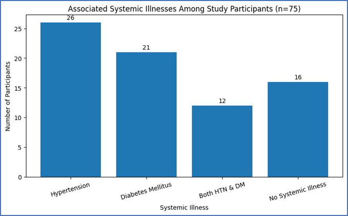
Figure 1: Associated Systemic Illnesses Among Study Participants (n = 75)
Hypertension was the most common associated systemic illness, present in 34.7% of participants, followed by diabetes mellitus (28.0%). Both conditions coexisted in 16.0% of patients. Systemic comorbidities may influence glaucoma progression and intraocular pressure control.
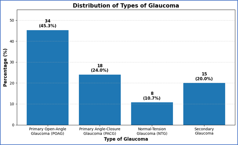
Figure 2: Distribution of Type of Glaucoma Diagnosed Among Study Participants (n = 75)
Primary Open-Angle Glaucoma (POAG) was the most common subtype, accounting for 45.3% of cases, followed by Primary Angle-Closure Glaucoma (24.0%). Secondary glaucoma and Normal-Tension Glaucoma were observed in smaller proportions. These findings are consistent with earlier hospital-based glaucoma studies.
Table 3: Baseline Intraocular Pressure (IOP) Values Among Study Participants (n = 75)
|
Baseline IOP (mmHg) |
Number (n) |
Percentage (%) |
Mean ± SD |
|
≤ 20 mmHg |
11 |
14.7 |
|
|
21–25 mmHg |
24 |
32.0 |
|
|
26–30 mmHg |
22 |
29.3 |
|
|
> 30 mmHg |
18 |
24.0 |
26.8 ± 5.7 mmHg |
Most patients had baseline intraocular pressure values between 21–30 mmHg, with a mean baseline IOP of 26.8 ± 5.7 mmHg. Elevated baseline IOP is a major risk factor for glaucoma progression and serves as an important parameter for treatment monitoring.
Table 4: Clinical Findings Among Study Participants (n = 75)
|
Clinical Finding |
Category |
Number (n) |
Percentage (%) |
|
Optic Disc Cupping (Cup–Disc Ratio) |
<0.5 |
12 |
16.0 |
|
0.5–0.7 |
38 |
50.7 |
|
|
>0.7 |
25 |
33.3 |
|
|
Visual Acuity |
Normal/Mild Impairment |
29 |
38.7 |
|
Moderate Impairment |
31 |
41.3 |
|
|
Severe Impairment |
15 |
20.0 |
|
|
Visual Field Defects |
Present |
49 |
65.3 |
|
Absent |
26 |
34.7 |
Most patients demonstrated moderate optic disc cupping, with a cup–disc ratio of 0.5–0.7 seen in 50.7% of cases. Moderate visual impairment was observed in 41.3% of participants, while visual field defects were present in 65.3%, indicating functional loss associated with glaucoma progression.
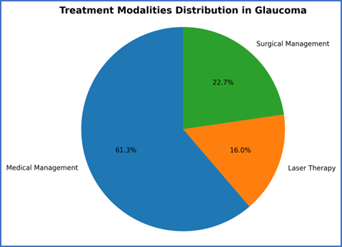
Figure 3: Treatment Modality Adopted Among Study Participants (n = 75)
Medical management was the most commonly adopted treatment modality, used in 61.3% of patients. Surgical intervention was required in 22.7%, while laser therapy was utilized in 16.0% of cases for intraocular pressure control.
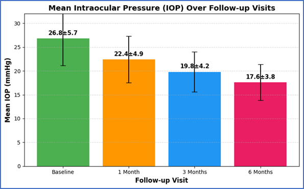
Figure 4: Follow-up Intraocular Pressure (IOP) Readings During Subsequent Visits
A progressive reduction in mean intraocular pressure was observed during follow-up visits. The mean IOP decreased from 26.8 ± 5.7 mmHg at baseline to 17.6 ± 3.8 mmHg at 6 months, indicating effective pressure control with treatment interventions.
Table 5: Assessment of Intraocular Pressure (IOP) Control Over the Study Period (n = 75)
|
IOP Control Status |
Number (n) |
Percentage (%) |
|
Well Controlled (<21 mmHg) |
48 |
64.0 |
|
Partially Controlled (21–24 mmHg) |
17 |
22.7 |
|
Poorly Controlled (>24 mmHg) |
10 |
13.3 |
During the study period, adequate intraocular pressure control was achieved in the majority of patients, with 64.0% demonstrating well-controlled IOP. Partial control was observed in 22.7%, while 13.3% had poorly controlled IOP despite treatment, indicating the need for closer monitoring and therapeutic adjustment.
Table 6: Comparison of Baseline and Follow-up Intraocular Pressure Using Paired t-test (n = 75)
|
Time Point |
Mean IOP (mmHg) ± SD |
Mean Difference |
t-value |
p-value |
|
Baseline |
26.8 ± 5.7 |
— |
— |
— |
|
6 Months Follow-up |
17.6 ± 3.8 |
9.2 |
11.84 |
<0.001 |
A statistically significant reduction in mean intraocular pressure was observed between baseline and 6-month follow-up (p < 0.001), indicating effective treatment response.
Table 7: Association Between Type of Glaucoma and IOP Control Status (Chi-square Test) (n = 75)
|
Type of Glaucoma |
Well Controlled |
Partially Controlled |
Poorly Controlled |
Total |
|
POAG |
24 |
7 |
3 |
34 |
|
PACG |
11 |
4 |
3 |
18 |
|
NTG |
7 |
1 |
0 |
8 |
|
Secondary Glaucoma |
6 |
5 |
4 |
15 |
|
Total |
48 |
17 |
10 |
75 |
|
Statistical Test Value |
Chi-square (χ²) |
8.92 |
p-value |
0.031 |
A significant association was observed between glaucoma subtype and IOP control status (p < 0.05), suggesting that treatment outcomes may vary according to glaucoma type.
Table 8: Logistic Regression Analysis for Predictors of Poor IOP Control (n = 75)
|
Variable |
Odds Ratio (OR) |
95% Confidence Interval |
p-value |
|
Age > 60 years |
1.84 |
1.02 – 3.41 |
0.041 |
|
Diabetes Mellitus |
2.31 |
1.18 – 4.52 |
0.018 |
|
Hypertension |
1.76 |
0.94 – 3.29 |
0.072 |
|
Baseline IOP > 30 mmHg |
3.12 |
1.45 – 6.74 |
0.004 |
|
Cup–Disc Ratio > 0.7 |
2.67 |
1.21 – 5.88 |
0.013 |
Higher baseline IOP, advanced optic disc cupping, diabetes mellitus, and older age were significant predictors of poor intraocular pressure control. Baseline IOP >30 mmHg showed the strongest association with poor treatment response.
DISCUSSION:
The present prospective study evaluated the clinical profile and intraocular pressure (IOP) control among glaucoma patients in a tertiary care setting. Glaucoma remains a leading cause of irreversible blindness worldwide, and early diagnosis with effective IOP control is essential to prevent visual disability. The findings of this study provide insight into demographic characteristics, clinical presentation, and treatment outcomes in glaucoma patients.
In this study, glaucoma predominantly affected older individuals, with a mean age of 56.4 ± 11.2 years. This is consistent with previous studies showing increased prevalence with advancing age due to age-related structural and vascular changes affecting the optic nerve [10]. Age remains one of the most significant non-modifiable risk factors for glaucoma, particularly primary open-angle glaucoma. A male predominance (57.3%) was observed, similar to other hospital-based studies, possibly due to differences in healthcare-seeking behavior and accessibility.
A significant proportion of patients (65.3%) belonged to rural areas, indicating disparities in access to ophthalmic care and awareness. Studies from developing countries have similarly shown that rural populations often present at advanced stages due to delayed diagnosis and limited screening facilities [11]. This highlights the importance of improving awareness and accessibility of eye care services. Blurred vision was the most common presenting complaint, with an average symptom duration of 8.6 ± 5.2 months, reflecting delayed presentation. Glaucoma is often asymptomatic in its early stages, leading to late detection after significant visual field loss has occurred [12]. This delay contributes to irreversible optic nerve damage and underscores the need for routine screening, especially in high-risk populations.
Family history of glaucoma was present in 18.7% of patients, reinforcing its role as an important risk factor. Genetic predisposition influences optic nerve vulnerability and aqueous humor dynamics, increasing susceptibility to glaucoma [13]. Screening of first-degree relatives is therefore essential for early detection and improved prognosis. Systemic comorbidities such as hypertension (34.7%) and diabetes mellitus (28.0%) were commonly observed. These conditions may contribute to glaucomatous damage through vascular mechanisms such as impaired ocular perfusion and microvascular dysfunction [14]. Although the association remains complex, their presence may influence disease progression and response to treatment. Primary Open-Angle Glaucoma (POAG) was the most common subtype (45.3%), consistent with global trends identifying it as the predominant form of glaucoma in adults [15]. Secondary glaucoma accounted for 20% of cases, emphasizing the importance of identifying underlying causes such as trauma, steroid use, or inflammation. This distribution reflects both demographic and environmental influences.
The mean baseline IOP in the present study was 26.8 ± 5.7 mmHg, indicating a moderate to severe disease burden at presentation. Elevated IOP remains the most important modifiable risk factor for glaucoma progression. Sustained pressure elevation causes mechanical stress on the optic nerve head, leading to retinal ganglion cell loss and optic neuropathy [16]. Many patients presented with IOP between 21–30 mmHg, suggesting delayed diagnosis.
Clinical examination revealed that a considerable number of patients had advanced optic disc cupping and visual field defects (65.3%), indicating established disease at presentation. These findings are consistent with studies demonstrating a strong correlation between structural optic nerve changes and functional visual field loss [17]. Advanced cupping is a key indicator of disease severity and progression risk. Medical management was the most commonly used treatment modality (61.3%), followed by laser and surgical interventions. This aligns with standard practice where medical therapy is the first-line approach due to its effectiveness and accessibility [18]. However, treatment adherence is crucial, as poor compliance may lead to inadequate IOP control and continued progression.
A key finding of this study was the significant reduction in IOP during follow-up. Mean IOP decreased from 26.8 ± 5.7 mmHg at baseline to 17.6 ± 3.8 mmHg at six months (p < 0.001). This is consistent with previous studies demonstrating that effective treatment significantly reduces IOP and slows disease progression [19]. Maintaining target IOP is essential in preventing further optic nerve damage. The study also found that 64% of patients achieved adequate IOP control, while 13.3% had poor control. Predictors of poor IOP control included high baseline IOP, advanced optic disc cupping, older age, and diabetes mellitus. These findings are in agreement with earlier studies identifying similar risk factors for disease progression [20]. Identification of such predictors is important for individualized patient management.
Visual field defects were present in nearly two-thirds of patients, highlighting their importance as a functional marker for disease monitoring. Baseline visual field loss has been shown to predict future progression, making regular perimetric evaluation essential [21]. The study also supports the concept that glaucoma is a multifactorial disease. In addition to elevated IOP, factors such as vascular dysregulation and impaired ocular blood flow contribute to disease progression. Emerging evidence suggests that retinal microcirculatory changes may play a role in optic nerve damage, indicating that management strategies should consider both ocular and systemic factors [22].
Overall, the findings of this study are consistent with existing literature and emphasize the importance of early diagnosis, comprehensive clinical evaluation, and sustained IOP control. The study also provides valuable regional data, aiding in better understanding of disease patterns and improving patient management strategies.
CONCLUSION:
The present prospective study demonstrated that glaucoma predominantly affects middle-aged and elderly individuals, with Primary Open-Angle Glaucoma being the most common subtype. Elevated baseline intraocular pressure, optic disc cupping, visual field defects, and associated systemic illnesses such as hypertension and diabetes mellitus were frequently observed. Significant reduction in IOP was achieved during follow-up, indicating effectiveness of treatment modalities. However, a subset of patients continued to exhibit inadequate pressure control, emphasizing the need for individualized management and close monitoring. Early diagnosis, regular follow-up, and prompt intervention remain essential to prevent disease progression and preserve visual function.
REFERENCES:
1. Konstas AGP, Quaranta L, Mikropoulos DG, et al. 24-Hour Intraocular Pressure Control Obtained with Evening- versus Morning-Dosed Travoprost in Primary Open-Angle Glaucoma. Ophthalmology. 2006;113(3):446–450.
2. Denis P, Baudouin C, Bron A, et al. First-line latanoprost therapy in ocular hypertension or open-angle glaucoma patients: a 3-month efficacy analysis stratified by initial intraocular pressure. BMC Ophthalmology. 2010;10:4.
3. Gračner T. Intraocular Pressure Response of Capsular Glaucoma and Primary Open-Angle Glaucoma to Selective Nd:YAG Laser Trabeculoplasty: A Prospective Comparative Clinical Trial. European Journal of Ophthalmology. 2002;12(4):287–292.
4. Pillunat KR, Herber R, Kocket GA, et al. Efficacy of selective laser trabeculoplasty on lowering intraocular pressure fluctuations and nocturnal peak intraocular pressure in treated primary open-angle glaucoma patients. Graefe's Archive for Clinical and Experimental Ophthalmology. 2023;261:1979–1985.
5. Shin YI, Jeoung JW, Kim YK, et al. Longitudinal evaluation of advanced glaucoma: ten-year follow-up cohort study. Scientific Reports. 2024;14:50512.
6. Sihota R, Sood A, Gupta V, Dada T, Agarwal HC. A prospective long-term study of primary chronic angle closure glaucoma. Acta Ophthalmologica Scandinavica. 2004;82(2):209–213.
7. Weinreb RN, Aung T, Medeiros FA. The Pathophysiology and Treatment of Glaucoma. JAMA. 2014;311(18):1901–1911.
8. European Glaucoma Society Terminology and Guidelines for Glaucoma, 5th Edition. British Journal of Ophthalmology. 2021;105(Suppl 1):1–169.
9. Caprioli J, Coleman AL. Intraocular Pressure Fluctuation: A Risk Factor for Visual Field Progression at Low Intraocular Pressures in the Advanced Glaucoma Intervention Study. Ophthalmology. 2008;115(7):1123–1129.
10. Rossetti L, Goni F, Denis P, Bengtsson B, Martinez A, Heijl A. Focusing on glaucoma progression and the clinical importance of progression rate measurement. Eye. 2010;24(Suppl 1):S1–S7.
11. Chan KKW, Tang F, Tham CCY, Young AL, Cheung CY. Retinal vasculature in glaucoma: a review. BMJ Open Ophthalmology. 2017;1:e000032.
12. Davis BM, Crawley L, Pahlitzsch M, Javaid F, Cordeiro MF. Glaucoma: the retina and beyond. Acta Neuropathologica. 2016;132(6):807–826.
13. Hamid SAT, Abdulmajeed SM, Chowdhury IH, Hasan MM. Risk Factors and Genetic Markers Associated with the Development and Progression of Glaucoma: A Review. Journal of Current Glaucoma Practice. 2025;19(4):216–222.
14. Grzybowski A, Och M, Kanclerz P, Leffler C, De Moraes CG. Primary Open Angle Glaucoma and Vascular Risk Factors: A Review of Population-Based Studies from 1990 to 2019. Journal of Clinical Medicine. 2020;9(3):761.
15. Banou L, Dastiridou A, Giannoukas A, Kouvelos G, Baros C, Androudi S. The Role of Color Doppler Imaging in the Diagnosis of Glaucoma: A Review of the Literature. Diagnostics. 2023;13(4):588.
16. Huang X, Islam MR, Akter S, Ahmed F, Kazami E, Yousefi S. Artificial intelligence in glaucoma: opportunities, challenges, and future directions. BioMedical Engineering OnLine. 2023;22:126.
17. Jasty U, Harris A, Siesky B, Rowe LW, Vercellin AC, Mathew S, Pasquale LR. Optic disc haemorrhage and primary open-angle glaucoma: a clinical review. British Journal of Ophthalmology. 2020;104(11):1488–1491.
18. Triolo G, Rabiolo A. Optical coherence tomography and optical coherence tomography angiography in glaucoma: diagnosis, progression, and correlation with functional tests. Therapeutic Advances in Ophthalmology. 2020;12:1–14.
19. Almarzouki N. Impact of Environmental Factors on Glaucoma Progression: A Systematic Review. Clinical Ophthalmology. 2024;18:2705–2720.
20. Berchuck SI, Mwanza JC, Warren JL. Diagnosing Glaucoma Progression with Visual Field Data Using a Spatiotemporal Boundary Detection Method. Journal of the American Statistical Association. 2018.
21. Huang X, Poursoroush A, Sun J, Boland MV, Johnson C, Yousefi S. Identifying factors associated with fast visual field progression in patients with ocular hypertension. 2023.
22. Chan KKW, Tang F, Tham CCY, Young AL, Cheung CY. Retinal vasculature in glaucoma: a review. BMJ Open Ophthalmology. 2017;1:e000032.