Unravelling the Central Role of Interleukin-6 in Cardiac Inflammation: A One of a Kind Cross-Disease Comparative Analysis across Tertiary Care Hospitals in Mumbai.
- Yasmeen Chand Patel , Associate Professor, Dept of General Medicine, Arundhati Institute of Medical Sciences, Hyderabad, Telngana, India.
- Snehal Bhosale , Senior Resident, Dept of Obstetrics and Gynaecology, Bharati Vidyapeeth Deemed University Medical College and Hospital, Sangli, Maharashtra, India.
- Smita Deokar , Professor (Addl), Dept of Biochemistry, HBT Medical College and Dr. RN Cooper Municipal General Hospital, Juhu, Mumbai, Maharashtra, India.
- Mahima Manoj Saptarshi , House Officer, Dept of Biochemistry, HBT Medical College, Dr.R.N. Cooper Municipal General Hospital, Juhu, Mumbai, Maharashtra, India.
- Nishant Anand Ratwani , House Officer, Dept of Biochemistry, HBT Medical College, Dr.R.N. Cooper Municipal General Hospital, Juhu, Mumbai, Maharashtra, India.
Article Information:
Abstract:
Background: Cardiovascular diseases (CVDs) continue to dominate global mortality statistics with inflammation recognized as a crucial contributor to their pathophysiology. Interleukin-6 (IL-6), a cytokine with diverse immunomodulatory effects, plays a central role in the inflammatory processes underlying various cardiac diseases. Objectives: to fill this gap by systematically measuring serum IL-6 levels in well-characterized patient groups with different cardiac pathologies and comparing them with healthy controls. Materials and Methods: This study enrolled 458 participants comprising 229 patients with diverse cardiac diseases and 229 healthy controls to quantitatively assess serum IL-6 levels using enzyme-linked immunosorbent assay (ELISA). Patients included those with myocardial infarction (MI), heart failure (HF), rheumatic heart disease (RHD), complete heart block (CHB), and related cardiac disorders. Results: Results demonstrated significantly elevated IL-6 levels across all patient groups compared to controls, with the highest median values in MI and HF cohorts. IL-6 concentrations correlated positively with clinical severity indicated by NYHA functional class and inversely with left ventricular ejection fraction. Conclusion: These findings reinforce IL-6's role as a unifying inflammatory biomarker across multiple cardiac disease states and support its potential utility in diagnosis, risk stratification, and therapeutic targeting.
Keywords:
Article :
INTRODUCTION:
Cardiovascular diseases (CVDs) remain the most prevalent cause of death globally, with a steadily rising burden in both developing and developed nations. The World Health Organization estimates that by 2030, more than 23 million individuals annually will succumb to CVDs, largely driven by ischemic heart disease and heart failure. [1-3] Increasing evidence underscores the integral role of inflammation in the initiation and progression of cardiac diseases. Inflammatory cytokines contribute significantly to atherosclerosis, plaque instability, myocardial injury, remodelling, and fibrosis, which ultimately impair cardiac function. [4-6]
One cytokine of particular interest is interleukin-6 (IL-6), a pleiotropic molecule produced by a variety of cells, including monocytes, macrophages, endothelial cells, vascular smooth muscle cells, and ischemic myocytes. IL-6 functions both as a pro-inflammatory and an anti-inflammatory cytokine and orchestrates acute-phase responses, immune cell activation, and cardiac metabolism regulation [7,8]. Elevated IL-6 levels have been documented in coronary artery disease, congestive heart failure, and rheumatic heart disease, often correlating with disease severity and adverse outcomes. [9-11] Despite extensive study in specific cardiac conditions such as heart failure, comprehensive analyses comparing IL-6 levels across multiple cardiac disease groups remain scarce.
This study aims to fill this gap by systematically measuring serum IL-6 levels in well-characterized patient groups with different cardiac pathologies and comparing them with healthy controls. We hypothesize that IL-6 elevations reflect ongoing cardiac inflammation and differ in magnitude across disease states, offering potential as a biomarker of disease activity and prognosis.
MATERIALS AND METHODS:
Study design and setting
This prospective case–control study was conducted at a tertiary care medical center.
Study population
The study enrolled 229 patients with diagnosed cardiac diseases and 229 age- and sex-matched healthy controls. Patients were consecutively recruited from the cardiology department. Controls were selected from individuals without known cardiovascular disease and with normal clinical examination and electrocardiographic findings.
Case definition and classification
Patients were categorized into the following diagnostic groups: myocardial infarction (MI; n = 169), heart failure (HF; n = 22), rheumatic heart disease (RHD; n = 20), complete heart block (CHB; n = 16), and other cardiac conditions (n = 2). Diagnoses were established based on clinical assessment, electrocardiography, echocardiography, and standard consensus criteria relevant to each condition.
Inclusion and exclusion criteria
Adult patients (≥18 years) with a confirmed diagnosis of cardiac disease were included. Patients with acute systemic illness, chronic inflammatory conditions, malignancy, or incomplete clinical data were excluded. Controls with a history of cardiovascular disease or systemic illness were also excluded.
Data collection
Demographic and clinical data were recorded using a standardized case record form. Diagnostic evaluations were performed by qualified cardiologists using calibrated equipment according to institutional protocols.
Ethical considerations
The study protocol was reviewed and approved by the Institutional Ethics Committee of the participating center. Written informed consent was obtained from all participants prior to enrolment. The study was conducted in accordance with the principles of the Declaration of Helsinki.
Statistical Analysis
Data distribution was assessed using the Shapiro–Wilk test and demonstrated non-normality. Accordingly, continuous variables are presented as median with interquartile range (IQR), and categorical variables as frequencies and percentages. Comparisons between two groups were performed using the Mann–Whitney U test. Comparisons among multiple groups were conducted using the Kruskal–Wallis test followed by Dunn’s post hoc test for pairwise comparisons.
Correlations between interleukin-6 (IL-6) levels and clinical parameters, including New York Heart Association (NYHA) functional class and left ventricular ejection fraction (LVEF), were assessed using Spearman’s rank correlation coefficient. Receiver operating characteristic (ROC) curve analysis (Figure 1) was used to evaluate the discriminatory ability of IL-6 to differentiate cardiac patients from healthy controls, with calculation of sensitivity, specificity, and area under the curve (AUC). A two-tailed p value < 0.05 was considered statistically significant. Statistical analyses were performed using standard statistical software.
RESULTS:
Demographic and Clinical Characteristics
The cardiac patient and control groups were comparable with respect to age (54.49 ± 11.71 vs. 54.03 ± 12.35 years; p = 0.636) and sex distribution (male: 70.3% vs. 74.7%; p = 0.295), indicating appropriate matching. No significant differences were observed in baseline demographic or clinical variables, including body mass index and systolic blood pressure. Detailed characteristics of the study population are presented in Tables 1 and 2.
Table 1: Demographic Characteristics of Cardiac Patients and Controls
|
Table 2: Clinical Distribution of Cardiac Disease Groups
|
Group |
N |
Median Age (IQR) |
Male % |
NYHA Class Distribution (%) |
|
Myocardial Infarction |
169 |
56 (48-64) |
68 |
Class II: 40%, III: 35%, IV: 25% |
|
Heart Failure |
22 |
57 (50-65) |
73 |
Class II: 15%, III: 45%, IV: 40% |
|
Rheumatic Heart Disease |
20 |
52 (46-59) |
55 |
Class II: 50%, III: 30%, IV: 20% |
|
Complete Heart Block |
16 |
54 (48-62) |
68 |
Class I/II: 60%, III: 40% |
Serum IL-6 Concentrations
Serum interleukin-6 (IL-6) levels were significantly higher in cardiac patients compared with healthy controls. Median (IQR) IL-6 concentration in cases was 5.5 (11.3) pg/mL versus 0.0 (3.5) pg/mL in controls (p < 0.001).
Among cardiac subgroups, patients with myocardial infarction (MI) and heart failure (HF) demonstrated the highest IL-6 levels [MI: 10.1 (8.3) pg/mL; HF: 12.0 (9.1) pg/mL], which were significantly greater than those observed in rheumatic heart disease (RHD) [5.6 (4.7) pg/mL] and complete heart block (CHB) [6.2 (5.2) pg/mL] (p < 0.01). Overall intergroup differences were statistically significant by Kruskal–Wallis analysis (p < 0.001). These findings are summarized in Table 3.
Table 3: Serum IL-6 Levels Across Cardiac Disease Groups and Controls (pg/mL)
|
Group |
Median (IQR) |
p-value vs controls |
|
Myocardial Infarction |
10.1 (8.3) |
<0.0001 |
|
Heart Failure |
12.0 (9.1) |
<0.0001 |
|
Rheumatic Heart Disease |
5.6 (4.7) |
<0.0001 |
|
Complete Heart Block |
6.2 (5.2) |
<0.0001 |
|
Controls |
0.0 (3.5) |
- |
Association of IL-6 with Clinical Severity
Spearman’s correlation analysis revealed a strong positive association between IL-6 levels and New York Heart Association (NYHA) functional class (ρ = 0.68, p < 0.001), indicating increasing inflammatory activity with worsening functional status. In contrast, IL-6 levels showed a moderate inverse correlation with left ventricular ejection fraction (LVEF) (ρ = −0.54, p < 0.001), suggesting higher inflammatory burden in patients with more severe systolic dysfunction.
Diagnostic Performance of IL-6
Receiver operating characteristic (ROC) curve analysis demonstrated that an IL-6 cut-off value of 2.5 pg/mL effectively differentiated cardiac patients from healthy controls, with a sensitivity of 80.80% and specificity of 76.4%. The area under the curve (AUC) was 0.830 (95% CI 0.791 – 0.869), indicating excellent diagnostic accuracy (Figure 1).
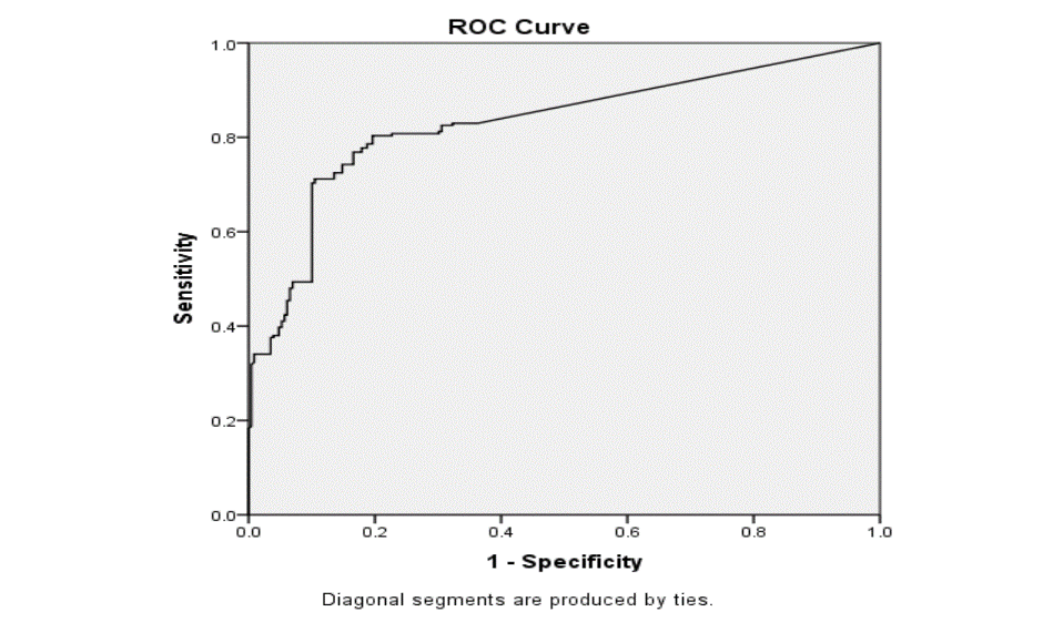
Fig.1: Receiver Operator Curve (ROC) for IL-6 (pg/mL)
The analysis for IL 6 for Receiver Operator Curve was done with 458 observations
The area under the curve for the ROC curve is 0.830 (95% CI 0.791 – 0.869)
|
Cut-Off point* for IL6 |
Sensitivity |
1- Specificity |
|
2.1 |
0.830 |
0.354 |
|
2.5 |
0.808 |
0.240 |
|
2.7 |
0.803 |
0.201 |
|
3.0 |
0.777 |
0.179 |
|
3.5 |
0.725 |
0.140 |
|
4.0 |
0.646 |
0.100 |
|
5.1 |
0.528 |
0.100 |
|
6.0 |
0.454 |
0.066 |
|
7.0 |
0.410 |
0.052 |
|
8.0 |
0.380 |
0.044 |
|
9.2 |
0.362 |
0.035 |
|
10.0 |
0.354 |
0.035 |
*Positive if greater than or equal to the given value
The optimum level of the estimate for cut-off levels of IL-6 levels was found to be 2.5 pg with a sensitivity of 80.8% and a specificity of 76.4%.
DISCUSSION:
This analysis confirms the association between elevated IL-6 levels and cardiac pathologies, such as MI, HF, RHD and CHB. MI and HF were associated with the highest elevations of IL-6 levels, due to widespread inflammation. Elevated levels of IL-6 can be correlated with monocyte and macrophage activation and endothelial dysfunction. A positive association was also noticed between NYHA functional classes, the ejection fraction and the serum IL-6 levels. This finding can be useful in assessing a patient’s functional status and prognosis.
The results obtained are in tandem with prior studies focusing on similar aspects, thus helping to broaden the potential of IL-6 as a cardiac biomarker. The role of IL-6 as a pro and anti-inflammatory cytokine makes it possible for clinicians to use it to assess clinical outcomes.
LIMITATIONS:
The observational design of the study does not permit causality. Serial IL-6 levels were also not measured and thus, temporal changes cannot be assessed. These pitfalls pave the way for future prospective studies that could evaluate the association between IL-6 levels and response to therapies. These findings can also be used change the way cardiac pathologies are managed, with therapies having IL-6 specific actions.
CONCLUSION:
Elevated serum IL-6 is a common and significant feature across multiple cardiac diseases, positively correlating with clinical severity and systolic dysfunction. IL-6 holds promise as an accessible biomarker for comprehensive cardiac risk assessment, with potential roles in guiding targeted therapies and improving patient outcomes.
REFERENCES:
1. Huffman MD, Prabhakaran D. Heart failure: epidemiology and prevention in India. Natl Med J India. 2010;23(5):283-288.
2. Cardiovascular diseases (CVDs). World Health Organization. Published July 31 2025. https://www.who.int/news-room/fact-sheets/detail/cardiovascular-diseases-(cvds)
3. Wang H, Jing ZC. Inflammation and cardiovascular diseases. Chronic Dis Transl Med. 2020;6(4):215-218. Published 2020 Nov 3. doi:10.1016/j.cdtm.2020.09.003
4. Mehta NN, deGoma E, Shapiro MD. IL-6 and Cardiovascular Risk: A Narrative Review. Curr Atheroscler Rep. 2024;27(1):12. Published 2024 Nov 26. doi:10.1007/s11883-024-01259-7
5. Libby P. Inflammation and cardiovascular disease mechanisms. Am J Clin Nutr. 2006;83(suppl):456S-60S.
6. Vasan RS. Biomarkers of cardiovascular disease: Molecular basis and practical considerations. Circulation. 2006;113(19):2335–62
7. Tanaka T, Narazaki M, Kishimoto T. IL-6 in inflammation, immunity, and disease. Cold Spring Harb Perspect Biol. 2014;6(10):a016295. Published 2014 Sep 4. doi:10.1101/cshperspect.a016295
8. Gabay C. Interleukin-6 and chronic inflammation. Arthritis Res Ther. 2006;8 Suppl 2(Suppl 2):S3. doi:10.1186/ar1917
9. Kanda T, Takahashi T. Interleukin-6 and cardiovascular diseases. Jpn Heart J. 2004;45(2):183-193. doi:10.1536/jhj.45.183
10. Deokar SA, Dandekar SP, Shinde GA, et al. Role of serum interleukin-6 in heart failure. Int J Adv Med. 2018;5(4):936-940.
11. Bartekova M, Radosinska J, Jelemensky M, et al. Role of cytokines and inflammation in heart function during health and disease. Heart Fail Rev. 2018;23(5):733-758. doi:10.1007/s10741-018-9716-x.