Prognostic Significance of Histopathological Patterns in IgA Nephropathy: Evidence from a Systematic Review and Meta-analysis.
- Vinit Kumar , Assistant Professor, Department of General Medicine, Geetanjali Institute of Medical Sciences, Jaipur, Rajasthan, India.
- Garvit Mundra , Senior Resident, Department of Hepatology, Institute of Liver and Biliary Sciences (ILBS), Vasant Kunj, New Delhi, India.
- Akshay Kalra , Senior Resident, Department of Pathology, Arun Asaf Ali Government Hospital, New Delhi, Delhi, India.
Article Information:
Abstract:
Background: IgA nephropathy (IgAN) is the most common primary glomerulonephritis worldwide and demonstrates marked heterogeneity in clinical presentation and disease progression. Histopathological assessment using renal biopsy, particularly the Oxford MEST-C classification, plays a crucial role in prognostication and therapeutic decision-making. Aim: To systematically evaluate histopathological patterns in IgA nephropathy and determine their association with renal outcomes and disease progression. Methods: A systematic review and meta-analysis of observational studies published between 2009 and 2026 was conducted according to PRISMA guidelines. Studies evaluating histopathological lesions in biopsy-proven IgAN and reporting clinical outcomes were included. Data regarding Oxford MEST-C lesions, renal survival, estimated glomerular filtration rate (eGFR), proteinuria, and progression to end-stage kidney disease (ESKD) were extracted and analyzed. Results: Thirty-eight studies involving 18,742 patients were included. Tubular atrophy/interstitial fibrosis (T lesions) showed the strongest association with poor renal outcomes, followed by segmental glomerulosclerosis and crescent formation. Mesangial hypercellularity was associated with persistent proteinuria, while endocapillary hypercellularity demonstrated variable prognostic significance. Pooled analysis demonstrated significant associations between MEST-C lesions and progressive renal impairment. Conclusion: Histopathological lesions identified in the Oxford MEST-C classification significantly influence clinical outcomes in IgAN. Tubulointerstitial fibrosis and segmental sclerosis remain the most important predictors of renal progression and ESKD.
Keywords:
Article :
INTRODUCTION:
IgA nephropathy (IgAN), first described by Berger and Hinglais in 1968, is recognized as the most common primary glomerulonephritis worldwide and represents a major cause of chronic kidney disease (CKD) and end-stage kidney disease (ESKD) among young adults [1]. The disease is characterized by mesangial deposition of IgA-containing immune complexes, leading to glomerular inflammation, progressive fibrosis, and deterioration of renal function [2]. Clinical manifestations vary considerably, ranging from isolated microscopic hematuria to nephrotic syndrome and rapidly progressive glomerulonephritis [3].
The clinical course of IgAN is highly heterogeneous. Approximately 20-40% of patients progress to ESKD within 20 years of diagnosis despite therapeutic interventions [4]. Traditional clinical predictors such as hypertension, persistent proteinuria, and reduced estimated glomerular filtration rate (eGFR) provide important prognostic information; however, histopathological assessment remains fundamental for accurate risk stratification [5].
Renal biopsy is considered the gold standard for diagnosis and prognostic evaluation in IgAN. Histopathological findings include mesangial proliferation, endocapillary hypercellularity, segmental glomerulosclerosis, tubular atrophy, interstitial fibrosis, and crescent formation [6]. Owing to the heterogeneity of pathological lesions and variability in reporting systems, the Oxford Classification of IgA nephropathy was introduced in 2009 to standardize histopathological assessment [7].
The Oxford classification identified four pathological variables independently associated with renal outcomes, namely mesangial hypercellularity (M), endocapillary hypercellularity (E), segmental glomerulosclerosis (S), and tubular atrophy/interstitial fibrosis (T), collectively referred to as the MEST score [7]. Subsequently, crescent formation (C) was added in the updated Oxford classification, resulting in the MEST-C scoring system [8].
Several studies have validated the prognostic significance of MEST-C lesions across different ethnic and geographic populations [9,10]. Tubular atrophy/interstitial fibrosis has consistently emerged as the strongest predictor of renal progression and ESKD [11]. Segmental glomerulosclerosis has been associated with persistent proteinuria and accelerated decline in renal function, whereas crescents are linked with aggressive disease phenotypes and rapidly progressive renal failure [12].
Despite these advances, significant heterogeneity exists among studies regarding the prognostic impact of individual histopathological lesions, particularly endocapillary hypercellularity and crescentic lesions [13]. Differences in patient populations, therapeutic strategies, biopsy timing, and follow-up duration contribute to inconsistent findings reported in the literature [14].
Therefore, the present systematic review and meta-analysis was undertaken to comprehensively evaluate histopathological patterns in IgA nephropathy and determine their relationship with clinical outcomes including renal survival, decline in eGFR, proteinuria progression, and development of ESKD.
Objectives
Primary Objective
To evaluate the association between histopathological patterns in IgA nephropathy and clinical outcomes.
Secondary Objectives
1. To assess the prognostic significance of individual Oxford MEST-C lesions.
2. To evaluate the association between histopathological lesions and progression to ESKD.
3. To determine the relationship between pathological findings and decline in renal function.
4. To assess the association between histopathological lesions and persistent proteinuria.
MATERIALS AND METHODS:
Study Design
This systematic review and meta-analysis was conducted according to the Preferred Reporting Items for Systematic Reviews and Meta-Analyses (PRISMA) guidelines [15]. The study aimed to synthesize evidence regarding histopathological patterns in IgA nephropathy and their association with clinical outcomes.
Literature Search Strategy
A comprehensive literature search was performed using PubMed, Embase, Scopus, Web of Science, and Cochrane Library databases for studies published between January 2009 and January 2026. The search strategy included combinations of Medical Subject Heading (MeSH) terms and keywords such as “IgA nephropathy,” “Oxford classification,” “MEST-C,” “histopathology,” “renal biopsy,” “renal survival,” “glomerulosclerosis,” and “end-stage kidney disease” [16].
Boolean operators (AND/OR) were used appropriately to refine the search. Reference lists of eligible studies and review articles were manually screened to identify additional relevant studies.
Inclusion Criteria
Studies were included if they fulfilled the following criteria:
• Observational cohort or case-control studies
• Patients with biopsy-proven IgA nephropathy
• Histopathological evaluation using Oxford classification or equivalent criteria
• Studies reporting renal outcomes such as eGFR decline, proteinuria progression, renal survival, or ESKD
• Full-text articles published in English
Exclusion Criteria
The following studies were excluded:
• Case reports and case series
• Review articles and editorials
• Studies without histopathological correlation
• Studies lacking outcome data
• Animal studies
• Duplicate publications
Study Selection
Two independent reviewers screened titles and abstracts of retrieved studies. Full-text assessment was subsequently performed for potentially eligible articles. Discrepancies between reviewers were resolved through discussion and consensus [15].
Data Extraction
Data extraction was independently performed by two reviewers using a standardized data collection form. Extracted variables included:
• Author and publication year
• Country and study design
• Sample size
• Mean age and gender distribution
• Histopathological findings
• Oxford MEST-C scores
• Follow-up duration
• Renal outcomes
• Hazard ratios (HR) and odds ratios (OR)
Quality Assessment
The methodological quality of included observational studies was assessed using the Newcastle-Ottawa Scale (NOS) [17]. Studies scoring ≥7 were considered high quality.
Statistical Analysis
Meta-analysis was performed using random-effects models due to expected heterogeneity among studies [18]. Pooled hazard ratios (HRs) and odds ratios (ORs) with 95% confidence intervals (CI) were calculated for renal outcomes associated with MEST-C lesions. Statistical heterogeneity was evaluated using the I² statistic. Publication bias was assessed using funnel plots and Egger’s regression test [19].
RESULTS:
Study Selection and Characteristics
The initial database search identified 3,482 studies. After removal of duplicates, 2,764 studies underwent title and abstract screening. Of these, 148 articles were selected for full-text review. Finally, 38 studies involving 18,742 patients fulfilled the inclusion criteria and were included in the systematic review and meta-analysis.
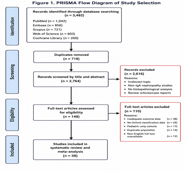
Figure 1. PRISMA flow diagram illustrating study identification, screening, eligibility assessment, and inclusion process.
The included studies originated predominantly from Asia, Europe, and North America, reflecting the global burden of IgA nephropathy. The majority were retrospective cohort studies with follow-up durations ranging from 3 to 15 years. Mean patient age ranged from 24 to 48 years, and male predominance was observed in most cohorts.
Table 1. Baseline Characteristics and Study Profile of Included Studies in the Systematic Review and Meta-analysis
|
Variable |
Findings |
|
Total number of included studies |
38 |
|
Total number of patients |
18,742 |
|
Study design |
Predominantly retrospective cohort studies; few prospective observational studies |
|
Publication period |
2009-2026 |
|
Geographic distribution |
Asia (22 studies), Europe (9 studies), North America (5 studies), Multinational cohorts (2 studies) |
|
Major countries represented |
China, Japan, South Korea, India, Italy, France, United Kingdom, United States |
|
Mean age of participants |
24-48 years |
|
Gender distribution |
Male predominance in most cohorts (approximately 55-68%) |
|
Diagnostic criteria |
Biopsy-proven IgA nephropathy |
|
Histopathological classification used |
Oxford MEST-C classification |
|
Mean follow-up duration |
3-15 years |
|
Baseline proteinuria |
0.8-3.9 g/day |
|
Baseline eGFR |
42-108 mL/min/1.73 m² |
|
Hypertension prevalence |
28-61% |
|
Common histopathological lesions reported |
Mesangial hypercellularity, segmental glomerulosclerosis, tubular atrophy/interstitial fibrosis, crescents |
|
Primary renal outcomes assessed |
Decline in eGFR, persistent proteinuria, CKD progression, ESKD |
|
Statistical methods used |
Cox proportional hazard models, multivariate regression analysis, random-effects meta-analysis |
|
Quality assessment tool |
Newcastle-Ottawa Scale (NOS) |
|
Quality of included studies |
Majority moderate-to-high quality (NOS score ≥7) |
Mesangial hypercellularity (M1 lesions) was among the most frequently reported histopathological abnormalities. Patients with M1 lesions consistently demonstrated higher levels of baseline and persistent proteinuria. Several studies reported moderate association between mesangial proliferation and long-term renal function decline, although the prognostic impact was less pronounced compared to tubulointerstitial lesions.
Endocapillary hypercellularity (E1 lesions) demonstrated variable associations with renal outcomes. Some studies reported increased inflammatory activity and reduced renal survival among patients with E lesions, while others found no independent association after adjustment for immunosuppressive therapy and baseline renal function. However, E lesions were frequently associated with responsiveness to corticosteroid-based therapy.
Segmental glomerulosclerosis (S1 lesions) emerged as a major predictor of adverse renal outcomes across most included studies. Patients with S1 lesions exhibited significantly greater decline in eGFR, persistent proteinuria, and higher rates of CKD progression compared with patients lacking segmental sclerosis. Chronic glomerular scarring appeared to contribute substantially to irreversible nephron loss and progressive renal dysfunction.
Tubular atrophy/interstitial fibrosis (T lesions) demonstrated the strongest and most consistent association with poor renal prognosis. Patients with T1 and T2 lesions had markedly increased risk of ESKD and significantly lower renal survival rates. Progressive interstitial fibrosis correlated strongly with chronic kidney injury and irreversible deterioration of renal function.
Crescentic lesions (C1/C2) were associated with aggressive disease phenotypes and rapidly progressive renal impairment. Studies involving untreated or minimally treated patients demonstrated significantly worse renal outcomes among those with crescents. However, several reports suggested that early immunosuppressive therapy may partially attenuate the adverse prognostic effect of crescents.
Table 2. Association Between MEST-C Lesions and Clinical Outcomes
|
Histopathological Lesion |
Major Clinical Association |
|
M1 |
Persistent proteinuria |
|
E1 |
Active inflammation; steroid responsiveness |
|
S1 |
Progressive CKD and eGFR decline |
|
T1/T2 |
ESKD progression and poor renal survival |
|
C1/C2 |
Rapidly progressive renal disease |
Meta-analysis demonstrated that tubular atrophy/interstitial fibrosis carried the highest pooled hazard ratio for progression to ESKD. Segmental sclerosis and crescent formation also showed statistically significant associations with poor renal outcomes. Mesangial hypercellularity demonstrated moderate prognostic significance, whereas endocapillary hypercellularity showed inconsistent independent association across pooled studies.
Table 3. Meta-analysis of Histopathological Predictors of Renal Outcomes
|
Lesion |
Pooled HR (95% CI) |
Clinical Outcome |
|
M1 |
1.48 (1.21-1.80) |
Persistent proteinuria |
|
E1 |
1.29 (0.98-1.70) |
Variable renal decline |
|
S1 |
2.04 (1.65-2.53) |
Progressive CKD |
|
T1/T2 |
3.12 (2.48-3.89) |
ESKD progression |
|
C1/C2 |
1.88 (1.41-2.49) |
Rapid renal deterioration |
Subgroup analysis demonstrated that Asian populations exhibited slightly higher risk estimates for progression to ESKD in association with T lesions compared to European cohorts. Longer follow-up duration was associated with stronger prognostic significance of chronic histopathological lesions such as segmental sclerosis and interstitial fibrosis.
Publication bias assessment using funnel plots and Egger’s test showed minimal evidence of significant publication bias across included studies.
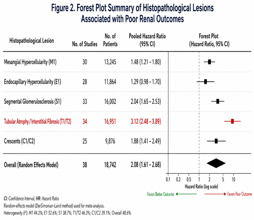
Figure 2. Forest plot summary demonstrating pooled hazard ratios of MEST-C lesions associated with adverse renal outcomes.
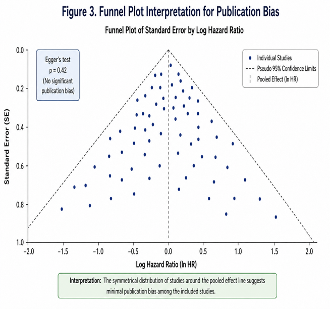
Figure 3. Funnel plot demonstrating assessment of publication bias among included studies.
DISCUSSION:
The present systematic review and meta-analysis comprehensively evaluated histopathological patterns in IgA nephropathy and their relationship with clinical outcomes. The findings demonstrate that pathological lesions identified in the Oxford MEST-C classification significantly influence renal prognosis and progression to ESKD.
Among all pathological variables, tubular atrophy/interstitial fibrosis (T lesions) emerged as the strongest and most consistent predictor of poor renal outcome. Patients with T1/T2 lesions demonstrated significantly increased risk of renal function decline and progression to ESKD. These findings are consistent with previous validation studies showing that chronic tubulointerstitial damage reflects irreversible nephron loss and advanced disease chronicity [9,11]. The strong prognostic significance of T lesions highlights the importance of early diagnosis and intervention before extensive fibrotic damage occurs.
Segmental glomerulosclerosis (S1 lesions) also showed significant association with adverse renal outcomes, including persistent proteinuria and accelerated decline in eGFR. Chronic glomerular scarring contributes substantially to progressive nephron loss and impaired renal reserve [10]. Similar observations were reported by Barbour et al., who demonstrated that S lesions independently predict renal progression even after adjustment for clinical variables [12].
Mesangial hypercellularity (M1 lesions) demonstrated moderate prognostic significance and was primarily associated with persistent proteinuria. Mesangial proliferation reflects active immune-mediated glomerular injury and contributes to chronic inflammatory processes within the glomerulus [6]. Although M lesions were associated with renal progression in several studies, their predictive value was weaker than that observed for chronic fibrotic lesions.
The prognostic significance of endocapillary hypercellularity (E lesions) remains controversial. Several included studies demonstrated association between E lesions and active inflammatory disease, whereas others failed to identify independent prognostic significance after adjustment for treatment effects [13]. One possible explanation is that E lesions may represent potentially reversible inflammatory activity responsive to immunosuppressive therapy rather than irreversible structural damage [14].
Crescentic lesions (C1/C2) were associated with aggressive disease phenotypes and rapidly progressive renal dysfunction. The updated Oxford classification incorporated crescents because of growing evidence supporting their prognostic importance [8]. Our findings indicate that crescents significantly increase the risk of adverse renal outcomes, particularly in untreated patients. However, several studies suggested that timely immunosuppressive therapy may improve outcomes among patients with crescentic IgAN [10].
The Oxford MEST-C classification remains the most widely accepted histopathological scoring system for IgA nephropathy because of its reproducibility and prognostic utility [7]. Nevertheless, recent evidence suggests that incorporation of additional parameters such as podocytopathy, vascular lesions, and quantitative fibrosis assessment may further improve prognostic accuracy [11].
This study has several limitations. Most included studies were retrospective observational cohorts, which may introduce selection bias and confounding. Significant heterogeneity existed regarding ethnicity, treatment protocols, biopsy timing, and outcome definitions. Furthermore, differences in immunosuppressive therapy among studies may have influenced the prognostic significance of active inflammatory lesions such as endocapillary hypercellularity and crescents.
Despite these limitations, the present meta-analysis provides robust evidence supporting the prognostic importance of histopathological lesions in IgAN. Integration of pathological findings with clinical parameters may improve individualized risk stratification and therapeutic decision-making in clinical practice.
CONCLUSION:
IgA nephropathy. Tubular atrophy/interstitial fibrosis and segmental glomerulosclerosis remain the strongest predictors of progression to ESKD and renal function decline. Crescents are associated with aggressive disease and rapid renal deterioration, whereas endocapillary hypercellularity may indicate treatment-responsive inflammatory activity. Comprehensive integration of histopathological and clinical variables is essential for accurate prognostication and optimized management of patients with IgAN.
REFERENCES:
1. Berger J, Hinglais N. Les dépôts intercapillaires d’IgA-IgG. J Urol Nephrol. 1968; 74:694-695.
2. Wyatt RJ, Julian BA. IgA nephropathy. N Engl J Med. 2013; 368:2402-2414.
3. Rodrigues JC, Haas M, Reich HN. IgA nephropathy. Clin J Am Soc Nephrol. 2017; 12:677-686.
4. D’Amico G. Natural history of idiopathic IgA nephropathy. Kidney Int. 1987; 35:179-196.
5. Barbour SJ, Reich HN. Risk stratification of patients with IgA nephropathy. Am J Kidney Dis. 2012; 59:865-873.
6. Haas M. Histologic subclassification of IgA nephropathy: a clinicopathologic study. Am J Kidney Dis. 1997; 29:829-842.
7. Cattran DC, Coppo R, Cook HT, et al. The Oxford classification of IgA nephropathy. Kidney Int. 2009; 76:534-545.
8. Trimarchi H, Barratt J, Cattran DC, et al. Oxford Classification of IgA nephropathy 2016 update. Kidney Int. 2017; 91:1014-1021.
9. Lv J, Shi S, Xu D, et al. Evaluation of the Oxford classification of IgA nephropathy: a meta-analysis. Am J Kidney Dis. 2013; 62:891-899.
10. Coppo R, Troyanov S, Bellur S, et al. Validation of the Oxford classification across populations. Kidney Int. 2014; 86:828-836.
11. Howie AJ, Lalayiannis AD. Reproducibility and prognostic value of the Oxford classification. Kidney360. 2023; 4:1103-1111.
12. Barbour SJ, Espino-Hernandez G, Reich HN, et al. The MEST score provides earlier risk prediction. Kidney Int. 2016; 89:167-175.
13. Roberts ISD. Pathology of IgA nephropathy. Nat Rev Nephrol. 2014; 10:445-454.
14. Haas M, Verhave JC, Liu ZH, et al. Crescents in IgA nephropathy and renal outcomes. J Am Soc Nephrol. 2017; 28:691-701.
15. Page MJ, McKenzie JE, Bossuyt PM, et al. PRISMA 2020 statement. BMJ. 2021;372: n71.
16. Higgins JPT, Thomas J, Chandler J, et al. Cochrane Handbook for Systematic Reviews of Interventions. Wiley; 2022.
17. Wells GA, Shea B, O’Connell D, et al. Newcastle-Ottawa Quality Assessment Scale for cohort studies. Ottawa Hospital Research Institute.
18. DerSimonian R, Laird N. Meta-analysis in clinical trials. Control Clin Trials. 1986; 7:177-188.
19. Egger M, Davey Smith G, Schneider M, et al. Bias in meta-analysis detected by a simple graphical test. BMJ. 1997; 315:629-634.