VENTRICULOPERITONEAL SHUNT AS A CONTENT OF INGUINAL HERNIA: A RARE CASE REPORT.
- Gauravkumar Govindbhai Patel , Senior Resident, Department of General Surgery, B. J. Medical College, Ahmedabad, Gujarat, India.
- Nina M. Shah , Professor (Additional) & Head of Unit Department, Department of General Surgery, B. J. Medical College, Ahmedabad, Gujarat, India.
- Hardik B. Bhatt , Associate Professor, Department of General Surgery, B. J. Medical College, Ahmedabad, Gujarat, India.
- Sanjeev Kumar , Senior Resident, Department of General Surgery, B. J. Medical College, Ahmedabad, Gujarat, India.
Article Information:
Abstract:
Background: Objective: To report a rare case of migration of a ventriculoperitoneal shunt into the inguinal canal and scrotal sac in a paediatric patient with hydrocephalus, presenting as right inguinoscrotal swelling associated with hydrocele, and to highlight the importance of timely diagnosis and surgical management. Materials and Methods: A 3-year-old male child with a known history of hydrocephalus treated with ventriculoperitoneal shunt placement presented with swelling in the right inguinoscrotal region. Clinical examination and radiological investigations were performed to evaluate the swelling and determine the position of the shunt catheter. Surgical management was carried out by open right inguinal herniotomy. Intraoperatively, the distal tip of the ventriculoperitoneal shunt was identified within the hernia sac and reduced back into the peritoneal cavity, followed by herniotomy repair. Results: Clinical and imaging findings confirmed migration of the distal ventriculoperitoneal shunt catheter into the right inguinal canal and scrotal sac with associated hydrocele. The patient underwent successful surgical reduction of the shunt into the peritoneal cavity through open right inguinal herniotomy. Postoperative recovery was uneventful, with no evidence of shunt malfunction or recurrence during follow-up. Discussion: Migration of the distal ventriculoperitoneal shunt into the inguinal canal or scrotum is a rare but recognized complication in paediatric patients. Persistence of the processus vaginalis and increased intra-abdominal pressure are considered important contributing factors. Children are particularly susceptible due to the higher incidence of patent processus vaginalis. Patients commonly present with inguinoscrotal swelling, hydrocele, or signs of shunt dysfunction. Early diagnosis using clinical assessment and imaging is essential to avoid complications such as shunt obstruction, infection, and recurrence. Surgical management with herniotomy and repositioning of the catheter remains the treatment of choice and generally results in good outcomes.
Keywords:
Article :
INTRODUCTION:
An inguinal hernia occurs when intra-abdominal contents protrude through a weak point in the abdominal wall, most commonly through the inguinal canal.[5] Inguinal hernias demonstrate a bimodal age distribution, with peaks in early childhood and in elderly individuals.[5] Approximately two-thirds of paediatric inguinal hernias are indirect in nature and are associated with persistence of the processus vaginalis.[6]
Ventriculoperitoneal shunt implantation is the most commonly performed surgical procedure for the treatment of hydrocephalus.[1] Although generally safe and effective, several complications related to ventriculoperitoneal shunts have been reported, including infection, ascites, bowel perforation, catheter obstruction, and distal catheter migration.[1,7] Migration of the distal catheter into the scrotum through a patent processus vaginalis is an uncommon but recognised complication, particularly in the paediatric age group because of the relatively small peritoneal cavity and persistence of the processus vaginalis.[2,8]
CASE PRESENTATION
A 3-year-old male child presented with complaints of swelling in the right inguinoscrotal region for a duration of one week. The onset was sudden and associated with dull aching pain. There was no history of vomiting, constipation, abdominal distension, fever, or skin discoloration.
The child was a known case of hydrocephalus for which a ventriculoperitoneal shunt had been placed in December 2022.
Clinical Examination
On examination, the right side of the scrotum appeared enlarged compared to the left side. Palpation revealed a long, stick-like hard structure measuring approximately 4 cm × 0.3 cm, which was palpated separately from the testes. The upper border of the structure could not be reached on palpation. The swelling increased in size on straining and coughing. Bilateral testes were palpable separately.
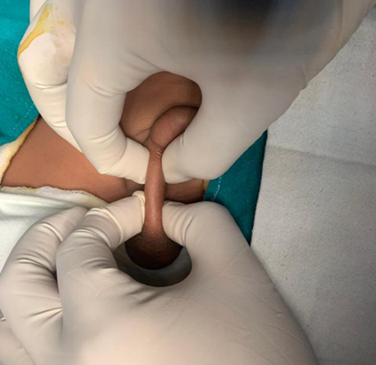
Figure 1: Clinical photograph showing right-sided inguinoscrotal swelling in a child with ventriculoperitoneal shunt migration.
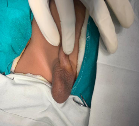
Figure 2: Preoperative appearance of the enlarged right scrotum prior to surgical exploration.
Laboratory and Radiological Findings
Ultrasonography suggested herniation of the ventriculoperitoneal shunt into the right scrotal region associated with hydrocele. Plain radiograph confirmed migration of the distal catheter into the inguinal canal and scrotal region.
Operative Findings and Management
The patient underwent open right inguinal herniotomy in August 2023. Intraoperatively, the distal tip of the ventriculoperitoneal shunt was identified as the content of the inguinal hernia sac. The catheter was carefully reduced back into the abdomen (peritoneal cavity), and herniotomy was completed successfully.
The postoperative period was uneventful, and the patient recovered well without evidence of shunt malfunction or recurrence during follow-up.
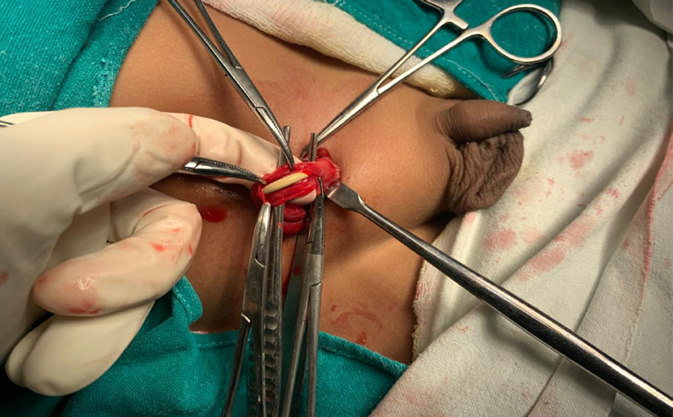
Figure 3: Close-up intraoperative photograph showing the ventriculoperitoneal shunt catheter identified as the content of the inguinal hernia sac during herniotomy.
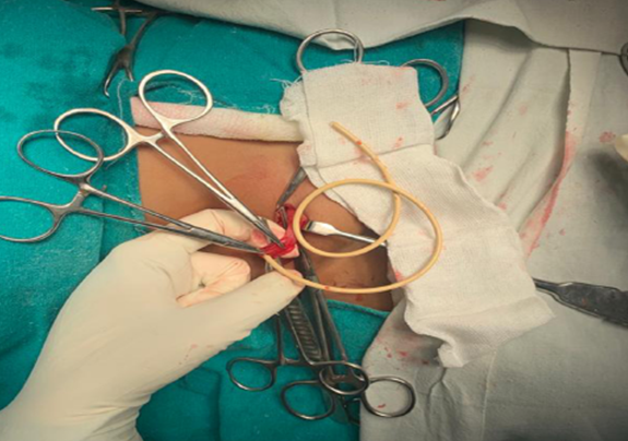
Figure 4: Intraoperative image demonstrating the distal ventriculoperitoneal shunt catheter herniating through the inguinal canal into the scrotal sac.
DISCUSSION:
Migration of a ventriculoperitoneal shunt into the inguinal canal or scrotum is a rare but important complication encountered predominantly in paediatric patients.[1,2] The proposed mechanism involves persistence of the processus vaginalis, increased intra-abdominal pressure, and the relatively small peritoneal cavity seen in children.[8]Most reported cases in the literature have occurred in infants and young children due to persistence of the processus vaginalis and increased intra-abdominal pressure following ventriculoperitoneal shunt placement.[2,8]
Among paediatric patients with ventriculoperitoneal shunts and newly diagnosed inguinal hernias, the incidence of shunt catheter incarceration has been reported to be as high as 20%.[7] Failure of spontaneous reduction may result in catheter obstruction, shunt malfunction, bowel complications, and recurrent hydrocele formation.[2,7]
Clinically, patients may present with inguinoscrotal swelling, hydrocele, shunt dysfunction, or symptoms related to raised intracranial pressure.[3] Imaging modalities such as ultrasonography and plain radiography are useful in confirming distal catheter migration and identifying associated complications.[3,8]
Early surgical intervention is recommended to avoid complications and preserve shunt function.[4] Herniotomy with reduction or repositioning of the distal catheter is generally sufficient in uncomplicated cases.[4] Some authors have also advocated simultaneous repair of contralateral patent processus vaginalis in selected paediatric patients due to the risk of bilateral occurrence.[6]
The present case is notable because of the rare occurrence of a ventriculoperitoneal shunt acting as the content of an inguinal hernia in a young child. Prompt diagnosis and timely surgical management resulted in a favourable outcome.
CONCLUSION:
Ventriculoperitoneal shunt migration into an inguinal hernia sac is an uncommon complication in paediatric patients with hydrocephalus. A high degree of clinical suspicion is required in children presenting with inguinoscrotal swelling following ventriculoperitoneal shunt placement. Appropriate imaging and early surgical management are essential to prevent shunt malfunction and recurrence.Early identification and prompt surgical intervention can prevent shunt-related complications and ensure favorable outcomes.
Conflict of Interest: None declared
Funding: None
Declaration of Patient Consent: The authors certify that they have obtained all appropriate patient consent forms.
Author Contributions: All authors contributed equally and approved the final manuscript.
REFERENCES:
1. Lee BS, Vadera S, Gonzalez-Martinez JA. Rare complication of ventriculoperitoneal shunt: early onset of distal catheter migration into scrotum in an adult male. Int J Surg Case Rep. 2015;6:198-202.
2. Bawa M, Garge S, Garg R, Narasimha Rao KL. Scrotal migration of tubing: an unusual complication after ventriculoperitoneal shunt. Asian J Neurosurg. 2017;12:738-740.
3. Grosfeld JL, Cooney DR. Inguinal hernia after ventriculoperitoneal shunt for hydrocephalus. J Pediatr Surg. 1974;9:311-315.
4. Clarnette TD, Lam SK, Hutson JM. Ventriculo-peritoneal shunts in children reveal the natural history of closure of the processus vaginalis. J Pediatr Surg. 1998;33(3):413-416.
5. Burcharth J, Pedersen M, Bisgaard T, Pedersen C, Rosenberg J. Nationwide prevalence of groin hernia repair. PLoS One. 2013;8(1):e54367.
6. Brandt ML. Pediatric hernias. Surg Clin North Am. 2008;88(1):27-43.
7. Kwok CK, Yue CP, Wen HL. Bilateral inguinal hernia after ventriculoperitoneal shunt for hydrocephalus. J Pediatr Surg. 1986;21(1):46-47.
8. Ozveren B, Cetin H, Emre S, et al. Migration of ventriculoperitoneal shunt catheter into the scrotum: case report and review of the literature. Pediatr Neurosurg. 1999;31(6):313-315.