Low-Grade Appendiceal Mucinous Neoplasm: A Clinicopathological Study Highlighting the Pathology that Changes Surgical Destiny.
- Meena N Jadhav , Professor, Department of Pathology, D. Y. Patil Medical College, Kolhapur, Maharashtra, India.
- Sandeep Yadav , Assistant Professor, Department of Pathology, D. Y. Patil Medical College, Kolhapur, Maharashtra, India.
- Aishwarya Kolte , JR3, Department of Pathology, D. Y. Patil Medical College Kolhapur, Maharashtra, India.
- Suchita Deshmukh , HOD, Department of Pathology, D. Y. Patil Medical College, Kolhapur, Maharashtra, India.
Article Information:
Abstract:
Background: Low-grade appendiceal mucinous neoplasm (LAMN) is a rare epithelial tumor of the appendix characterized by mucin-producing epithelium with low-grade cytologic atypia and progressive luminal distension. Despite its relatively indolent appearance, LAMN may disseminate mucin into the peritoneal cavity and cause pseudomyxoma peritonei. Aim: To analyze the clinicopathological characteristics of low-grade appendiceal mucinous neoplasm and highlight its surgical implications. Materials and Methods: A retrospective analysis of eight cases of LAMN was conducted. Appendectomy specimens were fixed in formalin, processed routinely, and stained with hematoxylin and eosin for histopathological examination. Clinical data, imaging findings, operative details, gross features, and microscopic findings were reviewed. Results: All specimens showed dilated appendiceal lumen filled with mucinous material. Histologically, tumors demonstrated low-grade mucinous epithelium with flattened or villous architecture, nuclear stratification, and mucin dissecting through the appendiceal wall. Acellular extra-appendiceal mucin was noted in some cases. No invasive carcinoma was identified. Conclusion: LAMN is an uncommon appendiceal tumor with important surgical implications. Accurate pathological diagnosis helps guide appropriate surgical management and prevent overtreatment.
Keywords:
Article :
INTRODUCTION:
Low-grade appendiceal mucinous neoplasm (LAMN) is a rare epithelial tumor arising from mucin-secreting cells of the appendix and represents a distinct pathological entity among appendiceal mucinous tumors. It is characterized by mucinous epithelial proliferation with low-grade cytologic atypia and progressive accumulation of mucin within the appendiceal lumen¹. Although histologically indolent, LAMN has the potential to disseminate mucin and neoplastic cells into the peritoneal cavity, leading to pseudomyxoma peritonei, a condition associated with significant morbidity and complex surgical management².
Appendiceal tumors are relatively uncommon and are detected in approximately 1% of appendectomy specimens. Among these tumors, mucinous neoplasms represent a small but clinically significant group that requires careful pathological evaluation³. LAMN typically affects adults in middle to older age groups, although cases have been reported across a wide age range. Clinically, patients often present with nonspecific symptoms such as right lower quadrant abdominal pain or features mimicking acute appendicitis, resulting in incidental diagnosis during appendectomy⁴.
The pathogenesis of LAMN involves abnormal proliferation of mucin-producing epithelial cells leading to progressive luminal dilatation and mucin accumulation. Persistent mucin production may weaken the appendiceal wall and predispose it to rupture, allowing mucin to disseminate into the peritoneal cavity⁵. Histopathologically, these tumors demonstrate low-grade cytologic atypia, villous or flattened mucinous epithelium, mural fibrosis, and extracellular mucin deposition within the appendiceal wall⁶.
Accurate pathological diagnosis is essential because management strategies differ from those used for other gastrointestinal malignancies. When the tumor remains confined to the appendix, simple appendectomy is usually adequate treatment. However, if extra-appendiceal mucin or peritoneal dissemination is present, more extensive surgical intervention may be required⁷. Therefore, recognition and appropriate classification of LAMN are critical for guiding surgical management and preventing unnecessary overtreatment.
MATERIALS AND METHODS:
The retrospective study was conducted at a tertiary care hospital. 80 cases of low-grade appendiceal mucinous neoplasm diagnosed in the pathology department were taken into consideration for the study. Ethical clearance was obtained from the Institutional Ethics Committee before starting the study.
Appendectomy specimens were fixed in 10% formalin for 24–48 hours. Representative sections were taken from areas showing luminal dilatation or mucin accumulation. Tissue sections of 4–5 μm thickness were prepared and stained with hematoxylin and eosin.
Clinical information including age, sex, presenting symptoms, radiological findings, operative notes, and specimen size were obtained from hospital records. Gross examination included evaluation of appendiceal size, wall thickness, mucin accumulation, and evidence of perforation. Microscopic examination assessed epithelial architecture, cytologic atypia, mural fibrosis, extracellular mucin, and extra-appendiceal spread.
CASE PRESENTATION
Eight patients diagnosed with LAMN were analyzed. Most patients presented with right lower quadrant abdominal pain suggestive of appendicitis.
Case 1: A 29-year-old male presented with abdominal pain for ten days. The appendectomy specimen measured 4 cm in length. The external surface was congested and the lumen was markedly dilated.
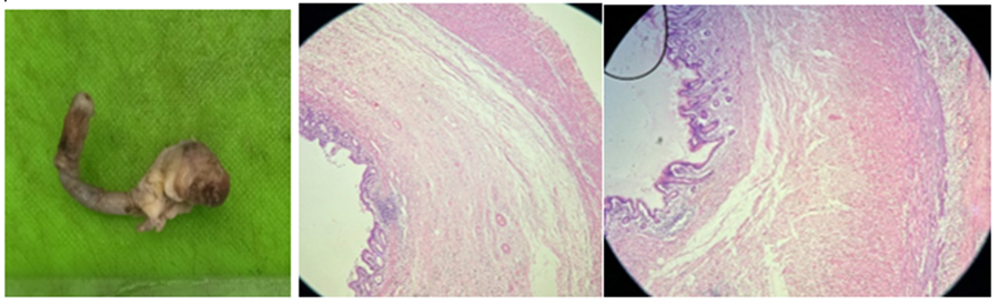
Figure 1 Figure 2: H & E staining, 4x; Figure 3: H & E staining, 4x
Figure 1: Gross photograph of appendicular mass at the tip.
Figure 2: Microscopic picture showing dilated lumen with flattened mucosa
Figure 3: Microscopic picture showing flattened mucosa with focal and undulating mucosa with mucin invading the muscularis propria and serosa.
Case 2: A 25-year-old female presented with abdominal pain for one month. The appendix measured 5 cm and showed mucinous material within the dilated lumen.
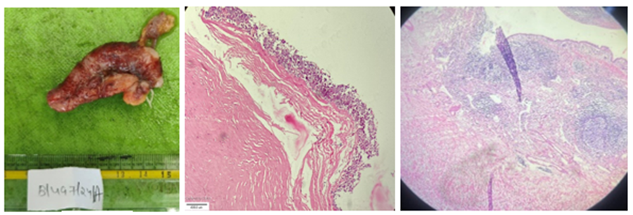
Figure 4 Figure 5: H & E staining, 4x; Figure 6: H & E staining, 4x
Figure 4: Gross photograph showing dilated appendicular mass
Figure 5: Microscopic picture showing disruption in muscularis mucosa
Figure 6: Microscopic picture showing mucin in the muscle layer
Case 3: A 32-year-old male presented with acute abdominal pain. Gross examination revealed a dilated appendicular mass measuring 4 × 3 × 3 cm filled with mucinous material.
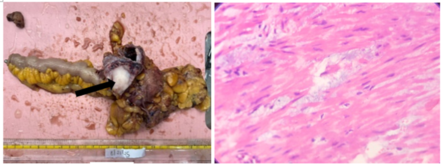
Figure 7 Figure 8: H & E staining, 40x
Fig.7: Gross photograph hemicolectomy specimen showing dilated appendicular mass filled with mucin(marked with arrow).
Fig.8: Microscopic picture showing mucin in the muscle layer
Case 4: A 35-year-old male underwent resection of an appendicular mass at the ileocecal junction measuring 4 × 3 × 3 cm.
Histopathological examination in all cases showed mucinous epithelial proliferation with nuclear stratification, mucin pools within the appendiceal wall, and disruption of muscularis mucosa. No invasive carcinoma was identified.
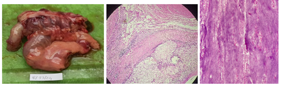
Figure 9; Figure 10: H & E staining, 4x; Figure11: PAS staining, 10x
Figure 9: Gross photograph showing appendicular mass with ileum, cecum and ascending colon.
Figure 10: Submucosa shows abundant mucin with atrophied lymphoid follicles.
Figure 11: PAS positive stained mucin material
Radiological and CT Findings
Radiological imaging played an important role in preoperative evaluation. Ultrasound in several patients revealed a cystic tubular structure in the right iliac fossa suggestive of appendiceal mucocele. Computed tomography demonstrated a dilated appendix filled with low-attenuation mucinous material. The appendiceal wall appeared smooth and mildly thickened. In some cases, CT imaging showed a localized appendicular mass near the ileocecal junction measuring approximately 4 × 3 × 3 cm without evidence of distant metastasis.
RESULTS:
Table 1: Clinicopathological Summary of Cases
|
Case |
Age |
Sex |
Clinical Presentation |
Specimen Size |
Gross Findings |
Histopathological Findings |
|
1 |
29 |
M |
Abdominal pain (10 days) |
4 cm appendix |
Dilated lumen |
Villous mucinous epithelium, mucin pools |
|
2 |
25 |
F |
Abdominal pain (1 month) |
5 cm appendix |
Mucinous lumen |
Flattened mucosa, nuclear stratification |
|
3 |
32 |
M |
Acute abdominal pain |
4×3×3 cm mass |
Dilated lumen |
Mucin dissecting muscularis |
|
4 |
35 |
M |
Abdominal mass |
4.5 cm appendix |
Appendicular mass |
Mucin pools with fibrosis |
|
5 |
– |
– |
Abdominal pain |
– |
Dilated appendix |
Low-grade mucinous epithelium |
|
6 |
– |
– |
Suspected appendicitis |
– |
Mucin-filled lumen |
Mural fibrosis |
|
7 |
– |
– |
Right iliac fossa pain |
– |
Dilated appendix |
Acellular mucin |
|
8 |
– |
– |
Appendiceal mass |
– |
Mucin-filled lumen |
LAMN features |
Gross examination showed dilated appendices filled with mucinous material in all cases. Microscopically, tumors demonstrated low-grade mucinous epithelium with villous or flattened architecture. Nuclear stratification and mucin dissecting through the appendiceal wall were observed. Acellular extra-appendiceal mucin was present in some cases, but none showed high-grade dysplasia or invasive carcinoma.
DISCUSSION:
Mucinous tumors of the appendix may exhibit features that indicate a risk of peritoneal dissemination. When mucin is identified grossly on the serosal surface or within the mesoappendix, the entire specimen should be thoroughly submitted for histopathological examination to determine the presence or absence of extra-appendiceal neoplastic epithelium.
On microscopy, these tumors characteristically show villous architecture with pushing or effacement of the muscularis mucosae, often accompanied by mural atrophy, fibrosis, and/or calcification. The lesion may produce diverticulum-like outpouchings or even result in perforation of the appendix. In such cases, important differential diagnoses include retention cyst, reactive or hyperplastic epithelium, and appendiceal diverticulum.
Low-grade appendiceal mucinous neoplasm (LAMN) must be distinguished from mucinous adenocarcinoma, as the prognosis and management differ significantly. Grossly, LAMN typically presents as a sausage-like dilatation of the appendix. Microscopically, it shows flat or villous mucinous epithelium with low-grade dysplasia. The underlying appendiceal wall is often fibrotic and may exhibit calcification. Inflammatory infiltrate is usually sparse, although mucin may extend into the omentum or pelvis.
The survival rate in such cases may extend from 5 to 10 years or longer after initial presentation. Additionally, certain invasive appendiceal lesions may demonstrate localized spread of mucin without identifiable neoplastic epithelium; these low-risk lesions should be carefully distinguished from LAMN.
Thus LAMN is a distinct appendiceal tumor characterized by mucin production and luminal dilatation. Despite low-grade histologic features, it may cause serious complications such as pseudomyxoma peritonei if mucin escapes into the peritoneal cavity.
Early and accurate diagnosis is therefore essential. For tumors confined to the appendix, appendectomy is usually sufficient treatment. However, cases with peritoneal dissemination may require cytoreductive surgery and intraperitoneal chemotherapy. Pathological evaluation plays a critical role in determining the extent of disease and guiding surgical management.
CONCLUSION:
Low-grade appendiceal mucinous neoplasm is an uncommon but clinically significant appendiceal tumor. Although histologically low grade, it carries the potential risk of pseudomyxoma peritonei. Careful pathological examination is essential for accurate diagnosis and for guiding appropriate surgical management. Early recognition helps prevent unnecessary extensive surgery and ensures proper follow-up.
REFERENCES:
1. Misdraji J. Low-grade appendiceal mucinous neoplasms: clinicopathologic features and prognosis. Am J Surg Pathol. 2003;27(8):1089-1103.
2. Carr NJ, Cecil TD, Mohamed F, Sobin LH, Sugarbaker PH, Gonzalez-Moreno S, et al. A consensus for classification and pathologic reporting of pseudomyxoma peritonei and associated appendiceal neoplasia. Am J Surg Pathol. 2016;40(1):14-26.
3. Xiao J, Li P, Liu W. Analysis of clinical characteristics of low-grade appendiceal mucinous neoplasm: a retrospective cohort study of 51 patients. J Investig Surg. 2021;34(7):721-727.
4. Zhuang CC, Yu J, Yang Q, Zhou S, Dong W. Unusual giant low-grade appendiceal mucinous neoplasm: a case report and literature review. Medicine (Baltimore). 2023;102(3): e32745.
5. Sugarbaker PH. Epithelial appendiceal neoplasms and pseudomyxoma peritonei: a clinical review. J Surg Oncol. 2010;101(8):731-738.
6. Pai RK, Longacre TA. Appendiceal mucinous tumors and pseudomyxoma peritonei: histologic classification and clinical implications. Arch Pathol Lab Med. 2005;129(8):1006-1012.
7. Rosai J. Rosai and Ackerman’s Surgical Pathology. 11th ed. Philadelphia: Elsevier; 2018.
8. Lester SC. Manual of Surgical Pathology E-Book: Manual of Surgical Pathology E-Book. Elsevier Health Sciences; 2010 Jul 23.
9. Fletcher CD. Diagnostic histopathology of tumors: 2-volume. Elsevier Health Sciences; 2007 Mar 29.