Perioperative Management of Pulmonary Embolism due to Bilateral Femur Fractures – A Case Report.
- Geetha J. , Professor, Department of Anaesthesia, Dhanalakshmi Srinivasan Medical College and Hospital, Siruvachur, Perambalur, Tamil Nadu, India.
- Ilayaraja B.U. , Postgraduate of Anaesthesiology, Dhanalakshmi Srinivasan medical college, Siruvachur, Perambalur, Tamil Nadu, India.
- Dileepan Prassanna M. , Postgraduate of Anaesthesiology, Dhanalakshmi Srinivasan medical college, Siruvachur, Perambalur, Tamil Nadu, India.
Article Information:
Abstract:
INTRODUCTION: Road traffic accidents are the major causes of musculoskeletal trauma and fractures of extremities form essential components of skeletal trauma of which femoral fractures account for around 13% of all adult fractures. 50% of these adult femur fractures occur around diaphysis.[1] Bilateral femoral fractures contribute to around 2–7% of femoral shaft fractures.[2] Patients with multiple long bone fractures are at an increased risk of systemic complications, especially pulmonary failure. Bilateral femur fractures have a high mortality rate of 9.8% without other associated injuries and 31.6% when associated with other injuries.[3] Aggressive and prompt management of such patients including early fracture fixation and mobilisation could reduce serious complications such as deep vein thrombosis, fat and pulmonary embolism. This case report describes a young patient with bilateral femur fractures and dyspnoea who was managed to yield a successful outcome.
Keywords:
Article :
HISTORY:
24 years old male presented to the casualty following a two-wheeler collision road traffic accident. He was driving and hit by high-speed collision of another two-wheeler. He was thrown out and brought by the bystanders.
He was pale, tachycardic, tachypnoeic and dyspnoeic, hypoxic, conscious and oriented. He was unable to move his legs and exact time of the RTA was not known. His thighs were swollen with abrasions both lower limbs. He was unmarried with no systemic comorbidities. He had passed urine after the accident.
CLINICAL FEATURES
The patient was conscious, oriented, GCS 15/15, pallor was present. His pulse rate was 126 per minute, blood pressure 90/60 mmHg and SpO₂ was 80% on room air.
Cardiovascular system showed tachycardia and fine creps were auscultatable in both bases. Abdomen was soft and bowel sounds were heard. Both thighs were swollen; blackish macules and abrasions were found. Both distal lower limb pulses were felt equally. Primary and secondary survey proved the same findings.
Propped up position and oxygenation by NRBM mask were instituted, followed by IV volume replacement with crystalloids via 18G needle. 2 units of packed cells were arranged and started. His bladder was catheterised to note hourly urine output.
Table 1: Investigations
|
Hb 10.1g |
ABG |
|
TLC 9700 |
pH 7.53 |
|
PT 15.9 |
pCO2 24.75 |
|
INR 1.8 |
PaO2 55.1 |
|
APTT 27.8 |
HCO3 20.43 |
The observations were summarised as follows: hypoxic, decompensated respiratory alkalosis with internal blood loss of around 2.5 litres, deranged coagulation profile.
His X-ray chest showed abrupt cut-off of right pulmonary artery, enlarged right descending pulmonary artery, peripheral hyperlucency secondary to oligemia, cardiothoracic ratio of 55 percent. No fracture ribs and trachea was in the midline.
X-ray of limbs showed bilateral fractures of shaft of femur. According to Winquist and Hansen classification, the fracture was type II on the right femur and type III of the left femur.
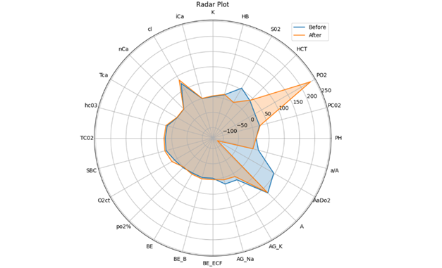
Figure 1: Radar plot of ABG before and after intervention
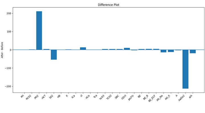
Figure 2: Difference plot of ABG before and after intervention
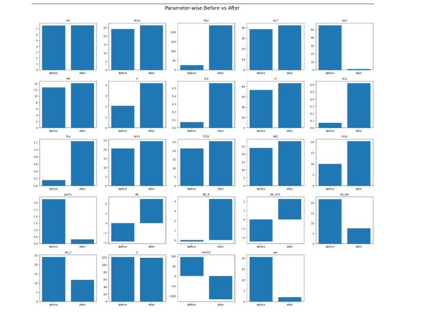
Figure 3: Box whisker plot of ABG before and after intervention
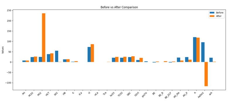
Figure 4: Bar diagram plot of ABG before and after intervention
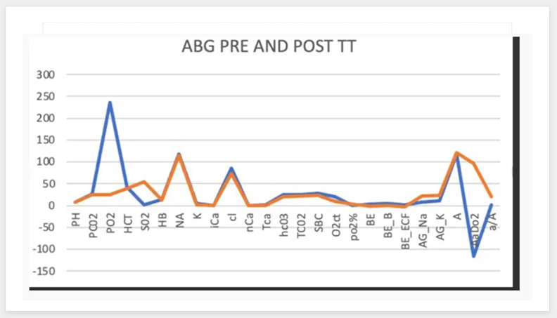
Figure 5: Graphical representation of ABG before and after intervention
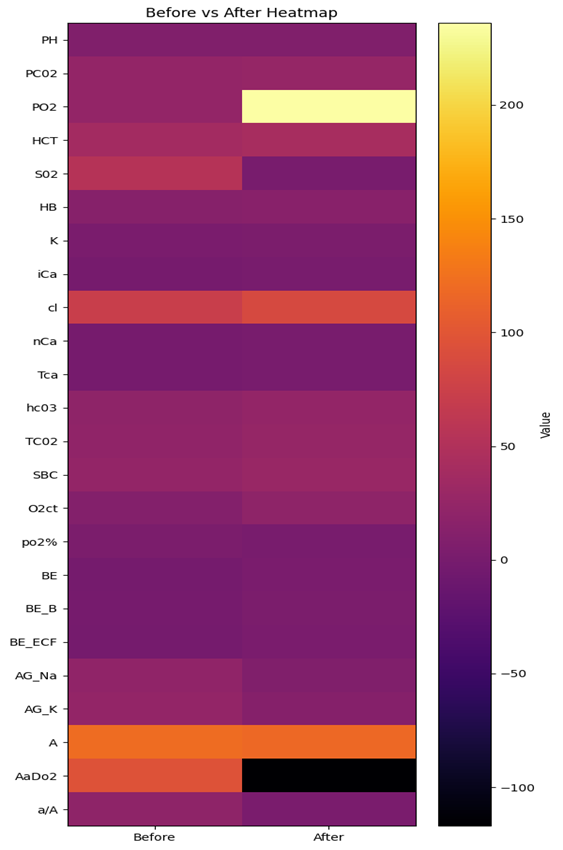
Figure 6: Heatmap of ABG before and after intervention
The fractures were of closed type with transverse minimal comminution on the right femur and oblique communited on the left femur at mid shaft level. There were no skin discolouration or purpuric spots on the fracture site.
The patient’s hypoxia improved by oxygenation, and in view of classical X-ray chest and clinical findings, an impending pulmonary embolism and haemorrhagic shock was concluded and resorted to early fixation of the fractures to avoid progression of the embolic syndrome. The presence of external abrasions and lacerations on the legs though fracture site was free, necessitated external fixation of the fractures.
Problems and risks
· Polytrauma
· Impending haemorrhagic shock
· Hypoxia
· Sepsis
· Altered coagulation profile
· Pulmonary embolism
· Immobilisation of bilateral fractures
Goals
The patient is hemodynamically unstable with impending haemorrhagic shock and pulmonary embolism. The foremost step is to prevent further haemorrhage and progress of embolism by fixing the fractures.
Plan
External fixation and general anaesthesia
Preparation and optimisation
1. Oxygen therapy – Gradual reduction of FiO₂ to maintain SpO₂ by Venti mask alone – maintained at 4 L/min.
2. Anti-coagulant therapy – Heparin 5000 units intravenously 8th hourly with periodic check of APTT and INR.
3. Monitor and maintain hourly urine output at 0.5 to 1 ml/kg/hour to detect haematuria or oliguria due to crush injury syndrome.
4. 2 units of packed cells and 2 units of fresh frozen plasma were transfused preoperatively to combat blood loss.
5. Emotional support, counselling and motivation to manage the stress situation and fear of future were offered.
6. Appropriate antibiotics and informed consent regarding blood loss, use of blood and blood products and elective ventilation was obtained.
Conduct of Anaesthesia
Patient was induced with Inj. Ketamine 50, Glycopyrrolate 0.2 mg, Propofol 100 mg, Fentanyl 100 µg and intubated with 8 mm cuffed oral endotracheal tube on paralysing with 50 mg of vecuronium. Heparin was withheld 6 hours before procedure to be restarted 6 hours after the procedure. Anaesthesia was maintained with vecuronium and intermittent positive ventilation with a PEEP of 6 cm of water. Following external fixation, the patient was electively ventilated for 24 hours. Good improvement in blood gas values including PaO₂ increased from 84 mmHg to 236 mmHg while SpO₂ increased from 55% to 99%. The patient’s respiratory shallow breathing index was less than 80 and patient was extubated and closely observed. Heparin therapy continued and after three days patient underwent definitive procedure namely interlocking intramedullary nailing under general anaesthesia.
The patient’s intraoperative period was uneventful and he was extubated on the table and closely observed through arterial blood gas measurements for two days. He was started on oral aspirin and discharged after one week in a well-being state.
This case is presented to highlight aggressive initiation of treatment following high degree of suspicion and graded manner of definitive surgery could save the patient from a stormy intra and postoperative period apart from reducing mortality.
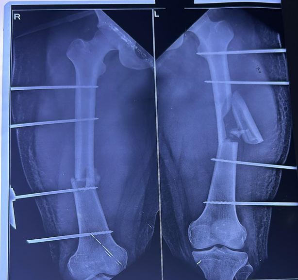
Figure 7: Post-op X-ray after External fixation Surgery
DISCUSSION:
Long bone fractures can be considered as one of the most common causes of non-fatal injuries sustained due to trauma.[4] The incidence of long bone fractures is roughly estimated to be 406 per 1 lakh population each year worldwide.[5] Femoral shaft fractures contribute to 13% of these. The management of isolated diaphyseal long bone fracture in adults involves definitive intramedullary fixation followed by early mobilisation. However, the damage caused by immediate definitive surgery may outweigh it benefits and causes serious systemic complications. In a retrospective study of Copeland et al,[6] reported an increased risk of mortality and acute respiratory distress syndrome (15.7%) in bilateral shaft femur fractures when compared to unilateral fractures with 11.7% and 7.7% respectively. Bonevaulle et al[7] evaluated 40 patients with bilateral femoral fractures out of which 3 patients developed fat embolism. This accounts for 7.5% whereas the incidence of ARDS in bilateral femur fractures is as high as 43% according to Kaman et al.[8]
The injury severity score may underestimate the severity of injury in patients with multiple extremity fractures as it does not take into account the other injuries in the same body region. The injuries of head, chest and abdomen if severe get the extremity fractures out of focus in various scoring systems. Various trauma scoring systems now in vogue such as[9] new injury severity score, revised trauma score and trauma severity combined injury severity score do not take into consideration the complexity of multiple extremity fractures and hence do not provide a reliable guide to time the definitive surgical fixation. Blood markers have been proposed to help in deciding a definitive surgery. Serum lactate levels mark tissue hypoperfusion in polytrauma and improvement could indicate the degree of resuscitation.[10] The concept of damage control orthopaedics concluded serum lactate measurements along with other clinical and laboratory adjuncts could help time the definitive surgery.[11]
Traffic light system - red, amber and green codes were devised by O’Toole et al[12] based on serial lactate measurements.
· Lactate more than 2.5 mmol/L - RED
· Means stop definitive surgery
· Lactate 2.5–2 mmol/L - AMBER
· Management depends on lactate trend
· Lactate less than 2 mmol/L - GREEN indicates early femoral nailing.
Intra-operative lactate levels could suggest an early sign of deterioration. Persistently elevated lactate levels is a predictor of increased mortality in trauma patients.[13]
Injury severity scoring systems have been designed to predict and correlate injuries to morbidity, mortality, hospital stay and timing of definitive surgery following initial resuscitation.
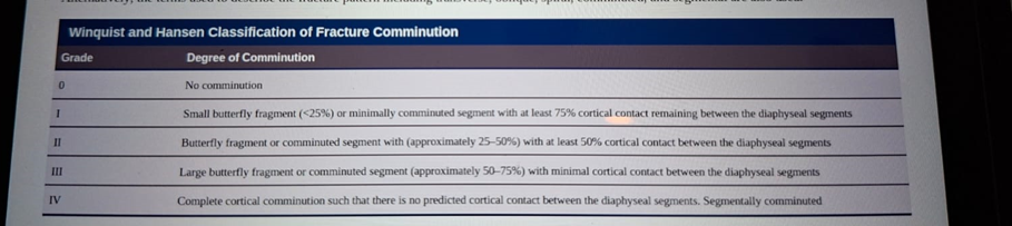
Figure 8: Winquist and Hansen Classification of Fracture Comminution
Scoring systems table
Anatomical Scoring Systems
* Abbreviated injury scale (AIS)
* Injury severity score (ISS)
* New injury severity score (NISS)
* Organ injury scale (OIS)
* Anatomic profile
* International Classification of Diseases (ICD-9) Injury Severity Score (ICISS)
Physiological Scoring Systems
* Revised trauma score
* Glasgow coma score
* APACHE scoring (Acute physiology and chronic health evaluation (APACHE I, II, III)
There are anatomical and physiological scoring systems. TRISS and ASCOT combine both anatomical and physiological scoring systems.
Injury Severity Score (ISS) takes values from 0 to 75. If an injury is assigned AIS of 6, it is unsurvivable injury, the ISS is 75. Major trauma is considered if ISS is more than 15.
Each injury is assigned an abbreviated injury scale (AIS) score and is allocated to six body regions. The highest AIS in each body region is used. The three most severely injured body regions have their score squared and added together to produce the ISS score.[14] Bolorunduro et al categorised and validated ISS as follows:[15]
· < 9: Mild
· 9–15: Moderate
· 16–24: Severe
· 25: Profound
Trauma and Injury Severity Scores TRISS
Determines the probability of survival Ps of a patient from ISS and RTS using the following formulae:
Ps=1/1+e-b
Where b is:
b = b0+b1 (RTS)+b2(ISS)+b3(Age Index)
b0, b1 and b2 are co-efficient derived from multiple regression analysis of major Trauma outcome study MTOS database.
Age Index is Zero if less than 54 years and one if more than 55.[16]
The New Injury Severity Score (NISS) simply takes the sum of squares of three most severe injuries regardless of body region injured.
Revised Trauma Score (RTS) is a physiological scoring system based on a set of data obtained from patients namely Glasgow Coma Scale, systolic blood pressure and respiratory rate.
The Orthopaedic Trauma Association classification is based on location and fracture pattern and appears more descriptive.[17]
The anatomic location is designated by two numbers with first number identifying the bone and second identifying location (proximal – one, midshaft – two and distal – three). Thus, a midshaft femur fracture is 32.
The fracture type is described based on the degree of comminution.
· Type A are simple
· Type B spiral / segmental
· Type C are complex with no cortical contact
Henceforth our patient could be classified as:
· 32 Type A Right or 32AR
· 32 Type C Left or 32CL
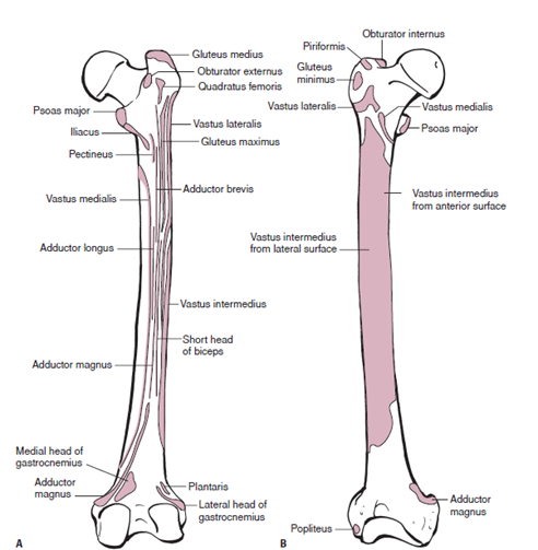
Figure 9: Muscular attachments of femur
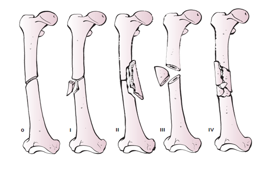
Figure 10: Winquist and Hansen classification of midshaft femoral fractures
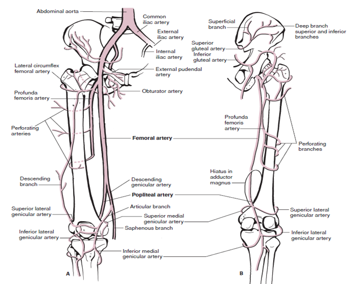
Figure 11: Vascular anatomy of thigh
The Patho anatomy of femoral fractures from figures 1, 2 and 3 indicate the severity, complexity and bleeding expected from major blood vessels.
External fixation is a well-established technique for rapid stabilisation of femoral shaft fracture in an unstable polytrauma patient.[18]
External Fixation of Femur Fractures: Indications and Contraindications are as follows:
Indications
· Severe soft tissue injuries with extensive contamination
· Evolving muscular crush that requires an extensive secondary debridement
· Medullary contamination
· Associated vascular injury requiring stabilization prior to repair
· Polytrauma or injuries that prevent other treatments; as a temporary bridge to femoral nailing (damage control orthopaedics)
· Infected non-unions requiring a metal holiday
Relative Contraindications
· No absolute contraindications exist but external fixation is uncommonly used as a definitive treatment
The patient with bilateral femur fracture was unstable and hypoxic and would not tolerate intramedullary nailing due to exacerbated bleeding and embolic phenomena.
Intramedullary nailing is the best surgical treatment to the advantage of this young patient with no chest or head injuries. Pulmonary issues and reaming would greatly affect the overall outcome.
Early mobility, limb strength and stability would be achieved by intramedullary nailing but the timing of fixation and reaming would have serious consequences.
Mooshed et al[19] noted reduction in mortality of almost 50% when delayed beyond 12 hours. They hypothesized the delay was for ensuring adequate resuscitation prior to definitive fixation. The concept of damage control orthopaedics grossly reduced postoperative mortality in diaphyseal fractures of the femur.
Approximately 5–10% of venous thromboembolism develops into pulmonary embolism. The presence of shock in the setting of pulmonary embolism is associated with five to sevenfold increase in mortality.[20]
Several cardiac causes such as cardiomyopathy, cardiac tamponade and aortic dissection may be the differential diagnosis, though the gold standard of diagnosis is the pulmonary angiogram, classical signs on X-ray chest PA and bedside 2-D echo could confirm the diagnosis. Initiation of therapy should not be delayed for confirmatory diagnostic testing.
Original and Simplified Pulmonary Embolism Severity Index (PESI)
Parameters utilized in each version of the PESI / sPESI
* Age: 1 point (if age older than 80 years)
* Male sex: +10 points / –
* Cancer: +30 points / 1 point
* Chronic heart failure: +10 points / 1 point
* Chronic pulmonary disease: +10 points / 1 point
* Pulse rate ≥110 bpm: +20 points / 1 point
* Systolic BP <100 mm Hg: +30 points / 1 point
* Respiratory rate >30 breaths/min: +20 points / –
* Temperature <36°C: +20 points / –
* Altered mental status: +60 points / –
* Arterial oxyhaemoglobin saturation <90%: +20 points / 1 point
Risk stratification in PESI
* Class I: Points ≤65; low 30-day mortality risk from 1% to 6%
* Class II: Points 66 to 85; low mortality risk from 1.7% to 3.5%
* Class III: Points 86 to 105; moderate mortality risk
* Class IV: Points 106 to 125; high mortality risk
* Class V: Points >125; high mortality risk from 10% to 24.5%
Risk stratification in sPESI
* If 0 points: 1.0% 30-day mortality risk
* If ≥1 points: 10.9% 30-day mortality risk
Complications associated with PE
* Recurrent thromboembolism
* Chronic thromboembolic pulmonary hypertension
* Right heart failure
Our patient could not afford an angiogram and had classical signs of pulmonary embolism on chest X-ray.
The specific signs include:
Westermark sign – focal oligemia
Hampton’s hump – wedge shaped opacity.
Palla’s sign – enlarged right descending pulmonary artery, often appearing as a sausage-shaped shadow.
Fleischner’s sign – enlargement of central pulmonary artery due to massive thrombus or pulmonary hypertension.
Knuckle sign – abrupt tapering or amputation of pulmonary artery.
Non-specific findings include pleural effusion, atelectasis, elevated diaphragm and cardiomegaly.
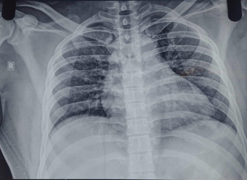
Figure 12: X-ray chest of our patient
Our patient had pulmonary oligemia, abrupt tapering of pulmonary artery, enlarged right descending pulmonary artery and cardiomegaly.
Mechanical shock can be precipitated by an array of syndromes that produce an acute loss of pulmonary vascular cross-sectional area, either by direct obstruction of pulmonary vasculature or through vasoconstriction by vasoactive mediators, the end result is rise in pulmonary vascular resistance that leads to right ventricular strain and failure.
The four major aetiologies of mechanical shock are massive pulmonary embolism, air embolism, fat embolism and amniotic fluid embolism.
The degree of hemodynamic compromise due to mechanical shock is determined by magnitude of pulmonary vascular obstruction or vasoconstriction, right ventricular performance and reserve and pre-existing cardiopulmonary disease.
The pulmonary circulation is normally a high capacitance, low resistance circuit. The right ventricle cannot acutely compensate for a mean pulmonary arterial pressure more than 40 mm Hg. In the absence of a cardiopulmonary derangement prior to the event, an increase in RV afterload and mean PAP is directly proportional to the magnitude of pulmonary vascular obstruction.
Post-traumatic pulmonary embolism following mid shaft fractures of femur are not only dislodgement of clots and bone marrow particles but also due to various chemical mediators released as metabolic response to trauma constitute the clinical presentation of pulmonary embolism.
Types of Pulmonary Embolism (PE)
Based on hemodynamic stability pulmonary embolism is categorized as:[21]
1. Massive PE or high-risk PE or hemodynamically unstable PE.
2. Sub massive PE or intermediate-risk PE where there is mild hypotension stabilized by fluid therapy.
3. Asymptomatic or low-risk PE.
Our patient belonged to the intermediate risk group with mild hypotension and mild right ventricular dysfunction.
Pathophysiology of PE
A thrombus entering the pulmonary circulation often enters lower lobes. Large emboli tend to obstruct main pulmonary artery causing saddle embolus with deleterious cardiovascular consequences. Smaller sized emboli block peripheral arteries and lead to pulmonary infarction manifested by intra-alveolar haemorrhage. Pulmonary capillary blood flow reduces. Ventilation perfusion mismatch ensues as alveolar ventilation remains the same. This leads to dead space ventilation and hypoxia.
Chemical mediators such as serotonin are released causing further vasospasm. Local accumulation of inflammatory mediators alters surfactant and stimulates respiratory drive resulting in tachypnoea, dyspnoea, hypocapnea leading to respiratory alkalosis.[22] If thromboembolic occlusion is more than 30% to 50% of the total cross-sectional area of the pulmonary arterial bed, pulmonary vascular resistance increases along with rise in RV afterload, impediment of RV filling and reduced cardiac output causing hypotension.[23]
The lung parenchyma receives its oxygen supply from three non-redundant sources:
· Deoxygenated blood from pulmonary arteries
· Oxygenated blood from bronchial arteries
· Direct oxygen diffusion from alveoli[24]
Any impediment to any of these sources results in pulmonary infarction.
Inflammatory mediators from ischemic parenchyma can further limit gas exchange following resultant vasoconstriction and bronchoconstriction.[25] When ischemia of lung tissue is not reversed promptly infarction ensues with 77 to 87% unilaterally with strong predilection to right lower lobe. This is thought as the influence of gravity on alveolar, pulmonary and bronchial arterial pressure.[26]
Evaluation of PE: The gold standard is CT pulmonary angiogram.
Arterial blood gas analysis: with alveolar arterial oxygen gradient, respiratory alkalosis and hypocapnia are common findings.
Brain natriuretic peptide: levels though with limited diagnostic value may suggest severity of right ventricular dysfunction.[27]
Troponin: Serum Troponin T levels are related to prognostic value. Higher the levels greater the risk of mortality.
D-Dimer levels: D-Dimer is the smallest end product of degradation of cross-linked fibrin by plasmin. Both ELISA and agglutination assays are available.
D-Dimer lacks specificity as it increases in: Venous thromboembolism, Arterial thrombosis, DIC, Pregnancy, Post surgery, Liver diseases, Malignancy. D-Dimer has high negative predictive value, that is to say that a negative D-Dimer indicates that morbidity and mortality risks are low and PE itself may be minimal or absent.[28] A negative ELISA D-Dimer and low clinical probability could exclude PE in approximately 30% of patients examined for PE. The specificity of D-Dimer increases with age. Age adjusted cut-off levels using the formula: Age × 10 ng/ml rather than standard D-Dimer cut-off of 500 ng/ml ruled out the possibility of pulmonary embolism without additional false negative findings.[29] The formula for D-Dimer is: Age × 10 ng/ml for patients older than 50. If age is 75 years: * Age adjusted D-Dimer is 750 ng/ml.
Electrocardiography: Often shows nonspecific abnormalities:[30] Sinus tachycardia, ST-T abnormalities, RV strain, RBBB, S1Q3T3.
Chest X-ray: May show many abnormalities including: Atelectasis, Effusion, Pulmonary infarction, Fracture ribs if present. The Westermark sign which is a sharp cut off pulmonary vessels with distal hypoperfusion in a segmental distribution within the lung and is especially specific in acute pulmonary embolism.[31]
CTPA
Computed tomographic pulmonary angiography is the diagnostic modality of choice with Sensitivity of 83% and Specificity of 90%. CTPA allows appropriate visualization of pulmonary arteries down to subsegmental level.[32] But the negative predictive value was only 58–60% with CTPA but a positive predictive value was 92–96%. Right ventricular enlargement which has significant predictive value in pulmonary embolism is also detectable by CTPA which could suggest overall outcome of the patients.
Pulmonary Angiography
The contrast is directly injected into right heart under fluoroscopy and imaging was done. Radiation risks and renal damage were two concerns in the above investigations. Magnetic resonance angiography is devoid of radiation risk but has low sensitivity and low availability in emergency settings and is not recommended as the first line test. MR angiography has a specificity of 92%.
Acute Pulmonary Embolism Diagnostic Criteria
Geneva scoring system offers points to clinical features and final scores are arrived and graded.
Geneva score
*Previous PE or DVT → 3 / 1 (original version / simplified version)
* Heart rate 75–94/min → 3 / 1
* 95/min → 5 / 2
* Surgery or fractures within one month → 2 / 1
* Haemoptysis → 2 / 1
* Active malignancy → 2 / 1
* Unilateral lower limb pain → 3 / 1
* Pain and unilateral lower limb edema → 4 / 1
* Age more than 65 years → 1 / 1
Three level score
* Low: 0–3 / 0–1
* Intermediate: 4–10 / 2–4
* High: More than 11 / more than 5
Two level score
* PE unlikely: 0–5 / 0–2
* PE likely: more than 6 and more than 3.
Our patient’s Geneva score was 11 and hence denoted pulmonary embolism
Wells criteria is another scoring system which is based on deep vein thrombosis. A Wells score more than 6 suggests high probability of pulmonary embolus.
Goals of management includes Hemodynamic stability, Oxygenation and Prevention of further dislodgement of thrombi by anticoagulation and thrombolysis. Heparin 5000 units intravenously every eighth hourly, catheter directed suction or rotational embolectomy, surgical embolectomy and vena cava filters have all been considered as modes of removing the embolus.
All patients with PE should be continued on anticoagulants for 3 to 6 months, periodic monitoring of coagulation profile, preventive measures for DVT are also essential. Original and simplified Pulmonary Embolism Severity Index.
Complications associated with Pulmonary Embolism Include
* Recurrent thromboembolism
* Chronic thromboembolic pulmonary hypertension
* Right heart failure
* Cardiogenic shock
Summary
This case report involves a 24-year-old male with bilateral femur fractures on the midshaft with anaemia, impending haemorrhagic shock, hypoxia and impending pulmonary embolism - each component carries a high risk for mortality. The patient had characteristic clinical features and X-ray findings for pulmonary embolism. The patient could not afford a pulmonary angiogram. Remembering that an elaborate diagnostic procedure should not deter initiation of therapy in a high-risk case, and damage control orthopaedics was concerned by the orthopaedic team. A type III and type II fracture of shaft of femur on the left and right thigh respectively was stabilized by external fixator under general anaesthesia without much delay. Patient was ventilated for 24 hours and he recovered. Definitive surgery of intramedullary nailing was again performed under GA 3 days later with patient still on anticoagulation therapy. The patient recovered well, was mobilized and discharged after two weeks thereafter.
CONCLUSION:
Bilateral femoral fractures pose high risk for pulmonary embolism which when left untreated has 30% mortality. Aggressive and prompt initiation of resuscitative measures, interprofessional cohesive management, collaboration of different specialties involved, effective communication and management in unison with progressive patient management and response from the time of admission would avoid gross morbidity, mortality and enhance early mobilization of the patient following good surgical outcome and early discharge from the hospital.
REFERENCES:
1. Ng AC, Drake MT, Clarke BL et al. Trends in subtrochanteric, diaphyseal and distal femur fractures 1984-2007. Osteoporos Int 2012;23(6):1721-26.
2. Willett K, Al-Khateeb H, Kornos R, et al. Risk of mortality: relationship with associated injuries and fracture treatment methods in patients with unilateral or bilateral femoral shaft fractures. J Trauma 2010;69(2):405-10.
3. O’Toole RV, Lindbloom BJ, Hail E, et al. Bilateral femoral fractures a marker of death? J Orthop Trauma 2014;28(2):77-81.
4. Mock C, Cherian MN. The global burden of musculoskeletal injuries: challenges and solutions. Clin Orthop Relat Res 2008;466(10):2306-16.
5. Melling T, Harboe K, Soreide K. Incidence of traumatic long bone fractures requiring in hospital management: prospective age and gender specific analysis of 4890 fractures. Injury 2009;40(11):1212-9.
6. Copeland CE, Mitchell KA, Brumback RJ, et al. Mortality in patients with bilateral femoral fractures. J Orthop Trauma 1998;12(5):315-9.
7. Bonnevialle P, Cauhepe C, Alqoh F, et al. Risks and results after simultaneous intramedullary nailing in bilateral femoral fractures – a retrospective analysis of 40 cases. Rev Chir Orthop Reparatrice Appar Mot 2000;86(6):598-607.
8. Raman R, Botham P, Page HC, et al. Systemic effects of bilateral tibial versus bilateral femoral shaft fractures. Acta Orthop Belg 2004;70(2):134-41.
9. Javali RH, Krishnamoorthy, Patil A, et al. Comparison of injury severity score, new injury severity score, revised trauma score for mortality prediction in elderly trauma patients. Int J Crit Care Med 2019;23(2):73-7.
10. Blow O, Magliore L, Claridge JA, et al. The golden hour and the silver day: detection and correction of occult hypoperfusion within 24 hours improves outcome from major trauma. J Trauma 1999;47(5):964-9.
11. Pape HC, Giannoudis PV, Krettek C, et al. Timing of fixation of major fractures in blunt polytrauma: role of conventional indicators in decision making. J Orthop Trauma 2005;19(8):551-62.
12. O’Toole RV, Lindbloom BJ, Hail et al. Are bilateral femoral fractures no longer a marker of death? J Orthop Trauma 2014;28(2):77-81.
13. Dezman ZD, Comer AC, Smith GS, et al. Failure to clear elevated lactate predicts 24-hour mortality in trauma patients. J Trauma Acute Care Surg 2015;79(4):580-5.
14. Champion HR, Copes WS, Buyer D, et al. Major trauma in geriatric patients. Am J Pub Health 1989;79(9):1278-82.
15. Bolorunduro OB, Villegas C et al. Validating ISS in different populations. J Surg Res 2011;166(1):40-4.
16. Boyd CR, Tolson MA, Copes WS. Evaluating trauma care: the TRISS method: Trauma and injury severity score. J Trauma 1987;27(4):370-8.
17. Marsh JL, Slongo TF et al. Fracture and dislocation compendium – 2007 Orthopaedic Trauma Association classification, database and outcome committee. J Orthop Trauma 2007;21(10 Suppl): S1-33.
18. Scalea TM, Boswell SA, Scott JD, et al. External fixation as a bridge to intramedullary nailing for patients with multiple injuries and femur fractures; damage control orthopedics. J Trauma 2000;48(4):613-21.
19. Moosed S, Miclau T, Bembon O, et al. Delayed internal fixation of femoral shaft reduces mortality in patients with multisystem trauma. J Bone and Joint Surg Am 2009;91(1):3-13.
20. Howard J. Huang. Mechanical causes of shock. Chap-6. In: Kollef MH, et al. The Washington Manual of Critical Care. 2nd edn. Wolters Kluwer/Lippincott Williams & Wilkins 2016: p. 2728.
21. Moser KM. Venous thromboembolism. Am Rev Resp Dis 1990;141(1):235-49.
22. Nakos G, Kitsiouli EI, Lekka ME. Bronchoalveolar lavage alterations in pulmonary embolism. Am J Respir Crit Care Med 1998;158(s Pt 1):1504-10.
23. Morrone D, Morrone V. Acute pulmonary embolism: focus on clinical picture. Korean Circ J 2018;48(5):365-81.
24. Tsao MS, Schraufnagel D, Wang NS. Pathogenesis of pulmonary infarction. Am J Med 1982;72(4):599-610.
25. Kroegel C, Reissig A. Principal mechanisms underlying venous thromboembolism: epidemiology, risk factors, pathophysiology and pathogenesis. Respiration 2017;93(4):271-8.
26. Choi SH, Chas J, Shon KM, et al. Clinical relevance of pleural effusion in patients with pulmonary embolism. Respiration 2017;93(4):271-8.
27. Henzler T, Roeger S, Meyer M, et al. Pulmonary embolism CT signs and cardiac biomarkers for predicting right ventricular dysfunction. Eur Respir J 2012;39(4):919-26.
28. Kawthalkar SM. Essentials of Hematology. Chap- 13, 3rd edn. JP Brothers Medical Pub 2020: p. 440.
29. Righini M, Van Es J, Den Exter PL, et al. Age adjusted D-Dimer cut off levels to rule out PE: the ADJUST-PE study. JAMA 2014;311(11):1117-24.
30. Rodger M, Makropoulos D, Turek M, et al. Diagnostic value of the electrocardiogram in suspected pulmonary embolism. Am J Cardiol 2000;86(7):807-9.
31. Worsley DF, Alavi A, Aronchick JM, et al. Chest radiographic findings in patients with acute pulmonary embolism: observations from PIOPED study. Radiology 1993;189(1):133-6.
32. Ghaye B, Szapiro D, Mastora I, et al. Peripheral pulmonary arteries: analysis by multi-detector CT angiography. Radiology 2001;219(3):629-36.