Comparison of Ultrasound-Guided and Landmark Techniques for Internal Jugular Vein Cannulation in Critically Ill Patients.
- Mitali Patel , Senior resident, Department of Anaesthesia, GMERS Medical College, Navsari, Gujarat, India.
- Bhavini Jagdishbhai Patel , Senior resident, Department of Anaesthesia, GMERS Medical College, Navsari, Gujarat, India.
Article Information:
Abstract:
Background: Internal jugular vein (IJV) cannulation is a commonly performed procedure in critically ill patients, traditionally done using anatomical landmark techniques. However, ultrasound guidance has emerged as a safer and more effective alternative. This study aimed to compare ultrasound-guided and landmark techniques for IJV cannulation in terms of success rate, number of attempts, procedure time, and complications. Methods: A hospital-based comparative study was conducted over one year at a tertiary care centre in India, including 60 critically ill patients requiring IJV cannulation. Patients were divided into two groups: ultrasound-guided (n=30) and landmark technique (n=30). Data regarding success rate, first-attempt success, number of attempts, time taken, and complications were recorded and analyzed using appropriate statistical methods. Results: The ultrasound-guided group showed a higher overall success rate (96.7% vs 83.3%) and significantly better first-attempt success (86.7% vs 60.0%) compared to the landmark group. The mean number of attempts was lower (1.2 ± 0.5 vs 2.1 ± 0.9), and the procedure time was significantly reduced (4.8 ± 1.2 minutes vs 8.6 ± 2.4 minutes). Complication rates were also lower in the ultrasound group (10.0% vs 40.0%), with fewer cases of arterial puncture and no pneumothorax observed. Conclusion: Ultrasound-guided IJV cannulation is superior to the landmark technique, offering higher success rates, fewer attempts, shorter procedure time, and reduced complications. It should be considered the preferred method for central venous access in critically ill patients.
Keywords:
Article :
INTRODUCTION:
Central venous catheterization is an essential procedure in the management of critically ill patients, providing reliable access for hemodynamic monitoring, administration of vasoactive drugs, parenteral nutrition, and rapid fluid resuscitation. Ultrasound guidance has increasingly been recommended as a safer alternative to conventional techniques, as it allows real-time visualization of vascular structures and improves procedural accuracy [1]. Despite these advancements, internal jugular vein (IJV) cannulation remains commonly performed using the anatomical landmark technique, particularly in settings where ultrasound facilities are not readily available [2].
The landmark technique relies on surface anatomical landmarks; however, variations in vascular anatomy can lead to procedural difficulty and increased risk of complications. Anatomical studies have demonstrated that the relationship between the internal jugular vein and carotid artery can vary significantly, predisposing patients to complications such as arterial puncture during blind cannulation [3]. Meta-analyses have shown that the use of ultrasound significantly reduces complications and improves success rates compared to landmark-based techniques [4].
The burden of critical illness is particularly high in resource-limited settings, where access to advanced technologies may be restricted, making the landmark technique still widely practiced [5]. However, clinical studies have consistently demonstrated that real-time ultrasound guidance improves first-attempt success rates to approximately 85–95%, compared to 60–70% with the landmark technique, and reduces complication rates significantly [6].
Training and adoption of point-of-care ultrasound in emergency and critical care settings have increased in recent years, but variability in skill and availability still influences clinical practice [7]. Early studies evaluating ultrasound-guided venous access also demonstrated its feasibility and safety, even in emergency situations, highlighting its potential benefits in critically ill patients [8].
Comparative studies have further shown that ultrasound guidance increases overall success rates and reduces the number of attempts required for cannulation compared to the landmark technique [9]. In addition, prospective studies have reported that complication rates such as arterial puncture, hematoma, and pneumothorax are significantly lower with ultrasound guidance, with reductions of up to 50% in complication rates compared to conventional techniques [10].
Given the clear advantages of ultrasound guidance in improving success rates and reducing complications, there is a growing shift toward its routine use. However, the landmark technique continues to be practiced in many clinical settings. Therefore, it is important to compare these two techniques in critically ill patients to evaluate their effectiveness, safety, and practical applicability. The present study aims to compare ultrasound-guided versus landmark techniques for internal jugular vein cannulation with respect to success rate, number of attempts, time required, and associated complications.
The present study aims to compare the effectiveness and safety of ultrasound-guided versus landmark techniques for internal jugular vein cannulation in critically ill patients. The specific objectives are to evaluate and compare the success rate, first-attempt success, number of attempts required, time taken for cannulation, and incidence of complications such as arterial puncture, hematoma, pneumothorax, and catheter malposition between the two techniques. Despite growing evidence supporting the superiority of ultrasound guidance, the landmark technique continues to be widely practiced, especially in resource-limited settings, creating a need for direct comparison in real-world clinical scenarios; hence, this study is justified to provide evidence-based insights into the most effective and safer method for central venous access. The findings of this study are expected to contribute to improved clinical practice by promoting safer cannulation techniques, enhancing patient outcomes, reducing procedure-related complications, and supporting the integration of ultrasound guidance into routine training and protocols, particularly in critical care and emergency settings.
MATERIALS AND METHODS:
This study was conducted as a hospital-based comparative observational study at a tertiary care centre in Gujarat over a period of one year. The study included a total of 60 critically ill patients requiring internal jugular vein cannulation for central venous access. Patients were divided into two groups of 30 each. Group A included patients who underwent ultrasound-guided internal jugular vein cannulation, while Group B included patients who underwent internal jugular vein cannulation using the conventional anatomical landmark technique.
Adult critically ill patients admitted to the intensive care unit or emergency department who required central venous catheterization through the internal jugular vein were included in the study. Patients with local infection at the puncture site, distorted neck anatomy, cervical trauma, previous neck surgery, coagulopathy, thrombosis of the internal jugular vein, or refusal to provide consent were excluded from the study.
After obtaining informed consent from the patient or legally authorized representative, demographic and clinical details were recorded. Baseline parameters such as age, sex, diagnosis, indication for central venous catheterization, hemodynamic status, and relevant laboratory parameters were documented. All procedures were performed under strict aseptic precautions by trained personnel.
In the ultrasound-guided group, the internal jugular vein was identified using a high-frequency linear ultrasound probe. The vein was assessed for patency, size, compressibility, and relation to the carotid artery. Under real-time ultrasound visualization, the needle was advanced into the vein, and venous placement was confirmed before guidewire insertion. Catheter placement was then completed using the standard Seldinger technique.
In the landmark group, cannulation was performed using the conventional anatomical landmark approach. The patient was placed in the Trendelenburg position with the head turned slightly to the opposite side. The apex of the triangle formed by the two heads of the sternocleidomastoid muscle and clavicle was used as the puncture site. The needle was advanced toward the ipsilateral nipple while aspirating until venous blood was obtained, followed by guidewire and catheter placement using the Seldinger technique.
The primary outcome assessed was successful internal jugular vein cannulation. Secondary outcomes included first-attempt success rate, number of attempts required, time taken for successful cannulation, incidence of arterial puncture, hematoma, pneumothorax, catheter malposition, and overall complication rate. Procedure time was recorded from skin puncture to successful guidewire placement. Post-procedure confirmation was done clinically and, where required, by chest radiograph or ultrasound.
Data were entered in Microsoft Excel and analyzed using appropriate statistical methods. Continuous variables were expressed as mean ± standard deviation, while categorical variables were presented as frequency and percentage. Comparison between the two groups was performed using the unpaired t-test for continuous variables and Chi-square test or Fisher’s exact test for categorical variables. A p-value of less than 0.05 was considered statistically significant.
RESULTS:
A total of 60 critically ill patients were included in the study, with 30 patients in the ultrasound-guided group and 30 in the landmark technique group. The demographic characteristics between the two groups were comparable in terms of age, gender distribution, and clinical indications for internal jugular vein cannulation (Table 1).
Table 1: Demographic Profile of Study Participants (n = 60)
|
Variable |
Ultrasound Group (n=30) |
Landmark Group (n=30) |
Total (%) |
|
Age (years) |
45.2 ± 12.3 |
47.6 ± 13.1 |
— |
|
Gender |
|||
|
Male |
18 (60.0%) |
17 (56.7%) |
35 (58.3%) |
|
Female |
12 (40.0%) |
13 (43.3%) |
25 (41.7%) |
|
Primary Diagnosis |
|||
|
Sepsis |
12 (40.0%) |
11 (36.7%) |
23 (38.3%) |
|
Shock |
10 (33.3%) |
9 (30.0%) |
19 (31.7%) |
|
Respiratory failure |
8 (26.7%) |
10 (33.3%) |
18 (30.0%) |
Table 2: Indications for Internal Jugular Vein Cannulation (n = 60)
|
Indication |
Ultrasound (n=30) |
Landmark (n=30) |
Total (%) |
|
Central venous pressure monitoring |
11 (36.7%) |
10 (33.3%) |
21 (35.0%) |
|
Drug administration (vasopressors) |
12 (40.0%) |
13 (43.3%) |
25 (41.7%) |
|
Fluid resuscitation |
7 (23.3%) |
7 (23.3%) |
14 (23.3%) |
The overall success rate of cannulation was higher in the ultrasound group (96.7%) compared to the landmark group (83.3%). First-attempt success was significantly greater with ultrasound guidance (86.7%) than with the landmark technique (60.0%). The mean number of attempts required for successful cannulation was lower in the ultrasound group (1.2 ± 0.5) compared to the landmark group (2.1 ± 0.9). Additionally, the time taken for successful cannulation was significantly reduced in the ultrasound group (4.8 ± 1.2 minutes) compared to the landmark group (8.6 ± 2.4 minutes).
Table 3: Success Rate and Cannulation Characteristics (Primary Outcomes)
|
Parameter |
Ultrasound (n=30) |
Landmark (n=30) |
p-value |
|
Overall success rate |
29 (96.7%) |
25 (83.3%) |
0.08 |
|
First attempt success |
26 (86.7%) |
18 (60.0%) |
0.02* |
|
Mean attempts |
1.2 ± 0.5 |
2.1 ± 0.9 |
<0.001* |
|
Time taken (minutes) |
4.8 ± 1.2 |
8.6 ± 2.4 |
<0.001* |
With regard to complications, the ultrasound-guided group demonstrated a lower overall complication rate (10.0%) compared to the landmark group (40.0%). Arterial puncture was observed in 3.3% of cases in the ultrasound group versus 20.0% in the landmark group. Hematoma formation was also less frequent in the ultrasound group (3.3%) compared to the landmark group (16.7%). No cases of pneumothorax were reported in the ultrasound group, whereas it occurred in 6.7% of patients in the landmark group. Catheter malposition was lower in the ultrasound group (3.3%) compared to the landmark group (13.3%). Overall, ultrasound-guided cannulation showed better performance across all outcome measures, including success rate, procedural efficiency, and safety.
Table 4: Complications Associated with Cannulation (Secondary Outcomes)
|
Complication |
Ultrasound (n=30) |
Landmark (n=30) |
p-value |
|
Arterial puncture |
1 (3.3%) |
6 (20.0%) |
0.04* |
|
Hematoma |
1 (3.3%) |
5 (16.7%) |
0.08 |
|
Pneumothorax |
0 (0%) |
2 (6.7%) |
0.15 |
|
Catheter malposition |
1 (3.3%) |
4 (13.3%) |
0.16 |
|
Any complication |
3 (10.0%) |
12 (40.0%) |
0.01* |
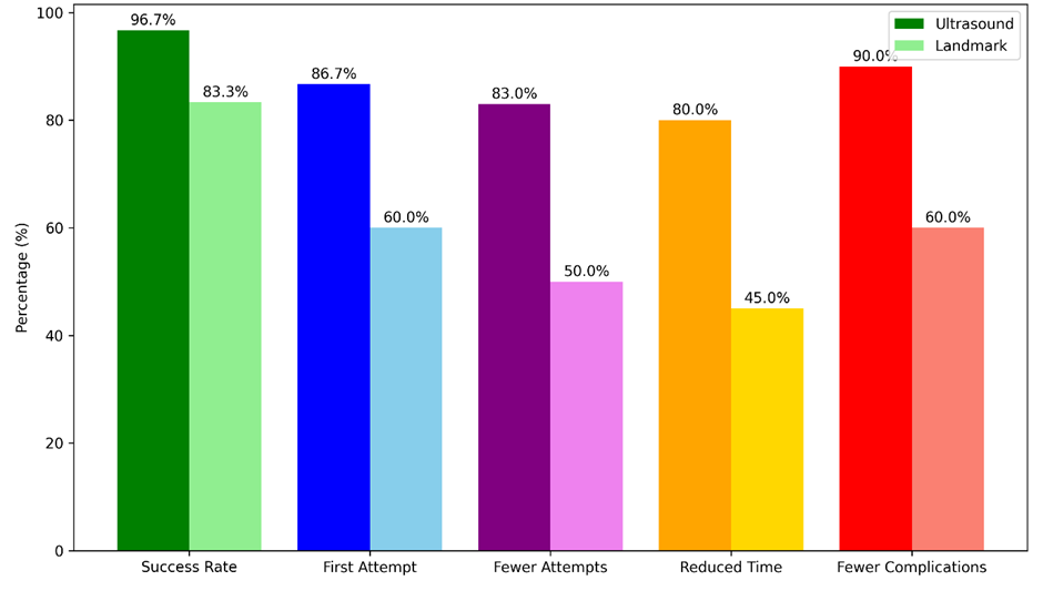
Figure 1: Comparison of Ultrasound Vs Landmark Technique Outcomes
DISCUSSION:
In the present study, ultrasound-guided internal jugular vein (IJV) cannulation demonstrated a higher overall success rate (96.7%) compared to the landmark technique (83.3%). Similar findings were reported by McGee and Gould (2003) [11], who highlighted that conventional landmark-based catheterization is associated with higher failure rates and increased complications, emphasizing the need for safer techniques. The higher success rate observed with ultrasound guidance in this study aligns with the growing evidence supporting its superiority in clinical practice.
The first-attempt success rate in this study was significantly higher in the ultrasound group (86.7%) compared to the landmark group (60.0%). Troianos et al. (1991) [12] reported comparable findings, where ultrasound-guided cannulation significantly improved first-attempt success rates (89% vs 67%) and reduced the need for multiple attempts. This similarity reinforces the reliability of ultrasound guidance in improving procedural efficiency.
The mean number of attempts required for successful cannulation was lower in the ultrasound group (1.2 ± 0.5) compared to the landmark group (2.1 ± 0.9). Rando et al. (2006) [13] reported a similar trend, with the ultrasound group requiring fewer attempts (1.1 vs 2.8 attempts), demonstrating that real-time visualization significantly reduces procedural difficulty and improves accuracy. These findings are consistent with the present study, highlighting the advantage of ultrasound guidance in minimizing repeated needle insertions.
The time required for successful cannulation was also significantly reduced in the ultrasound group (4.8 ± 1.2 minutes) compared to the landmark group (8.6 ± 2.4 minutes). Hrics et al. (1998) [14] reported that ultrasound-assisted IJV cannulation reduces procedure time compared to the landmark technique, particularly in emergency settings. Faster cannulation is crucial in critically ill patients, where timely vascular access can be lifesaving.
In terms of complications, the present study showed a significantly lower overall complication rate in the ultrasound group (10.0%) compared to the landmark group (40.0%). Specifically, arterial puncture was reduced (3.3% vs 20.0%), and no cases of pneumothorax were observed in the ultrasound group. Dolu et al. (2015) [15] reported similar findings, where complication rates were significantly lower in the ultrasound group (7% vs 28%) compared to the landmark technique. This reduction in complications is attributed to direct visualization of vascular anatomy, which minimizes accidental injury to adjacent structures.
Overall, the findings of this study are consistent with previous literature, demonstrating that ultrasound-guided IJV cannulation provides higher success rates, fewer attempts, reduced procedure time, and lower complication rates compared to the landmark technique. These advantages are particularly important in critically ill patients, where rapid and safe vascular access is essential. The results strongly support the routine use of ultrasound guidance for central venous catheterization in clinical practice.
Limitations: This study has certain limitations, including a relatively small sample size (n=60), which may limit the generalizability of the findings. Being a single-centre study conducted at a tertiary care hospital, the results may not be applicable to all clinical settings, especially resource-limited areas. Additionally, operator experience and skill level, which can influence procedural outcomes, were not standardized across all cases. The study also did not evaluate long-term catheter-related complications or cost-effectiveness, which are important considerations in clinical practice. Recommendations: Based on the findings, it is recommended that ultrasound guidance should be routinely used for internal jugular vein cannulation to improve success rates and reduce complications. Training and skill development in point-of-care ultrasound should be emphasized for healthcare providers, particularly in critical care and emergency departments. Healthcare institutions should ensure the availability of ultrasound equipment to facilitate its use. Further large-scale, multicentric studies are recommended to validate these results and to assess long-term outcomes, cost-effectiveness, and applicability in different healthcare settings.
CONCLUSION:
The present study demonstrates that ultrasound-guided internal jugular vein cannulation is significantly more effective and safer than the conventional landmark technique in critically ill patients, as evidenced by a higher overall success rate (96.7% vs 83.3%), better first-attempt success (86.7% vs 60.0%), fewer attempts, reduced procedure time, and markedly lower complication rates (10.0% vs 40.0%). These findings highlight that real-time visualization improves procedural accuracy and minimizes complications, making ultrasound guidance a superior technique for central venous access, particularly in emergency and critical care settings where rapid and safe vascular access is crucial.
REFERENCES:
1. Brass P, Hellmich M, Kolodziej L, Schick G, Smith AF. Ultrasound guidance versus anatomical landmarks for internal jugular vein catheterization. Cochrane Database Syst Rev. 2015 Jan 9;1(1):CD006962. doi: 10.1002/14651858.CD006962.pub2. PMID: 25575244; PMCID: PMC6517109.
2. Wyatt C. Vascular access. In: Tintinalli JE, editor. Tintinalli’s Emergency Medicine. 8th ed. New York: McGraw-Hill Education; 2016. p. 200–203.
3. Bannon MP, Heller SF, Rivera M. Anatomic considerations for central venous cannulation. Risk Manag Healthc Policy. 2011;4:27-39. doi: 10.2147/RMHP.S10383. Epub 2011 Apr 13. PMID: 22312225; PMCID: PMC3270925.
4. Hind D, Calvert N, McWilliams R, Davidson A, Paisley S, Beverley C, et al. Ultrasonic locating devices for central venous cannulation: Meta-analysis. BMJ. 2003;327:361.
5. Geiling J, Burkle FM Jr, Amundson D, Dominguez-Cherit G, Gomersall CD, Lim ML, et al. Task Force for Mass Critical Care. Resource-poor settings: infrastructure and capacity building: care of the critically ill and injured during pandemics and disasters: CHEST consensus statement. Chest. 2014 Oct;146(4 Suppl):e156S-67S. doi: 10.1378/chest.14-0744. PMID: 25144337; PMCID: PMC6679686.
6. Leung J, Duffy M, Finckh A. Real-time ultrasonographically guided internal jugular vein catheterization in the emergency department increases success rates and reduces complications: A randomized prospective study. Ann Emerg Med. 2006;48:540–547.
7. Thomas, Vimal & Abraham, Siju & Balakrishnan, Jayaraj & Krishnan S, Vimal & Amalakat, Ajay & Palatty, et al. (2017). Point‑of‑care ultrasound training in Indian emergency medicine programs: A resident's perspective. International Journal of Academic Medicine. 3. 10.4103/IJAM.IJAM_77_16.
8. Hilty WM, Hudson PA, Levitt MA, Hall JB. Real-time ultrasound-guided femoral vein catheterization during cardiopulmonary resuscitation. Ann Emerg Med. 1997;29:331–336.
9. Miller AH, Roth BA, Mills TJ, Woody JR, Longmoor CE, Foster B. Ultrasound guidance versus the landmark technique for the placement of central venous catheters in the emergency department. Academic Emergency Medicine. 2002;9(8):800-805. doi: 10.1197/aemj.9.8.800
10. Denys BG, Uretsky BF, Reddy PS. Ultrasound-assisted cannulation of the internal jugular vein. A prospective comparison to the external landmark-guided technique. Circulation. 1993 May;87(5):1557-62. doi: 10.1161/01.cir.87.5.1557. PMID: 8491011.
11. McGee DC, Gould MK. Preventing complications of central venous catheterization. N Engl J Med. 2003;348(12):1123–1133 DOI: 10.1056/NEJMra011883.
12. Rando K, Castelli J, Pratt JP, Scavino M, Rey G, Rocca ME, et al. Ultrasound- guided internal jugular vein catheterization: a randomized controlled trial. Heart Lung Vessel. 2014;6(1):13-23. PMID: 24800194; PMCID: PMC4009593.
13. Paul Hrics, Scott Wilber, Michelle P Blanda, Ugo Gallo, Ultrasound-assisted internal jugular vein catheterization in the ED, The American Journal of Emergency Medicine.1998, 16 (4): 401-403, https://doi.org/10.1016/S0735-6757(98)90140-1.
14. Dolu H, Goksu S, Sahin L, Ozen O, Eken L. Comparison of an ultrasound-guided technique versus a landmark-guided technique for internal jugular vein cannulation. J Clin Monit Comput. 2015;29(2):177–182.