Diagnostic Accuracy of Diaphragmatic Ultrasonography Parameters (Diaphragmatic Thickening Fraction and Diaphragmatic Excursion) in Predicting Extubation Outcomes in Critically Ill Patients.
- Soorya R Vimal , Associate Consultant, Department of Critical Care Medicine, Sree Gokulam Medical College and Research Foundation, Venjaramoodu, Thiruvananthapuram, Kerala, India.
- Deepak V , Senior Consultant and Regional Director, Department of Critical Care Medicine, Kimshealth, Thiruvananthapuram, Kerala, India
- Sreeja Sadan , Junior Consultant, Department of Critical Care Medicine, Lisie Medicity, Kochi,Kerala, India.
- Amal R , Senior Resident, Department of Orthopaedics, SUT Academy of Medical Sciences, Thiruvananthapuram, Kerala, India.
- Madhavan Unni , Senior Consultant and Chief Coordinator, Department of Imaging and Interventional Radiology, Kimshealth, Thiruvananthapuram, Kerala, India.
- Shameem A , Associate Consultant, Department of Critical Care Medicine, Kimshealth, Thiruvananthapuram, Kerala, India.
Article Information:
Abstract:
Background: Weaning from mechanical ventilation and predicting extubation success remain challenging in critically ill patients. Conventional weaning indices have limited accuracy, particularly in assessing diaphragmatic function. Diaphragmatic ultrasonography, including diaphragmatic excursion (DE) and diaphragmatic thickening fraction (DTF), has emerged as a promising bedside tool for evaluating extubation readiness. Objectives: To evaluate the diagnostic accuracy of diaphragmatic ultrasonography parameters (Diaphragmatic Excursion and Diaphragmatic Thickening Fraction) in predicting extubation outcomes in critically ill patients. Methods: This prospective observational study was conducted in a tertiary care ICU over a period of one year. A total of 86 adult patients requiring mechanical ventilation for more than 24 hours and undergoing spontaneous breathing trial (SBT) were included. Diaphragmatic ultrasonography was performed before and after SBT to measure diaphragmatic excursion (DE )and diaphragmatic thickening fraction (DTF). Extubation outcomes were categorized as successful or failed based on the need for ventilatory support within 48 hours. Diagnostic accuracy was assessed using sensitivity, specificity, positive predictive value (PPV), negative predictive value (NPV), and receiver operating characteristic (ROC) curve analysis. Results: Out of 86 patients, 68 (79.1%) had successful extubation, while 18 (20.9%) experienced failed extubation. The mean diaphragmatic excursion after SBT was 18.68 ± 4.45 mm, and the mean DTF after SBT was 38.36 ± 5.26%. DE after SBT demonstrated excellent diagnostic accuracy with an AUC of 0.967 (95% CI: 0.914–1.000, p<0.001). A cut-off value of ≥13.5 mm yielded 100% sensitivity and 88.9% specificity. DTF after SBT showed superior diagnostic performance with an AUC of 0.987 (95% CI: 0.961–1.000, p<0.001). A cut-off value of ≥35% provided 98.5% sensitivity and 100% specificity. Conclusion: Diaphragmatic ultrasonography parameters, particularly DTF and DE measured after SBT, are highly accurate predictors of extubation outcomes in critically ill patients. DTF demonstrated slightly superior diagnostic performance compared to DE. Incorporation of diaphragmatic ultrasound into routine weaning protocols may improve clinical decision-making and reduce extubation failure rates.
Keywords:
Article :
INTRODUCTION:
Weaning from mechanical ventilation (MV) is one of the most critical and complex processes encountered in intensive care unit (ICU) management. It involves the gradual transition of a patient from ventilatory support to spontaneous breathing, ultimately leading to extubation. This process requires careful clinical judgment and multidisciplinary coordination to determine the optimal timing. Both premature and delayed extubation are associated with adverse outcomes, including weaning failure, need for reintubation, ventilator-associated pneumonia (VAP), prolonged ICU stay, increased healthcare costs, and higher mortality rates [1,2].
Extubation failure, commonly defined as the need for reintubation or requirement of non-invasive ventilatory support within 48–72 hours after extubation, remains a significant clinical challenge. It is associated with increased morbidity, longer ICU stays, higher risk of nosocomial infections, and poor long-term outcomes [3]. Despite advances in critical care, predicting successful weaning and extubation remains difficult due to the heterogeneity of ICU patients and the multifactorial nature of respiratory failure.
Current guidelines recommend several bedside indices to guide weaning decisions, among which the spontaneous breathing trial (SBT) is considered the cornerstone. SBT assesses the patient’s ability to maintain spontaneous breathing with minimal ventilatory support. However, even after a successful SBT, extubation failure rates can be as high as 20%, particularly in high-risk populations, indicating the limitations of conventional assessment methods [2,4].
The diaphragm plays a pivotal role in the weaning process as it is the primary muscle of respiration, contributing up to 75% of the work of breathing at rest. Diaphragmatic dysfunction, particularly ventilator-induced diaphragmatic dysfunction (VIDD), is commonly observed in critically ill patients and is strongly associated with weaning failure. Factors such as sepsis, hypoxia, malnutrition, and prolonged mechanical ventilation contribute to diaphragmatic weakness, thereby impairing respiratory performance [5–7,13].
Traditional weaning indices such as respiratory rate, tidal volume, and the Rapid Shallow Breathing Index (RSBI) primarily reflect global respiratory mechanics and may not accurately assess diaphragmatic function. RSBI, in particular, may yield false-positive results when accessory respiratory muscles compensate for diaphragmatic weakness, thereby masking underlying dysfunction [13].
In recent years, diaphragmatic ultrasonography has emerged as a promising, non-invasive bedside tool for the assessment of diaphragmatic function. It enables real-time visualization of diaphragmatic movement and muscle thickness. Two key parameters derived from ultrasonography—diaphragmatic excursion (DE) and diaphragmatic thickening fraction (DTF)—have been shown to correlate with extubation outcomes. These parameters provide a more direct and physiological assessment of respiratory muscle performance compared to conventional indices [8–11,14].
Diaphragmatic ultrasound is simple, reproducible, and can be easily performed at the bedside without exposing patients to radiation. Typically, diaphragmatic excursion is measured using a low-frequency curvilinear probe in the subcostal region, while diaphragmatic thickening is assessed using a high-frequency linear probe at the zone of apposition in the mid-axillary line [2,4].
Given the limitations of conventional weaning parameters and the increasing evidence supporting diaphragmatic ultrasonography, the present study was undertaken to evaluate the utility of diaphragmatic excursion and diaphragmatic thickening fraction in predicting extubation outcomes among critically ill patients.
MATERIALS AND METHODS:
Study Design and Setting
This prospective observational study was conducted in the Department of Critical Care Medicine at KIMSHEALTH, Thiruvananthapuram, Kerala, over a period of one year.
Study Population
Adult critically ill patients admitted to the medical intensive care unit (MICU) requiring mechanical ventilation for more than 24 hours were screened for eligibility. Patients with a Clinical Frailty Score ≤3 were included, indicating good pre-illness functional status.
Inclusion Criteria
Patients were included if they fulfilled the following criteria:
· Age ≥18 years
· Conscious and oriented
· Able to tolerate spontaneous breathing trial (SBT) for 2 hours
· Afebrile for at least 24 hours
· Minimal tracheobronchial secretions with effective cough reflex
Exclusion Criteria
Patients were excluded if they had:
· Hemodynamic instability or significant arrhythmias
· Diastolic dysfunction
· Spinal cord injury
· Pneumothorax
· Recent thoracic or abdominal surgery
· Chest tube or conditions interfering with ultrasonography
· Pleural disease or history of pleurodesis
· Neuromuscular disorders
· Pregnancy
· Tracheostomy
· Poor ultrasound window
Sample Size
The sample size was calculated based on a previous study (Saravanan et al.), assuming a diagnostic accuracy of 94%, with a 95% confidence level and 5% absolute precision. The final calculated sample size was 86 patients.
Study Procedure
All eligible patients were prospectively enrolled after obtaining written informed consent. An observer-blinded design was adopted, wherein the intensivist performing extubation was blinded to ultrasonographic findings.
Weaning Protocol
Patients were assessed for readiness for weaning using standard ICU criteria:
· Adequate mentation (GCS ≥10T)
· Hemodynamic stability (MAP 60–110 mmHg , pulse rate 60-100/min)
· Adequate respiratory effort (RR <20/min, tidal volume >8 mL/kg)
· Muscle strength ( sustained head lift , strong handgrip)
· Low pain score (VAS <3 )
· Acceptable arterial blood gas parameters
Eligible patients underwent a spontaneous breathing trial (SBT). The Rapid Shallow Breathing Index (RSBI) was calculated as:
RSBI=Respiratory Rate/(Tidal Volume (L))
Patients meeting standard extubation criteria were extubated.
Diaphragmatic Ultrasonography
Ultrasound assessment was performed twice:
· Before SBT
· Immediately after SBT
Diaphragmatic Excursion (DE)
· Measured using a 3–5 MHz curvilinear probe
· Subcostal approach in semi-recumbent position
· M-mode used to record maximal excursion (mm)
Diaphragmatic Thickening Fraction (DTF)
· Measured using an 8–13 MHz linear probe
· Mid-axillary line (8th–9th intercostal space)
· Thickness measured at end-inspiration and end-expiration
DTF=(Thickness_inspirationⓜ-Thickness_expiration )/(Thickness_expiration )×100
All measurements were performed by an experienced intensivist, and images were archived.
Outcome Measures
· Diagnostic accuracy of diaphragmatic ultrasonographic parameters (DE and DTF) in predicting extubation success
Definition of Outcomes
· Successful extubation: Spontaneous breathing maintained for ≥48 hours without ventilatory support
· Failed extubation: Requirement of reintubation, non-invasive ventilation, or tracheostomy within 48 hours
Statistical Analysis
Data were analyzed using SPSS version 26.0. Continuous variables were summarized as mean ± standard deviation (SD), while categorical variables were presented as frequencies and percentages. The diagnostic performance of the study parameters was assessed using sensitivity, specificity, positive predictive value (PPV), and negative predictive value (NPV). Receiver operating characteristic (ROC) curve analysis was performed to evaluate the predictive accuracy of the variables. A p-value of less than 0.05 was considered statistically significant.
Ethical Considerations
The study was approved by the Institutional Ethics Committee. Written informed consent was obtained from all participants. The study adhered to standard ICU care protocols without additional risk to patients.
RESULTS:
Table 1: Baseline Characteristics of Study Subjects (N=86)
|
Variable |
Mean± SD/ n(%) |
|
Mean Age (years) |
39.6 ± 11.0 |
|
Age Range |
20–61 |
|
Male |
45 (52.3%) |
|
Female |
41 (47.7%) |
The study population had a mean age of 39.6 ± 11 years, with participants ranging from 20 to 61 years, indicating a predominantly middle-aged critically ill cohort. Gender distribution was nearly equal, with 52.3% males and 47.7% females, minimizing gender-related bias and enhancing generalizability of results [Table 1].
Table 2: Mechanical Ventilation and Clinical Parameters (N=86)
|
Variable |
Mean ± SD |
Range |
|
Duration of MV (days) |
3.6 ± 0.7 |
2–6 |
|
Respiratory Rate (breaths/min) |
20.28 ± 3.03 |
14–28 |
|
Tidal Volume (mL) |
369.79 ± 54.62 |
246–500 |
|
RSBI |
55.57 ± 9.21 |
40–93 |
|
DE Before SBT (mm) |
18.06 ± 4.41 |
10–28 |
|
DE After SBT (mm) |
18.68 ± 4.45 |
10–28 |
|
DTF Before SBT (%) |
37.04 ± 5.22 |
25–48 |
|
DTF After SBT (%) |
38.36 ± 5.26 |
27–48 |
The mean duration of mechanical ventilation was 3.6 ± 0.7 days, with most patients undergoing SBT within 3–4 days, indicating early weaning practices. The mean RSBI was 55.57 ± 9.21, which is significantly below the standard threshold of 105, suggesting that the majority of patients had favourable spontaneous breathing patterns. Diaphragmatic parameters showed stable and preserved function, with DE increasing slightly from 18.06 mm to 18.68 mm and DTF increasing from 37.04% to 38.36% after SBT, indicating maintained diaphragmatic contractility during the weaning process [Table 2].
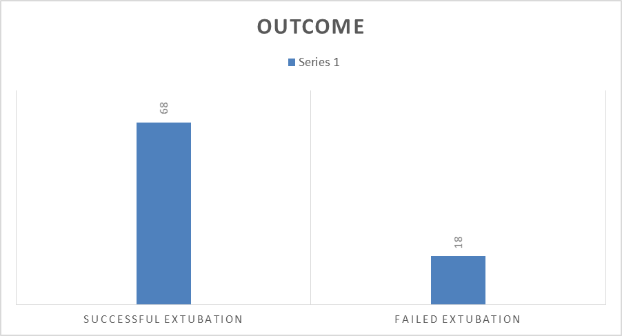
Figure 1: Extubation Outcomes (N=86)
Out of 86 patients, 68 (79.1%) had successful extubation, while 18 (20.9%) experienced failed extubation, requiring reintubation or NIV within 48 hours.
This reflects a high success rate, consistent with the observed favourable respiratory and diaphragmatic parameters.[Figure 1]
Table 3: Diagnostic Accuracy of Diaphragmatic Ultrasonography Parameters
|
Parameter |
AUC |
95% CI |
p-value |
Cut-off |
Sensitivity (%) |
Specificity (%) |
PPV (%) |
NPV (%) |
|
DE after SBT (mm) |
0.967 |
0.914–1.000 |
<0.001 |
13.5 |
100 |
88.9 |
93.2 |
100 |
|
DTF after SBT (%) |
0.987 |
0.961–1.000 |
<0.001 |
35.0 |
98.5 |
100 |
100 |
94.7 |
Both diaphragmatic parameters demonstrated excellent diagnostic accuracy in predicting extubation outcome.
DE after SBT showed an AUC of 0.967 (95% CI: 0.914–1.000, p<0.001), indicating near-perfect discrimination.
At a cut-off of 13.5 mm, it achieved 100% sensitivity and 88.9% specificity, with PPV 93.2% and NPV 100%, making it highly reliable for ruling out extubation failure.
DTF after SBT demonstrated an even higher AUC of 0.987 (95% CI: 0.961–1.000, p<0.001), suggesting almost perfect predictive ability. At a cut-off of 35%, it yielded 98.5% sensitivity and 100% specificity, with PPV 100% and NPV 94.7%, confirming it as an excellent predictor of extubation success.
Overall, DTF performed slightly better than DE, though both parameters are clinically robust and valuable bedside tools.[Table 3]
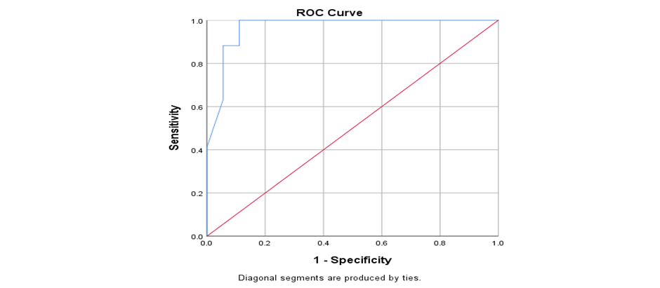
Figure 2 : ROC Curve of DE After SBT
The ROC curve for diaphragmatic excursion after SBT demonstrated an AUC of 0.967, reflecting excellent diagnostic performance. The curve lies close to the upper left corner, indicating high sensitivity and specificity. The optimal cut-off value of 13.5 mm provides perfect sensitivity (100%), ensuring no missed extubation failures, along with high specificity (88.9%).[Figure 2]
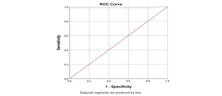
Figure 3: ROC Curve of DTF After SBT
The ROC curve for diaphragmatic thickening fraction after SBT showed an AUC of 0.987, indicating near-perfect discrimination.
The curve demonstrates superior diagnostic performance compared to DE, with an optimal cut-off of 35%, achieving 98.5% sensitivity and 100% specificity, making it the most reliable parameter for predicting extubation success in this study.[Figure 3]
Diaphragmatic ultrasonography parameters, particularly DTF and DE after SBT, demonstrated excellent diagnostic accuracy (AUC >0.95, p<0.001) in predicting extubation outcomes.
DTF showed superior performance, with higher specificity and overall accuracy compared to DE, establishing it as the most reliable bedside predictor of extubation success among critically ill patients.
DISCUSSION:
The present study evaluated the role of diaphragmatic ultrasonography, specifically diaphragmatic excursion (DE) and diaphragmatic thickening fraction (DTF), in predicting extubation outcomes among critically ill patients. A total of 86 patients undergoing weaning from mechanical ventilation were assessed using both conventional weaning parameters and ultrasound-based diaphragmatic indices before and after spontaneous breathing trial (SBT). The findings of this study demonstrated that both DE and DTF are highly reliable predictors of successful extubation , with excellent diagnostic accuracy.
The demographic profile of the study population revealed a relatively younger cohort, with a mean age of 39.6 ± 9.4 years, predominantly clustered in the 35–44-year age group. This is in contrast to previously published studies such as Blumhof et al. (2016) [9] and Baess et al. (2016) [15], where the mean ages were 62 and 59.1 years, respectively. Similarly, Haaksma et al. (2021) [16] reported a much older population with a median age exceeding 72 years. The younger age group in the present study likely contributed to the higher proportion of patients categorized as “fit” or “very fit” (86.1%) based on the clinical frailty scale, indicating better baseline physiological reserve and functional status.
The gender distribution in the present study was nearly equal, which is consistent with findings from Osman et al. (2017) [17] and Farghaly & Hasan (2017) [10], both of whom reported balanced male-to-female ratios. The mean duration of mechanical ventilation in this study was 3.6 ± 0.7 days, with the majority of patients extubated by day 3 or 4. This duration differs from that reported by Chen Gang et al. (2023) [18], where patients were ventilated for just over 24 hours in a surgical ICU setting, and from Blumhof et al. [9], where the duration exceeded 52 hours. These differences in demographic and clinical characteristics suggest that the study population was relatively stable and appropriately selected for weaning, which may have contributed to the high extubation success rate of 79.1%.
A key finding of the present study is the excellent diagnostic accuracy of diaphragmatic ultrasonography parameters. Both DE and DTF demonstrated high discriminative ability in predicting extubation outcomes, with DTF showing slightly superior performance. This may be explained by the fact that DTF directly reflects diaphragmatic muscle contraction and thickening during inspiration, making it a more sensitive indicator of diaphragmatic function compared to excursion alone. These findings are consistent with previous studies that have highlighted the superiority of ultrasound-based diaphragmatic assessment over conventional indices.
The present study identified DE ≥13.5 mm and DTF ≥35% as optimal cut-off values for predicting successful extubation, with high sensitivity, specificity, positive predictive value, and negative predictive value. These values are in close agreement with previously published literature. Chen Gang et al. (2023) [18] reported a DTF cut-off of ≥30%, while Mawla et al. (2022) [19] and Raj & Kumar Nagalaji (2023) [20] reported a similar threshold of ≥35%. McCool et al. (2020) [21] also suggested a DTF ≥30% with a positive predictive value of 90.9%. For diaphragmatic excursion, Osman et al. (2017) [17] reported a cut-off of ≥20.3 mm, while Blumhof et al. (2016) [9] suggested a lower threshold of ≥10 mm. Despite minor variations, there is a clear consensus across studies regarding effective threshold ranges, supporting the reliability of these parameters in predicting extubation outcomes.
Conventional weaning parameters such as respiratory rate, tidal volume, and Rapid Shallow Breathing Index (RSBI) have long been used to guide extubation decisions. However, these indices primarily reflect global respiratory mechanics and may not accurately assess diaphragmatic function, which plays a crucial role in sustaining spontaneous breathing. In contrast, diaphragmatic ultrasonography provides a direct and real-time evaluation of diaphragmatic contractility. The findings of the present study further support the growing body of evidence suggesting that ultrasound-based parameters may offer superior predictive value compared to conventional indices.
The utility of diaphragmatic ultrasonography has also been demonstrated in special populations. Thabit et al. (2022) [22] reported that a DTF ≥24% in mechanically ventilated pediatric patients had high predictive accuracy, with a positive predictive value of 80.3% and negative predictive value of 89.7%. Similarly, Osman et al. (2017) [17] demonstrated the effectiveness of both DE and DTF in predicting extubation outcomes in children following major surgery. These findings highlight the adaptability and applicability of diaphragmatic ultrasound across diverse clinical settings and patient groups.
From a clinical perspective, diaphragmatic ultrasonography offers several advantages. It is a non-invasive, bedside, radiation-free, and repeatable modality that can be easily integrated into routine ICU practice. Given its high diagnostic accuracy, incorporating DE and DTF into standard weaning protocols may help reduce extubation failure rates, decrease the need for reintubation, and improve overall patient outcomes. Additionally, it may enhance ICU efficiency by facilitating timely and evidence-based extubation decisions.
However, certain limitations must be acknowledged. This was a single-center study with a relatively small sample size, which may limit the generalizability of the findings. The study population was also relatively younger and less frail compared to other studies, which may have influenced the high success rate observed. Furthermore, diaphragmatic ultrasonography is operator-dependent, and variability in measurement techniques may affect reproducibility.
CONCLUSION:
In conclusion, diaphragmatic ultrasonography, particularly DTF and DE measured after spontaneous breathing trial, is a highly reliable and accurate predictor of extubation outcomes in critically ill patients. DTF demonstrated slightly superior performance compared to DE. Incorporation of these parameters into routine weaning protocols has the potential to improve clinical decision-making, reduce complications, and enhance patient care in the intensive care setting.
REFERENCES:
1. Parada-Gereda HM, Tibaduiza AL, Rico-Mendoza A, Molano-Franco D, Nieto VH, Arias-Ortiz WA , et al. Effectiveness of diaphragmatic ultrasound as a predictor of successful weaning from mechanical ventilation: a systematic review and meta-analysis. Critical Care. 2023 May 5;27(1):174.
2. Al-Husinat LI, Jouryyeh B, Rawashdeh A, Robba C, Silva PL, Rocco PR , et al. The role of ultrasonography in the process of weaning from mechanical ventilation in critically ill patients. Diagnostics. 2024 Feb 12;14(4):398.
3. DiNino E, Gartman EJ, Sethi JM, McCool FD. Diaphragm ultrasound as a predictor of successful extubation from mechanical ventilation. Thorax. 2014 May 1;69(5):431-5.
4. Saravanan R, Nivedita K, Karthik K, Venkatraman R. Role of diaphragm ultrasound in weaning mechanically ventilated patients: A prospective observational study. Indian Journal of Anaesthesia. 2022 Aug 1;66(8):591-8.
5. Matamis D, Soilemezi E, Tsagourias M, Akoumianaki E, Dimassi S, Boroli F, et al. Sonographic evaluation of the diaphragm in critically ill patients. Technique and clinical applications. Intensive care medicine. 2013 May;39:801-10.
6. Zambon M, Beccaria P, Matsuno J, Gemma M, Frati E, Colombo S , et al. Mechanical ventilation and diaphragmatic atrophy in critically ill patients: an ultrasound study. Critical care medicine. 2016 Jul 1;44(7):1347-52.
7. Ferrari G, De Filippi G, Elia F, Panero F, Volpicelli G, Aprà F. Diaphragm ultrasound as a new index of discontinuation from mechanical ventilation. Critical ultrasound journal. 2014 Dec;6:1-6.
8. Bansal P, Jakhar B, Arya RC, Sultania NS, Puhal S, Bansal K et al. Evaluation of abdominal expiratory muscle thickness pattern, diaphragmatic excursion, diaphragmatic thickness fraction and lung ultrasound score in critically ill patients and their association with weaning patterns: A prospective study. Journal of Anaesthesiology Clinical Pharmacology. 2025 Apr 1;41(2):257-64.
9. Blumhof S, Wheeler DW, Thomas K, Greening P, Dibb W. Diaphragmatic ultrasound as a predictor of successful extubation from mechanical ventilation. Thorax. 2016;71(5):436–7.
10. Farghaly S, Hasan AA. Diaphragm ultrasound as a new method to predict extubation outcome in mechanically ventilated patients. Australian Critical Care. 2017 Jan 1;30(1):37-43.
11. Santana PV, Cardenas LZ, Albuquerque AL, Carvalho CR, Caruso P. Diaphragmatic ultrasound: a review of its methodological aspects and clinical uses. Jornal Brasileiro de Pneumologia. 2020 Nov 20;46:e20200064.
12. Downey R. Anatomy of the normal diaphragm. Thoracic surgery clinics. 2011 May 1;21(2):273-9.
13. Banerjee A, Mehrotra G. Comparison of lung ultrasound-based weaning indices with rapid shallow breathing index: are they helpful?. Indian journal of critical care medicine: peer-reviewed, official publication of Indian Society of Critical Care Medicine. 2018 Jun;22(6):435.
14. Bianchini A, Pintus L, Vitale G, Mazzotta E, Felicani C, Zangheri E , et al. Lung Ultrasound in Mechanical Ventilation: A Purposive Review. Diagnostics. 2025 Mar 28;15(7):870.
15. Baess F, Similowski T, Demoule A. Diaphragm ultrasound to predict extubation outcome: A prospective observational study. Ann Intensive Care. 2016;6:38.
16. Haaksma ME, Tuinman PR, Heunks LMA. Predicting extubation failure using diaphragmatic ultrasound in critically ill patients: A prospective cohort study. Respir Res. 2021;22(1):1–8.
17. Osman AM, Hashim RM. Diaphragmatic and lung ultrasound application as new predictive indices for the weaning process in ICU patients. The Egyptian Journal of Radiology and Nuclear Medicine. 2017 Mar 1;48(1):61-6.
18. Chen G, Wang L, Yu C, Liu S. Diaphragm thickening fraction as a predictor of extubation success in thoracic surgery patients: A randomized controlled trial. J Thorac Dis. 2023;15(3):870–8.
19. Mawla AA, Al-Qadheeb N, Ahmed R, ElSaka A, Elsharkawy A. Diaphragmatic ultrasound as a predictor of extubation outcome in patients with COPD exacerbation. J Intensive Care Med. 2022;37(5):579–86.
20. Raj RK, Kumar R, Nagalaji B. Role of diaphragmatic ultrasound in predicting successful extubation in pediatric ICU patients. Indian J Crit Care Med. 2023;27(2):105–11.
21. McCool FD, Tzelepis GE. Dysfunction of the diaphragm. New England Journal of Medicine. 2012 Mar 8;366(10):932-42.
22. Thabit M, El-Shafey EM, Ali NA. Ultrasound assessment of diaphragm function to predict extubation outcome in pediatric patients. Egypt J Hosp Med. 2022;89(1):3165–70.