SURVIVAL OUTCOMES IN PATIENTS UNDERGOING CURATIVE TREATMENT FOR CARCINOMA GALLBLADDER: EXPERIENCE FROM A TERTIARY CANCER INSTITUTE OF NORTH-EAST INDIA.
- Deep Jyoti Kalita , Professor Department of Surgical Oncology, Dr. B.Borooah Cancer Institute, Guwahati Assam.
- Ashutosh Sahewalla , Assistant Professor Department of surgical oncology, State Cancer Institute ,Gauhati Medical college and Hospital, ,Guwahati, Assam.
- Dibyajyoti Deka , Assistant Professor, Department of surgical oncology, Dr. B.Borooah Cancer Institute, Guwahati, Assam.
- Manthan Thakkar , Assistant Professor, Department of surgical oncology, Dr. B.Borooah Cancer Institute, Guwahati, Assam.
- Aditya Borgaonkar , Senior resident, Department of surgical oncology, Dr. B.Borooah Cancer Institute, Guwahati, Assam.
- Gaurav Das , Professor, Department of surgical oncology, Dr. B.Borooah Cancer Institute, Guwahati, Assam.
- Shivaji Sharma , Asso. Prof, Department of surgical oncology, Dr. B.Borooah Cancer Institute, Guwahati, Assam.
Article Information:
Abstract:
Background: Gallbladder cancer is the most common malignancy of the biliary tract and the sixth most common gastrointestinal cancer, with significant geographic variation and an overall incidence of 3 per 100,000. It carries a poor prognosis due to aggressive tumor biology, late presentation, and advanced disease at diagnosis. At our institute, it is a prevalent cancer, with 20–25 patients treated annually with curative intent through radical cholecystectomy. Despite this, loco-regional and distant recurrence rates remain high. This study analyzes survival outcomes and recurrence patterns in patients who underwent curative surgery. Methods and Materials: This retrospective study included 95 patients who underwent radical cholecystectomy between January 2016 and December 2021. Inclusion criteria were patients aged 18–75 years, localized disease (cT1–T2, N0), locally advanced disease post-NACT (cT3–T4/N+), and incidental carcinoma gallbladder. Patients with metastatic disease or benign histopathology were excluded. Results: Out of 218 patients evaluated, 95 underwent curative surgery. Median overall survival (OS) was 66 months. Stage I patients had the highest OS (82 months), while stage IVB had the lowest (10 months, p = 0.07). Patients with incidental carcinoma had better OS (68 months) compared to those with gallbladder mass (31 months, p = 0.013). Node-negative patients had significantly better OS (66 months) than node-positive patients (16 months, p = 0.002). A total of 56.8% had ≥6 lymph nodes removed, with a median OS of 68 months vs. 35 months in those with <6 nodes (p = 0.67). Conclusion: Despite curative surgery, gallbladder cancer outcomes remain poor. Survival is significantly influenced by stage and nodal status. Adequate lymphadenectomy may improve outcomes, highlighting the importance of thorough surgical resection
Keywords:
Article :
INTRODUCTION:
Gallbladder cancer is the sixth most common malignancy of the gastrointestinal system and the most common cancer of the biliary tract. (1) It has an overall incidence of 3 per 100000 people with wide geographic variations in prevalence. (2) Gallbladder cancer is also the only gastrointestinal malignancy which is more common in women than men. (3) It accounts for 1.7% of all cancer mortalities which suggests its poor prognosis. Gallbladder cancer is associated with a poor prognosis due to aggressive tumour biology, late presentation, complicated anatomy , and advanced stage at diagnosis. (4)
Gallbladder cancer is found in 0.2%-3% of all cholecystectomies. (5) Gallbladder cancer is suspected preoperatively in only 30% of all patients while in the remaining 70% of cases it is diagnosed as incidental gallbladder cancer following cholecystectomy performed due to gallstone disease. (2) Approximately 15%-47% of preoperatively known gallbladder carcinomas are suitable for resection as the majority of symptomatic patients have an incurable tumour. (6) Gallbladder carcinoma is associated with poor outcomes with a 5-year overall survival rate being less than 5%.
Gallbladder cancer is relatively asymptomatic in its early stages. It has three different presentations: identified in histopathology following cholecystectomy; discovered during the index surgery; and suspicion before surgery due to atypical symptoms. (7) Because of such vague presentations it is often diagnosed at an advanced stage. Histopathological types include adenocarcinoma which is seen in more than 90% of cases; other types include papillary, mucinous, squamous or adeno-squamous carcinomas. (8)
Gallbladder cancer is rare in developed countries. In the United States, it accounts for 0.5% of all gastrointestinal malignancies with less than 5,000 cases occurring yearly. (9) India has one of the highest incidences worldwide, with the disease mostly seen in the north and northeast regions of the country. (10, 11) Evidence suggests that gallbladder cancer arises due to chronic inflammation progressing to dysplasia, carcinoma‐in‐situ, and finally, invasive cancer even though the etiology remains unknown. (11)
Incidence rates vary among different geographic areas and ethnicities, reaching extremely high rates in North and South American Indians (particularly Chilean Mapuche Indians). (12) Other high-risk regions include Eastern Europe (14/100,000 in Poland), northern India (as high as 21.5/100,000 for women from Delhi), south Pakistan (11.3/100,000), Israel (5/100,000), and Japan (7/100,000). (13) However in the rest of the world, the occurrence of gallbladder cancer is low (<2/100,000).
In our institute it is one of the most prevelant cancer . On an average we encounter 350-400 new cases of carcinoma gall bladder per year .But like rest of the world scenario , it presents very late. Around 85-90% present as metastatic or unresectable disease which finally gets palliative intent treatment. We treat on an average 20-25 cases of carcinoma gall bladder with curative intent i.e radical cholecystectomy. But even after treating patients with curative intent the loco regional and distant failure rates are very high.
Aim of the Study:
This study aims to analyze the survival rates and recurrence patterns in patients with carcinoma gallbladder who underwent curative intent surgery.
MATERIALS AND METHODS:
This was a retrospective study done on patients who had undergone radical cholecystectomy for carcinoma gallbladder between January 2016 and December 2021. The total number of patients enrolled was ninety-five. The inclusion criteria included patients with age ≥ 18 years and <75 years, patient who underwent curative resection at our institute, patients with localized disease (cT1 – T2, N0), patients with locally advanced disease post-NACT (cT3/N+) and patients with incidental carcinoma gallbladder (cT0). The exclusion criteria were patients with metastatic disease, GB mass with benign histopathology and ages <18 years and >75 years
Statistical analysis:
Patient and tumour characteristics were described using counts and percentages for categorical variables and ranges for continuous variables. All analyses for patients who underwent radical cholecystectomy were conducted using the TNM stage as assessed after the primary resection. The Chi-square or Fisher's exact tests were used to assess differences in two groups and Kaplan–Meier curves were used to calculate the median survival times.
Survival was defined as the time in days from the date of presentation to our hospital after carcinoma gall bladder diagnosis until the date of death from any cause or the date of last follow-up. Log-rank testing and Cox regression analysis were used to compare survival between groups of patients. Cox regression analysis was used to calculate hazard ratios (HRs) for potential prognostic factors in patients who underwent radical cholecystectomy. p values < 0.05 were considered statistically significant. Statistical analyses were conducted using the SPSS 29.0 statistical package.
RESULTS
Out of two hundred and eighteen patients who were planned for radical cholecystectomy a total of ninety-five patients underwent radical cholecystectomy for gall bladder cancer during the study period as the remaining were found to have metastatic disease on staging laparoscopy or had unresectable disease . Out of them, thirty patients were males and sixty-five were females which showed the incidence of carcinoma gall bladder being two times more common in females in our study. The median age was 53 years (range 30 to 74 years). (Table 1)
The most common presentation was abdominal pain. Thirty-nine patients were diagnosed with incidental carcinoma gallbladder. Twelve patients presented with jaundice of which five patients required biliary drainage.
Table 1: Preoperative characteristics of patients
|
Characteristics |
Number of patients (N = 95) |
|
Age (years) ≤ 50 years >50 years |
53 (30 – 74years) 43 52 |
|
Sex ratio (F: M) |
2.1:1 |
|
Presentation Abdominal pain Jaundice Mass abdomen Post simple cholecystectomy (incidental carcinoma gallbladder) |
78 12 7 39 |
|
Radical cholecystectomy Upfront Incidental Post NACT |
50 35 10 |
All these patients were evaluated and then planned for definitive management. Patients were evaluated with contrast-enhanced CT scans of the thorax, abdomen and pelvis. Thirty-five patients had normal CA19.9 levels while twenty-one patients had elevated levels and in the remaining thirty-nine patients reports could not be retrieved.
Patients who received neoadjuvant chemotherapy had undergone a biopsy for confirmation of malignancy before starting chemotherapy. A total of ten patients received neoadjuvant chemotherapy of which four patients were incidental carcinoma gallbladder and the remaining six patients had locally advanced disease.
The remaining eight five patients underwent upfront curative resection. The median duration of surgery was 195 minutes and the median blood loss was 150 ml. (Table 2)
The median duration of ICU stay was one day while the median duration of hospital stay was 7.5 days. Fourteen patients (14.7%) had complications of which bile leak was seen in six patients. Three patients required pigtail insertion for bile leak, one patient required re-laparotomy and the remaining two patients were managed conservatively.
Two patients had electrolyte imbalance and paralytic ileus which were managed conservatively and gradually they recovered. Four patients had wound infection which was managed with regular dressings and secondary suturing later. One patient with a bile leak expired due to septicemia.
Table 2: Intra-operative and Post-operative characteristics
|
Characteristic |
Value |
|
Median blood loss (ml) |
150ml (100 –400ml) |
|
Median duration of surgery |
195 mins (175–430 mins) |
|
Median duration of ICU stay (days) |
1 day (1 – 5 days) |
|
Median duration of hospital stay (days) |
7.5 days (5 – 36 days) |
|
30 day mortality |
1 |
|
Postoperative complications Bile leak Wound infection Electrolyte imbalance Paralytic ileus |
6 4 2 2 |
The histopathological features of the surgical specimens are listed in Table 3. The most common histology was adenocarcinoma which was seen in ninety patients (94.7%). Nodal positivity was seen in twenty three patients (24.2%). Thirty-seven patients (38.9%) had stage I disease, thirty-five patients (36.8%) had stage III disease , eighteen patients (18.9%) had stage II disease and 5.2 %had stage IV disease( not M1 but stage IV in view of N2 nodal status).
Table 3: Histopathological characteristics of the specimen
|
Characteristics |
Number of patients (N = 95) |
|
Histology Adenocarcinoma Signet ring cell carcinoma Mucinous Squamous cell carcinoma |
90 1 3 1 |
|
Degree of differentiation Well-differentiated Moderately differentiated Poorly differentiated |
61 18 12 |
|
Nodal status pN+/ ypN+ pN0/ ypN0 |
23 72 |
|
Lymphovascular invasion Yes No Not reported |
3 42 50 |
|
Perineural invasion Yes No Not reported |
11 32 52 |
|
Stage I IIA IIB IIIA IIIB IV A IVB |
37 10 8 16 19 0 5 |
Out of ninety-four patients(one having expired in post operative period), fifty-three patients (56.4%) completed adjuvant chemotherapy while twenty-four patients (25.5%) defaulted and did not complete their adjuvant treatment. Data could not be retrieved in seventeen patients (18.1%) regarding their completion of adjuvant treatment.
The median overall survival was 66 months with an HR of 21.5 (95% CI 23.81 – 108.18). The overall survival was calculated from the first day of presentation to our hospital after diagnosis of carcinoma gall bladder till the date of the last follow-up. (Figure 1)
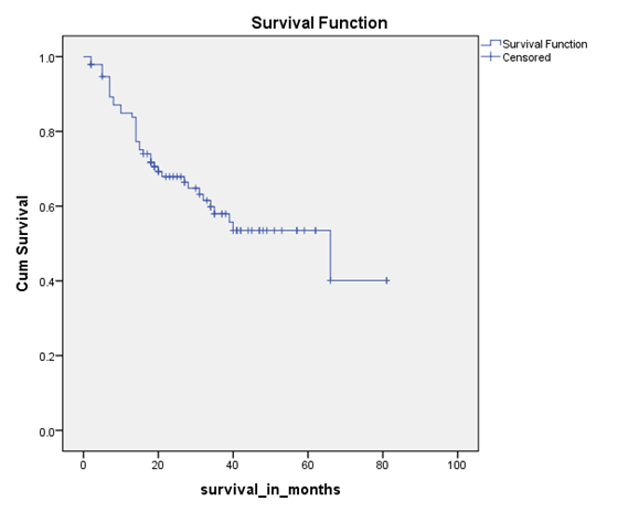
Fig 1: Kaplan Meier showing median overall survival
Stage-specific overall survival showed that stage I disease had a high median overall survival of 82 months while stage IVB had the lowest median overall survival of 10 months. This showed that with increased stage median overall survival decreased significantly with p value being 0.07. (Figure 2)
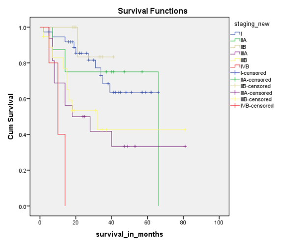
Fig 2: Kaplan Meier showing stage specific median overall survival
Patients who underwent radical cholecystectomy following diagnosis of incidental carcinoma gallbladder had a better median overall survival of 68 months compared to patients who had gallbladder mass with a median overall survival of 31 months with the result being statistically significant with p-value being 0.013. (Figure 3)
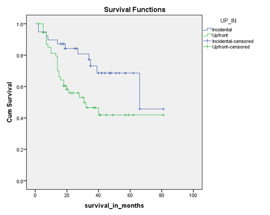
Fig 3: Kaplan Meier showing survival based on incidental vs. upfront gallbladder mass surgery
Patients who had node-negative disease had a median overall survival of 66 months with HR of 22.02 (95% CI 22.84 – 109.15) compared to node-positive patients who had a median overall survival of 16 months with HR of 2.33 (95% CI 11.43 – 20.57). (Figure 4) This showed that with nodal positivity survival decreased significantly with p value being 0.002.
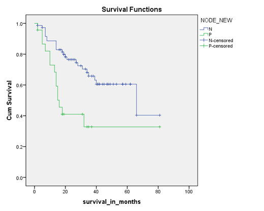
Fig 4: Kaplan Meier showing survival based on lymph node positivity
The median number of lymph nodes retrieved was six (Range 1-17). In our study, fifty-four patients (56.8%) had a total number of lymph nodes removed ≥6. Patients who had ≥6 nodes removed had a median overall survival of 68 months compared to patients who had <6 nodes removed which had a median overall survival of 35 months though the result was not statistically significant with p value of 0.67. (Figure 5)
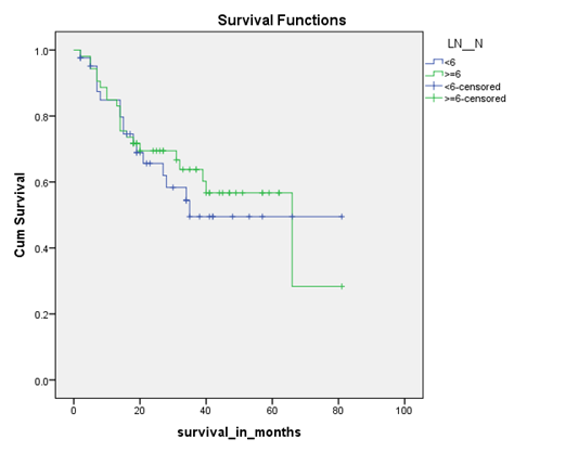
Fig 5: Kaplan Meier showing survival based on number of lymph nodes removed
Recurrence was seen in fifteen patients, which was most commonly seen in liver, followed by lymph nodes and peritoneum. At the time of study, forty patients were alive; thirty-seven patients were dead and seventeen patients are lost to follow-up.
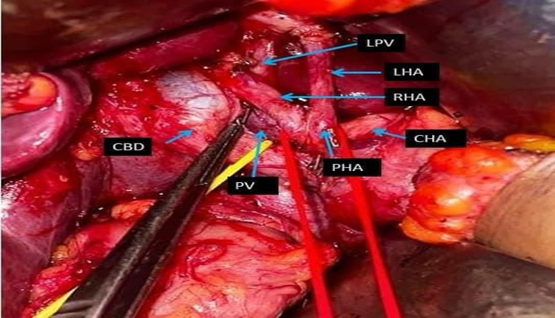
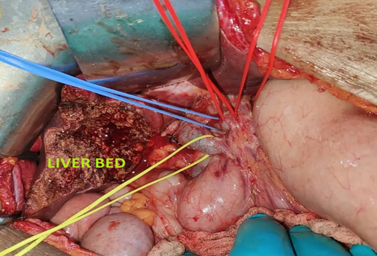
Fig 6 – Intraoperative images of radical cholecystectomy .
DISCUSSION:
Gallbladder carcinoma is one of the most aggressive malignancy of the biliary tract having very short-median survival. (14) Long-term survival is still poor in spite of improvement in the management of gallbladder cancer and is still dependent on curative surgical resection.(15) Radical curative surgery followed by adjuvant chemotherapy is a recommended treatment strategy.
Gallbladder cancer patients with regional lymph nodes (N1/N2) and distance metastasis have low survival rates (1.1% and 0.0%, respectively) and have higher mortality risk (HR = 1.468 and HR = 2.289, respectively). (16)
The present study showed that GBC predominantly affects mid aged females which is twice as common as in men. The median age in our study was 53 years. Multiple Indian studies had also found a higher female to male ratio of 2-3: 1 with younger onset of GBC in females as compared to males, at par with our study. (17, 18) Pain abdomen was the common clinical symptom seen in seventy-eight patients while jaundice was seen in twelve patients (12.6%). Jaundice may develop due to biliary obstruction by tumour infiltration of common bile duct or obstruction by enlarged porta hepatis nodes or sometimes due to liver dysfunction.
In this study, the median overall survival was 66 months with an HR of 21.5 (95% CI 23.81 – 108.18). Stage-specific survival revealed stage I had median overall survival of 82 months while stage IVB had 10 months. In a study by Yoshia Shiria , Jun sakata et al where they reported the survival outcome of 52 patients who underwent radical cholecystectomy it was seen that patients with pT1 and p T2 tumour (29 of 36) survived for more than 5 years, while most (10 of 12) patients with pT3 tumor and the only patient with pT4 tumor died within 5 years. (19) . This study proved that survival of patient with ca gall bladder depends on the stage of the disease. Survival becomes poor with advancement in stage.
Patients with node-negative disease had a median overall survival of 66 months compared to node-positive patients who had 16 months. Also patients who had ≥6 nodes removed had a median overall survival of 68 months compared to 35 months in patients who had <6 nodes removed. Thus demonstrating the fact that proper surgical resection is associated with better survival outcomes.In a study by Thorsten Oliver Goetze, Vittorio Paolucci et al the prognosis of the overall nodal-positive patients is bad.. None of the patients who had nodal-positive T1 carcinomas the 2 year survival mark. In T2 carcinoma cases, the 5 year survival of nodal-positive patients who undego a standardized lymph node dissection is only 9%. Patients with T3 carcinomas who had positive nodes did not reach 5 year survival. (20)
In our study routine frozen section of the cystic duct was done and in cases where it was positive extrahepatic biliary tree resection was done followed up with hepaticojejunostomy. Also routine frozen section of the interaortocaval node was done and in cases where it was found positive, further surgery was abandoned.
This study has several limitations firstly it being a retrospective study. Secondly, the follow-up period was less which could have affected our results. Also the follow-up data was not present in seventeen patients (18%) due to retrospective design and poor prognosis of this aggressive cancer. Moreover in forty-one patients (43.6%) either the patient's defaulted chemotherapy or data could not be retrieved regarding their adjuvant treatment which might have again affected the results.
The advantage of this study was that it was one of the first studies performed in this part of the country where gallbladder cancer incidence is very high. Such studies will help us to conduct other research activities in this field and help us identify other epidemiological and prognostic factors.
Neoadjuvant chemotherapy can be used for downstaging in T3 or highly suspicious cases and nodal positivity disease. Further studies in this regard may be undertaken to assess the response rates and outcomes.
CONCLUSION:
Despite curative intent treatment the overall survival of carcinoma gall bladder patients are very poor. The survival depends largely on tumour factors like disease stage and nodal status. Stage-specific survival revealed stage I had median overall survival of 82 months while stage IVB had 10 months . Patients with node-negative disease had a median overall survival of 66 months compared to node-positive patients who had 16 months.
Also patients who had ≥6 nodes removed had a median overall survival of 68 months compared to 35 months in patients who had <6 nodes removed , thus demonstrating the fact that proper surgical resection is associated with better survival outcomes . The future studies should focus on early detection techniques, with identification of possible risk factors in our region and effective methods of mass screening in high risk population. Nonetheless, much remains undefined in treatment of gallbladder cancer and further studies are needed.
Conflict of Interest- The author declare that they have no conflict of interest , financial or non financial ,related to the content of manuscript.
Funding- The author will like to declare that it was an investigator initiated study with no external financial support.
REFERENCES:
1. Hundal R, Shaffer EA. Gallbladder cancer: epidemiology and outcome. Clin Epidemiol. 2014;2014(6):99‐109.
2. Varshney S, Butturini G, Gupta R. Incidental carcinoma of the gallbladder. Eur J Surg Oncol. 2002;28:4-10.
3. Bray F, Ferlay J, Soerjomataram I, Siegel RL, Torre LA, Jemal A. Global cancer statistics 2018: GLOBOCAN estimates of incidence and mortality worldwide for 36 cancers in 185 countries. CA Cancer J Clin. 2018;68:394-424.
4. Kiran RP, Pokala N, Dudrick SJ. Incidence pattern and survival for gallbladder cancer over three decades--an analysis of 10301 patients. Ann Surg Oncol. 2007 Feb;14(2):827-32.
5. Toyonaga T, Chijiiwa K, Nakano K, Noshiro H, Yamaguchi K, Sada M, Terasaka R, Konomi K, Nishikata F, Tanaka M. Completion radical surgery after cholecystectomy for accidentally undiagnosed gallbladder carcinoma. World J Surg. 2003;27:266-271.
6. Mekeel KL, Hemming AW. Surgical management of gallbladder carcinoma: a review. J Gastrointest Surg. 2007;11:1188-1193.
7. Miller G, Jarnagin WR. Gallbladder carcinoma. Eur J Surg Oncol. 2008;34:306-312.
8. Okumura K, Gogna S, Gachabayov M, Felsenreich DM, McGuirk M, Rojas A, Quintero L, Seshadri R, Gu K, Dong XD. Gallbladder cancer: Historical treatment and new management options. World J Gastrointest Oncol 2021; 13(10): 1317-1335
9. Pandey M. Risk factors for gallbladder cancer: a reappraisal. Eur J Cancer Prev. 2003;12:15–24.
10. Dhir V, Mohandas KM. Epidemiology of digestive tract cancers in India IV. Gall bladder and pancreas. Indian J Gastroenterol. 1999;18(1):24‐28.
11. Lazcano‐Ponce EC, Miquel JF, Munoz N, et al. Epidemiology and molecular pathology of gallbladder cancer. CA Cancer J Clin. 2001;51(6):349‐364.
12. -Ponce EC, Miquel JF, Muńoz N, Herrero R, Ferrecio C, et al. Epidemiology and molecular pathology of gallbladder cancer. CA Cancer J Clin. 2001;51:349–364.
13. Randi G, Franceschi S, La Vecchia C. Gallbladder cancer worldwide: geographical distribution and risk factors. Int J Cancer. 2006;118:1591–1602.
14. Zhu AX, Hong TS, Hezel AF, et al. Current management of gallbladder carcinoma. Oncologist. 2010;15(2):168–181. doi: 10.1634/theoncologist.2009-0302.
15. Naveed S, Qari H, Thau CM, et al. Lymph node ratio is an important prognostic factor curatively resected gallbladder carcinoma, especially in node positive patients an experience from endemic region in a developing country. Euroasian J Hepto-Gastroentol. 2020;10(2):51–55.
16. Cai ZQ, Guo P, Si SB, Geng ZM, Chen C, Cong LL. Analysis of prognostic factors for survival after surgery for gallbladder cancer based on a Bayesian network. Sci Rep. 2017;7:293.
17. Pandey M, Pathak AK, Gautam A, Aryya NC, Shukla VK. Carcinoma of the gallbladder: a retrospective review of 99 cases. Dig Dis Sci 2001; 46: 1145-1151
18. Shukla VK, Khandelwal C, Roy SK, Vaidya MP. Primary carcinoma of the gall bladder: a review of a 16-year period at the University Hospital. J Surg Oncol 1985; 28: 32-35
19. Shirai Y, Sakata J, Wakai T, Ohashi T, Hatakeyama K. " Extended" radical cholecystectomy for gallbladder cancer: Long-term outcomes, indications and limitations. World Journal of Gastroenterology: WJG. 2012 Sep 9;18(34):4736.
20. Goetze TO, Paolucci V. The prognostic impact of positive lymph nodes in stages T1 to T3 incidental gallbladder carcinoma: results of the German Registry. Surgical endoscopy. 2012 May;26:1382-9.