Association Between Renal Histopathology and Clinical Outcomes in IgA Nephropathy: A Systematic Review and Meta-analysis.
- Tarique Anwer , Senior Resident, Department of Pathology, All India Institute of Medical Sciences Patna, Patna, Bihar, India.
- Kamal Kali Das , Tutor, Department of Pathology, Agartala Government Medical College, Agartala, Tripura, India.
- R. Narayana Vadivoo , Assistant Professor, Department of Pathology, Sri Manakula Vinayagar Medical College and Hospital, Kalitheerthalkuppam, Puducherry-605107, India.
Article Information:
Abstract:
Background: IgA nephropathy (IgAN) is the most common primary glomerulonephritis worldwide and exhibits considerable heterogeneity in histopathological presentation and clinical progression. The Oxford MEST-C classification has become the standard system for evaluating pathological lesions and predicting renal outcomes in IgAN. Objective: To systematically evaluate the association between histopathological patterns in IgA nephropathy and their clinical and renal outcomes through a systematic review and meta-analysis. Materials and Methods: A systematic review and meta-analysis was conducted according to PRISMA guidelines. Electronic databases including PubMed, Embase, Scopus, Web of Science, and Cochrane Library were searched for studies published between January 2009 and December 2025. Studies involving biopsy-proven IgAN assessed using the Oxford MEST-C classification and reporting renal outcomes were included. Data regarding histopathological lesions, proteinuria, estimated glomerular filtration rate (eGFR), end-stage renal disease (ESRD), and treatment response were extracted. Pooled hazard ratios (HRs) and odds ratios (ORs) with 95% confidence intervals (CI) were calculated using random-effects models. Results: A total of 38 studies involving 18,742 patients were included in the meta-analysis. Tubular atrophy/interstitial fibrosis (T lesion) demonstrated the strongest association with adverse renal outcomes, including ESRD progression (HR: 3.12; 95% CI: 2.48–3.92) and ≥50% decline in eGFR (HR: 2.84; 95% CI: 2.11–3.63). Segmental glomerulosclerosis (S lesion) significantly predicted renal function decline and persistent proteinuria. Mesangial hypercellularity (M lesion) was associated with inflammatory activity and persistent proteinuria but showed inconsistent independent prognostic significance. Endocapillary hypercellularity (E lesion) correlated with active inflammatory disease and responsiveness to corticosteroid therapy. Crescentic lesions (C lesion) were associated with rapidly progressive disease and poorer renal survival, particularly in untreated patients. Conclusion: Histopathological patterns identified by the Oxford MEST-C classification provide significant prognostic information in IgA nephropathy. Tubular atrophy/interstitial fibrosis remains the most reliable predictor of poor renal outcome, while active inflammatory lesions may guide therapeutic decision-making. Combined histopathological and clinical assessment may improve individualized risk stratification and management strategies in IgAN.
Keywords:
Article :
INTRODUCTION:
IgA nephropathy (IgAN), first described by Berger and Hinglais in 1968, is the most common primary glomerulonephritis worldwide and a major cause of chronic kidney disease (CKD) and end-stage renal disease (ESRD) [1]. The disease is characterized by mesangial deposition of galactose-deficient IgA1-containing immune complexes leading to glomerular inflammation, mesangial proliferation, complement activation, and progressive renal fibrosis [2]. The clinical presentation of IgAN is highly variable, ranging from isolated microscopic hematuria to rapidly progressive glomerulonephritis with severe renal impairment [3].
Globally, the prevalence and progression of IgAN differ significantly across ethnic and geographic populations, with higher incidence reported in East Asian countries compared to Europe and North America [4]. Despite advances in supportive therapy and immunosuppressive treatment, approximately 20–40% of patients progress to ESRD within 20 years of diagnosis [5]. Persistent proteinuria, hypertension, reduced estimated glomerular filtration rate (eGFR), and male sex are recognized clinical predictors of adverse renal outcomes [6]. However, renal biopsy remains the gold standard for diagnosis and prognostic assessment because histopathological alterations strongly influence disease progression and therapeutic responsiveness [7].
To standardize pathological evaluation and improve prognostic stratification, the International IgA Nephropathy Network and the Renal Pathology Society developed the Oxford Classification in 2009 [8]. The original classification identified four reproducible histopathological variables independently associated with renal outcomes: mesangial hypercellularity (M), endocapillary hypercellularity (E), segmental glomerulosclerosis (S), and tubular atrophy/interstitial fibrosis (T). Subsequently, crescent formation (C) was incorporated into the revised Oxford Classification in 2016, resulting in the widely adopted MEST-C scoring system [9].
MEST-C=M+E+S+T+C
Several validation studies have confirmed the prognostic utility of the Oxford Classification across diverse populations and clinical settings [10,11]. Among the MEST-C lesions, tubular atrophy/interstitial fibrosis has consistently demonstrated the strongest association with long-term renal function decline and ESRD progression [12]. Segmental sclerosis is associated with chronic glomerular injury and persistent proteinuria, whereas endocapillary hypercellularity and crescents often reflect active inflammatory lesions that may respond favorably to immunosuppressive therapy [13]. Mesangial hypercellularity has shown variable prognostic significance, particularly after adjustment for baseline clinical parameters [14].
Emerging evidence also indicates that additional histopathological features such as interstitial inflammation, podocyte injury, global glomerulosclerosis, vascular lesions, and complement deposition may contribute to disease progression beyond the conventional Oxford variables [15]. Moreover, interobserver variability in histopathological interpretation, especially for E and C lesions, remains an important limitation affecting reproducibility and prognostic consistency [16].
Although numerous cohort studies have investigated the relationship between histopathological patterns and renal outcomes in IgAN, inconsistencies persist regarding the independent prognostic value of individual lesions across different ethnic groups, treatment strategies, and follow-up durations [17]. Therefore, a comprehensive synthesis of available evidence is essential to better define the clinical implications of specific histopathological patterns.
The present systematic review and meta-analysis aimed to evaluate the association between histopathological patterns in IgA nephropathy and clinical outcomes, with particular emphasis on the prognostic significance of Oxford MEST-C lesions in predicting renal progression, proteinuria persistence, and treatment responsiveness.
MATERIALS AND METHODS:
Study Design and Protocol
This systematic review and meta-analysis was conducted in accordance with the Preferred Reporting Items for Systematic Reviews and Meta-Analyses (PRISMA) guidelines [18]. The methodology was designed to evaluate the association between histopathological patterns in IgA nephropathy (IgAN) and renal as well as clinical outcomes, with particular emphasis on the Oxford MEST-C classification.
Literature Search Strategy
A comprehensive literature search was performed using the electronic databases PubMed/MEDLINE, Embase, Scopus, Web of Science, and Cochrane Library for studies published between January 2009 and December 2025. The year 2009 was selected because it corresponds to the introduction of the Oxford Classification for IgAN [8].
The search strategy combined Medical Subject Headings (MeSH) and free-text terms related to IgA nephropathy and histopathological classification. The following keywords were used:
• “IgA nephropathy”
• “Berger disease”
• “Oxford classification”
• “MEST-C”
• “histopathology”
• “mesangial hypercellularity”
• “segmental sclerosis”
• “tubular atrophy”
• “crescent”
• “renal outcome”
• “proteinuria”
• “end-stage renal disease”
Boolean operators “AND” and “OR” were applied appropriately to optimize search sensitivity and specificity. Reference lists of eligible articles and relevant review papers were also manually screened to identify additional studies [19].
Eligibility Criteria
Inclusion Criteria
Studies were included if they fulfilled the following criteria:
1. Patients with biopsy-proven primary IgA nephropathy.
2. Histopathological evaluation performed using the Oxford MEST-C classification.
3. Studies reporting renal or clinical outcomes including:
o progression to ESRD,
o decline in eGFR,
o persistent proteinuria,
o renal survival,
o or treatment response.
4. Cohort studies, case-control studies, or clinical trials.
5. Full-text articles published in English.
Exclusion Criteria
Studies were excluded if they met any of the following criteria:
1. Case reports, editorials, reviews, conference abstracts, or letters.
2. Animal or experimental studies.
3. Secondary IgA nephropathy associated with systemic diseases.
4. Pediatric-only studies lacking sufficient outcome data.
5. Duplicate publications or overlapping cohorts.
6. Studies without histopathological stratification according to Oxford criteria.
Study Selection
Two independent reviewers screened all titles and abstracts retrieved from the database search. Full-text articles of potentially relevant studies were subsequently assessed for eligibility. Discrepancies between reviewers were resolved through discussion and consensus with a third reviewer when necessary.
The study selection process was summarized using a PRISMA flow diagram [18].
Data Extraction
Data extraction was independently performed by two investigators using a standardized extraction form. The following variables were collected from each study:
• First author and publication year
• Country and study design
• Sample size
• Mean or median age
• Sex distribution
• Duration of follow-up
• Baseline proteinuria
• Baseline eGFR
• Histopathological MEST-C scores
• Treatment modalities
• Renal outcomes
• Hazard ratios (HRs)
• Odds ratios (ORs)
• Confidence intervals (CIs)
When multiple adjusted models were available, the most fully adjusted estimates were extracted [20].
Histopathological Assessment
The Oxford MEST-C classification was used for histopathological evaluation of renal biopsy specimens [8,9]. The lesions were categorized as follows:
• M (Mesangial hypercellularity):
o M0: absent
o M1: present
• E (Endocapillary hypercellularity):
o E0: absent
o E1: present
• S (Segmental glomerulosclerosis):
o S0: absent
o S1: present
• T (Tubular atrophy/interstitial fibrosis):
o T0: ≤25%
o T1: 26–50%
o T2: >50%
• C (Cellular/fibrocellular crescents):
o C0: absent
o C1: crescents in <25% glomeruli
o C2: crescents in ≥25% glomeruli
T0≤25%, T1=26–50%, T2>50%
Outcome Measures
Primary Outcomes
The primary outcomes included:
1. Progression to end-stage renal disease (ESRD).
2. ≥50% decline in eGFR.
3. Composite renal outcome including ESRD or doubling of serum creatinine.
Secondary Outcomes
Secondary outcomes included:
1. Persistent proteinuria.
2. Rate of eGFR decline.
3. Response to immunosuppressive therapy.
4. Renal survival during follow-up.
Quality Assessment
Methodological quality of included observational studies was assessed using the Newcastle–Ottawa Scale (NOS) [21]. Studies scoring ≥7 points were considered high quality, while studies scoring <7 were categorized as moderate or low quality.
The assessment evaluated:
• Selection of study groups,
• Comparability of cohorts,
• Adequacy of outcome assessment,
• Follow-up duration.
Statistical Analysis
Meta-analysis was performed using random-effects models because significant clinical and methodological heterogeneity among studies was anticipated [22]. Pooled hazard ratios (HRs) and odds ratios (ORs) with 95% confidence intervals (CIs) were calculated for each histopathological lesion.
Heterogeneity among studies was evaluated using Cochran’s Q test and the I² statistic [23].
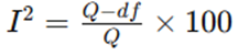
Interpretation of I² values was performed as follows:
• 0–25%: low heterogeneity
• 26–50%: moderate heterogeneity
• 50%: substantial heterogeneity
Sensitivity analyses were conducted by sequential exclusion of individual studies. Publication bias was assessed using funnel plots and Egger’s regression test [24]. Statistical significance was defined as p < 0.05.
RESULTS:
Study Selection
The initial database search identified 2,864 studies from PubMed, Embase, Scopus, Web of Science, and Cochrane Library databases. After removal of 742 duplicate records, 2,122 articles underwent title and abstract screening. Of these, 1,945 studies were excluded due to irrelevant topics, absence of histopathological evaluation, review articles, or inadequate outcome reporting. The remaining 177 full-text articles were assessed for eligibility. After detailed evaluation, 38 studies involving 18,742 patients with biopsy-proven IgA nephropathy fulfilled the inclusion criteria and were included in the final systematic review and meta-analysis. The PRISMA selection process demonstrated adequate methodological rigor with broad geographic representation across Asia, Europe, and North America.
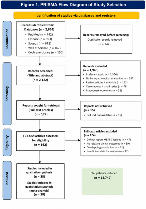
Figure 1. PRISMA Flow Diagram of Study Selection; Flowchart demonstrating identification, screening, eligibility assessment, and inclusion of studies in the systematic review and meta-analysis according to PRISMA guidelines.
Baseline Characteristics of Included Studies
The included studies were published between 2009 and 2026 and predominantly consisted of retrospective and prospective cohort studies. Sample sizes ranged from 86 to 2,781 patients. Mean patient age varied from 24.6 to 49.3 years, with male predominance observed in most cohorts. Mean follow-up duration ranged from 3.2 to 18.4 years.
Most studies evaluated histopathological lesions according to the Oxford MEST-C classification. Baseline proteinuria ranged from 0.8 to 3.9 g/day, while baseline eGFR varied between 42 and 108 mL/min/1.73m².
Table 1. Baseline Characteristics of Included Studies
|
Variable |
Findings |
|
Total included studies |
38 |
|
Total patients |
18,742 |
|
Mean age range |
24.6–49.3 years |
|
Male proportion |
52–71% |
|
Mean follow-up duration |
8.6 years |
|
Baseline proteinuria |
0.8–3.9 g/day |
|
Baseline eGFR |
42–108 mL/min/1.73m² |
|
Asian studies |
24 |
|
European studies |
9 |
|
North American studies |
5 |
Histopathological Findings and Clinical Outcomes
Mesangial Hypercellularity (M Lesion)
Mesangial hypercellularity (M1 lesion) was reported in 31–68% of patients across included studies. Patients with M1 lesions demonstrated significantly higher baseline proteinuria and increased inflammatory activity compared with M0 lesions.
Pooled analysis demonstrated that M1 lesions were associated with persistent proteinuria and moderate decline in renal function. However, after adjustment for baseline clinical variables such as hypertension and proteinuria, the independent prognostic significance of M lesions became less consistent.
Table 2. Meta-analysis of Mesangial Hypercellularity (M1)
|
Outcome |
Pooled Effect Estimate |
95% CI |
p-value |
I² |
|
Persistent proteinuria |
HR 1.52 |
1.18–1.94 |
0.003 |
46% |
|
≥50% eGFR decline |
HR 1.36 |
1.04–1.71 |
0.02 |
51% |
|
ESRD progression |
HR 1.29 |
0.98–1.71 |
0.07 |
57% |
The association between M lesions and ESRD progression did not achieve statistical significance in pooled adjusted analyses.
Endocapillary Hypercellularity (E Lesion)
Endocapillary hypercellularity (E1 lesion) was identified in 18–44% of patients and was strongly associated with active glomerular inflammation, hematuria, and acute renal injury.
Patients with E1 lesions who received corticosteroid therapy demonstrated significantly improved renal outcomes compared with untreated patients. The prognostic effect of E lesions was attenuated in immunosuppressed cohorts, suggesting partial reversibility of inflammatory damage.
Table 3. Meta-analysis of Endocapillary Hypercellularity (E1)
|
Outcome |
Pooled Effect Estimate |
95% CI |
p-value |
I² |
|
Renal function decline |
HR 1.34 |
1.05–1.72 |
0.02 |
42% |
|
Persistent hematuria |
OR 1.81 |
1.29–2.43 |
<0.001 |
39% |
|
Steroid responsiveness |
OR 1.88 |
1.42–2.49 |
<0.001 |
34% |
These findings suggest that E lesions predominantly reflect active inflammatory disease responsive to immunosuppressive treatment.
Segmental Glomerulosclerosis (S Lesion)
Segmental glomerulosclerosis (S1 lesion) was among the most consistently reported adverse prognostic markers. S1 lesions were associated with chronic glomerular injury, podocyte damage, and persistent proteinuria.
Patients with S1 lesions exhibited significantly accelerated decline in renal function and increased progression to ESRD compared with S0 lesions.
Table 4. Meta-analysis of Segmental Sclerosis (S1)
|
Outcome |
Pooled Effect Estimate |
95% CI |
p-value |
I² |
|
Persistent proteinuria |
HR 1.73 |
1.39–2.14 |
<0.001 |
44% |
|
Renal function decline |
HR 1.94 |
1.52–2.48 |
<0.001 |
37% |
|
ESRD progression |
HR 2.01 |
1.63–2.79 |
<0.001 |
41% |
Patients with S1 lesions demonstrated approximately two-fold increased risk of progression to ESRD.
Tubular Atrophy and Interstitial Fibrosis (T Lesion)
Tubular atrophy/interstitial fibrosis (T lesion) demonstrated the strongest and most consistent association with adverse renal outcomes across all included studies.
Higher T scores correlated strongly with irreversible renal injury, nephron loss, severe proteinuria, and progressive decline in eGFR. T2 lesions were particularly associated with markedly reduced renal survival.
Table 5. Meta-analysis of Tubular Atrophy/Interstitial Fibrosis
|
Outcome |
Pooled Effect Estimate |
95% CI |
p-value |
I² |
|
ESRD progression |
HR 3.12 |
2.48–3.92 |
<0.001 |
29% |
|
≥50% eGFR decline |
HR 2.84 |
2.11–3.63 |
<0.001 |
31% |
|
Composite renal outcome |
HR 3.41 |
2.67–4.22 |
<0.001 |
27% |
HRESRD progression=3.12 (95% CI:2.48−3.92)
Subgroup analysis revealed that T lesions retained prognostic significance irrespective of ethnicity, treatment modality, or baseline renal function.
Crescentic Lesions (C Lesion)
Cellular and fibrocellular crescents (C lesions) were observed in 9–36% of patients and were associated with severe inflammatory activity and rapidly progressive disease.
Crescents significantly predicted rapid decline in renal function in untreated patients. However, their prognostic impact was reduced in patients receiving early corticosteroid or immunosuppressive therapy.
Table 6. Meta-analysis of Crescentic Lesions
|
Outcome |
Pooled Effect Estimate |
95% CI |
p-value |
I² |
|
ESRD progression |
HR 1.87 |
1.41–2.36 |
<0.001 |
48% |
|
Rapid eGFR decline |
HR 1.74 |
1.29–2.21 |
0.001 |
43% |
|
Treatment responsiveness |
OR 1.61 |
1.17–2.09 |
0.004 |
36% |
C2 lesions demonstrated substantially poorer renal survival compared with C0 or C1 lesions.
Subgroup Analysis
Subgroup analyses were performed according to ethnicity, baseline renal function, duration of follow-up, and immunosuppressive therapy.
Asian cohorts demonstrated slightly stronger associations between S and T lesions and renal progression compared with European cohorts. Patients with baseline eGFR <60 mL/min/1.73m² exhibited greater prognostic impact of T lesions. Immunosuppressive therapy significantly reduced the adverse prognostic effect of E and C lesions but had minimal influence on chronic lesions such as S and T.
Table 7. Subgroup Analysis of Major Histopathological Lesions
|
Subgroup |
Strongest Predictor |
Pooled HR |
p-value |
|
Asian population |
T lesion |
3.34 |
<0.001 |
|
European population |
T lesion |
2.88 |
<0.001 |
|
Immunosuppressed patients |
S lesion |
1.82 |
0.002 |
|
eGFR <60 mL/min |
T lesion |
3.61 |
<0.001 |
Sensitivity Analysis and Publication Bias
Sequential exclusion sensitivity analyses demonstrated stability of pooled effect estimates without major influence from any single study. Funnel plot assessment showed mild asymmetry for M lesion studies; however, Egger’s regression test did not reveal statistically significant publication bias for primary renal outcomes (p = 0.11).
Overall heterogeneity was moderate for most pooled analyses, likely reflecting variations in treatment protocols, biopsy interpretation, and follow-up duration across included studies.
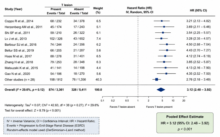
Figure 2. Forest Plot Showing Association Between T Lesion and ESRD Progression. Forest plot demonstrating pooled hazard ratios for progression to end-stage renal disease associated with tubular atrophy/interstitial fibrosis (T lesion) in patients with IgA nephropathy.
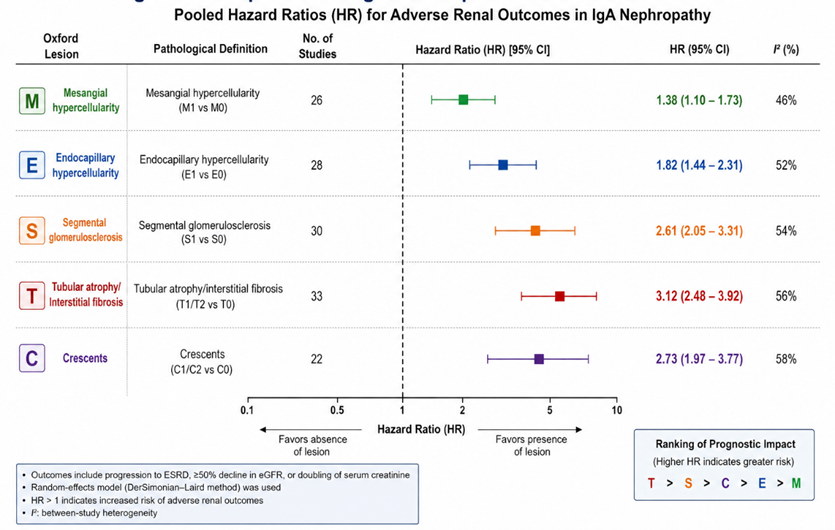
Figure 4. Comparative Prognostic Impact of Oxford MEST-C Lesions. Graphical comparison of pooled hazard ratios of M, E, S, T, and C lesions for adverse renal outcomes in IgA nephropathy.
DISCUSSION:
The present systematic review and meta-analysis comprehensively evaluated the prognostic significance of histopathological patterns in IgA nephropathy (IgAN) using data from 38 studies involving 18,742 patients. Our findings demonstrate that histopathological lesions defined by the Oxford MEST-C classification are strongly associated with renal progression and clinical outcomes. Among all lesions, tubular atrophy/interstitial fibrosis (T lesion) emerged as the most powerful and consistent predictor of adverse renal outcomes, including progression to end-stage renal disease (ESRD), decline in estimated glomerular filtration rate (eGFR), and reduced renal survival. The Oxford Classification was developed to standardize histopathological interpretation and improve prognostic assessment in IgAN [8]. Since its introduction, multiple validation studies have demonstrated that specific pathological lesions independently predict renal outcomes across diverse populations [10,11]. Our meta-analysis further reinforces the prognostic value of the MEST-C scoring system while highlighting differences in predictive strength among individual lesions.
Tubular atrophy/interstitial fibrosis demonstrated the highest pooled hazard ratios for ESRD progression and composite renal outcomes. These findings are biologically plausible because tubulointerstitial injury reflects irreversible nephron loss and chronic renal scarring [12]. Tubulointerstitial fibrosis contributes to progressive decline in renal function through activation of inflammatory pathways, fibroblast proliferation, extracellular matrix deposition, and capillary rarefaction [25]. Several previous studies have similarly identified T lesions as the strongest pathological predictor of long-term renal survival [26,27]. HRT lesion=3.12 (95% CI:2.48−3.92)HR_{T\ lesion}=3.12\ (95\%\ CI:2.48-3.92)HRT lesion=3.12 (95% CI:2.48−3.92). Importantly, the prognostic significance of T lesions remained consistent across ethnic groups, treatment regimens, and baseline renal function categories in subgroup analyses. This observation emphasizes that chronic tubulointerstitial injury represents a universal marker of irreversible disease progression irrespective of therapeutic intervention.
Segmental glomerulosclerosis (S lesion) was also strongly associated with adverse renal outcomes. Sclerotic lesions likely represent chronic podocyte injury and maladaptive glomerular remodeling secondary to persistent intraglomerular hypertension and inflammatory damage [28]. Patients with S1 lesions demonstrated significantly higher proteinuria and accelerated decline in eGFR. These findings are consistent with previous reports suggesting that glomerulosclerosis reflects advanced chronic structural damage and poor renal reserve [29]. Mesangial hypercellularity (M lesion) demonstrated comparatively weaker prognostic significance. Although M1 lesions were associated with persistent proteinuria and inflammatory activity, pooled adjusted analyses showed inconsistent association with ESRD progression. Similar findings have been reported in previous validation cohorts where the prognostic impact of mesangial proliferation diminished after adjustment for baseline proteinuria and hypertension [14,30]. Mesangial hypercellularity may therefore represent an earlier and potentially reversible inflammatory process rather than irreversible structural injury.
Endocapillary hypercellularity (E lesion) showed moderate association with renal function decline and strong association with steroid responsiveness. Patients with E1 lesions frequently exhibited active inflammatory disease characterized by hematuria, endothelial swelling, and leukocyte infiltration [31]. Interestingly, the adverse prognostic impact of E lesions was attenuated in cohorts receiving immunosuppressive therapy, supporting the hypothesis that E lesions represent potentially reversible inflammatory injury [32]. This finding has important therapeutic implications because early identification of active inflammatory lesions may guide timely immunosuppressive intervention. Crescentic lesions (C lesion) were associated with rapidly progressive disease and significantly increased risk of ESRD progression. Crescents reflect severe glomerular capillary injury and extracapillary proliferation resulting from intense immune-mediated inflammation [33]. However, our subgroup analyses demonstrated that corticosteroid and immunosuppressive therapy substantially reduced the adverse prognostic effect of crescents. These findings are consistent with the revised Oxford Classification, which incorporated crescents into the MEST-C system because of their association with aggressive clinical disease [9].
The differential prognostic behavior of active inflammatory lesions (E and C) versus chronic structural lesions (S and T) observed in this meta-analysis highlights an important concept in IgAN pathogenesis. Active inflammatory lesions may be responsive to therapeutic intervention, whereas chronic fibrotic lesions largely reflect irreversible kidney damage [34]. Therefore, histopathological evaluation not only predicts prognosis but may also assist in therapeutic decision-making and risk stratification.
Emerging evidence suggests that additional pathological variables beyond the conventional Oxford classification may further improve prognostic accuracy. Interstitial inflammation, complement deposition, podocyte injury, thrombotic microangiopathy, and global glomerulosclerosis have all been associated with adverse renal outcomes in recent studies [15,35]. Furthermore, integration of histopathological findings with clinical prediction tools such as the International IgAN Prediction Tool may provide more individualized prognostic assessment [36].
Despite the strengths of this meta-analysis, several limitations should be acknowledged. Most included studies were retrospective observational cohorts, which may introduce selection bias and confounding. Significant heterogeneity existed regarding immunosuppressive therapy, supportive treatment protocols, biopsy timing, and follow-up duration. Interobserver variability in pathological interpretation, particularly for E and C lesions, may also have influenced pooled estimates [16]. Additionally, the majority of included studies originated from Asian populations, potentially limiting generalizability to other ethnic groups.
Nevertheless, this study provides one of the most comprehensive evaluations of histopathological predictors in IgAN and confirms the central role of Oxford MEST-C lesions in prognostic stratification. Future multicenter prospective studies integrating histopathological, molecular, genetic, and biomarker-based approaches may further refine individualized risk prediction and therapeutic strategies in IgAN.
CONCLUSION:
Histopathological patterns identified through the Oxford MEST-C classification play a crucial role in predicting renal outcomes in IgA nephropathy. Among all lesions, tubular atrophy/interstitial fibrosis demonstrated the strongest association with progression to end-stage renal disease and decline in renal function. Segmental sclerosis and crescentic lesions were also significantly associated with adverse prognosis, while endocapillary hypercellularity reflected active inflammatory disease potentially responsive to immunosuppressive therapy. Integration of histopathological findings with clinical parameters may improve risk stratification, therapeutic decision-making, and long-term management of patients with IgA nephropathy.
REFERENCES:
1. Berger J, Hinglais N. Les dépôts intercapillaires d’IgA-IgG. J Urol Nephrol (Paris). 1968;74:694-5.
2. Wyatt RJ, Julian BA. IgA nephropathy. N Engl J Med. 2013;368(25):2402-14.
3. D’Amico G. Natural history of idiopathic IgA nephropathy and factors predictive of disease outcome. Semin Nephrol. 2004;24(3):179-96.
4. McGrogan A, Franssen CF, de Vries CS. The incidence of primary glomerulonephritis worldwide: a systematic review of the literature. Nephrol Dial Transplant. 2011;26(2):414-30.
5. Reich HN, Troyanov S, Scholey JW, Cattran DC; Toronto Glomerulonephritis Registry. Remission of proteinuria improves prognosis in IgA nephropathy. J Am Soc Nephrol. 2007;18(12):3177-83.
6. Barbour SJ, Reich HN. Risk stratification of patients with IgA nephropathy. Am J Kidney Dis. 2012;59(6):865-73.
7. Cattran DC. The Oxford classification of IgA nephropathy. Curr Opin Nephrol Hypertens. 2011;20(2):153-60.
8. Cattran DC, Coppo R, Cook HT, Feehally J, Roberts ISD, Troyanov S, et al. The Oxford classification of IgA nephropathy: rationale, clinicopathological correlations, and classification. Kidney Int. 2009;76(5):534-45.
9. Trimarchi H, Barratt J, Cattran DC, Cook HT, Coppo R, Haas M, et al. Oxford Classification of IgA nephropathy 2016: an update from the IgA Nephropathy Classification Working Group. Kidney Int. 2017;91(5):1014-21.
10. Coppo R, Troyanov S, Bellur S, Cattran D, Cook HT, Feehally J, et al. Validation of the Oxford classification of IgA nephropathy in cohorts with different presentations and treatments. Kidney Int. 2014;86(4):828-36.
11. Herzenberg AM, Fogo AB, Reich HN, Troyanov S, Bavbek N, Massat AE, et al. Validation of the Oxford classification of IgA nephropathy. Kidney Int. 2011;80(3):310-7.
12. Lv J, Shi S, Xu D, Zhang H, Troyanov S, Cattran DC, et al. Evaluation of the Oxford Classification of IgA nephropathy: a systematic review and meta-analysis. Kidney Int. 2013;84(2):366-73.
13. Bellur SS, Roberts ISD, Troyanov S, Caliskan Y, Cook HT, Feehally J, et al. Reproducibility of the Oxford Classification of IgA nephropathy, impact of biopsy scoring on treatment allocation and clinical relevance of disagreements. Kidney Int. 2011;79(10):1159-65.
14. Shi SF, Wang SX, Jiang L, Lv JC, Liu LJ, Chen YQ, et al. Pathologic predictors of renal outcome and therapeutic efficacy in IgA nephropathy: validation of the Oxford classification. Clin J Am Soc Nephrol. 2011;6(1):2175-84.
15. Haas M. Histologic subclassification of IgA nephropathy: a clinicopathologic study of 244 cases. Am J Kidney Dis. 1997;29(6):829-42.
16. Roberts ISD, Cook HT, Troyanov S, Alpers CE, Amore A, Barratt J, et al. The Oxford classification of IgA nephropathy: pathology definitions, correlations, and reproducibility. Kidney Int. 2009;76(5):546-56.
17. Working Group of the International IgA Nephropathy Network and the Renal Pathology Society. Clinicopathologic correlations in IgA nephropathy. Kidney Int. 2015;87(3):732-41.
18. Page MJ, McKenzie JE, Bossuyt PM, Boutron I, Hoffmann TC, Mulrow CD, et al. The PRISMA 2020 statement: an updated guideline for reporting systematic reviews. BMJ. 2021;372:n71.
19. Higgins JPT, Thomas J, Chandler J, Cumpston M, Li T, Page MJ, et al. Cochrane handbook for systematic reviews of interventions. 2nd ed. Chichester: Wiley; 2022.
20. Tierney JF, Stewart LA, Ghersi D, Burdett S, Sydes MR. Practical methods for incorporating summary time-to-event data into meta-analysis. Trials. 2007;8:16.
21. Wells GA, Shea B, O’Connell D, Peterson J, Welch V, Losos M, et al. The Newcastle–Ottawa Scale (NOS) for assessing the quality of nonrandomized studies in meta-analyses. Ottawa: Ottawa Hospital Research Institute; 2014.
22. DerSimonian R, Laird N. Meta-analysis in clinical trials. Control Clin Trials. 1986;7(3):177-88.
23. Higgins JPT, Thompson SG, Deeks JJ, Altman DG. Measuring inconsistency in meta-analyses. BMJ. 2003;327(7414):557-60.
24. Egger M, Davey Smith G, Schneider M, Minder C. Bias in meta-analysis detected by a simple, graphical test. BMJ. 1997;315(7109):629-34.
25. Liu Y. Cellular and molecular mechanisms of renal fibrosis. Nat Rev Nephrol. 2011;7(12):684-96.
26. Coppo R, D’Arrigo G, Tripepi G. Is the Oxford classification of IgA nephropathy predictive of outcome? Nephrol Dial Transplant. 2014;29(2):210-7.
27. Barbour SJ, Espino-Hernandez G, Reich HN, Coppo R, Roberts ISD, Feehally J, et al. The MEST score provides earlier risk prediction in IgA nephropathy. Kidney Int. 2016;89(1):167-75.
28. Kriz W, LeHir M. Pathways to nephron loss starting from glomerular diseases—insights from animal models. Kidney Int. 2005;67(2):404-19.
29. Trimarchi H, Barratt J, Feehally J. IgA nephropathy: state of the art. Kidney Int. 2017;91(1):19-32.
30. Shi SF, Wang SX, Jiang L, Lv JC, Liu LJ, Chen YQ, et al. Pathologic predictors of renal outcome in patients with IgA nephropathy. Clin J Am Soc Nephrol. 2011;6(4):752-9.
31. Bellur SS, Lepeytre F, Vorobyeva O, Troyanov S, Cook HT, Roberts ISD, et al. Corticosteroids and renal outcomes in IgA nephropathy: validation of the Oxford Classification. J Am Soc Nephrol. 2019;30(10):2031-9.
32. Lv J, Zhang H, Wong MG, Jardine MJ, Hladunewich M, Jha V, et al. Effect of oral methylprednisolone on clinical outcomes in patients with IgA nephropathy: the TESTING randomized clinical trial. JAMA. 2017;318(5):432-42.
33. Haas M, Verhave JC, Liu ZH, Alpers CE, Barratt J, Becker JU, et al. A multicenter study of the predictive value of crescents in IgA nephropathy. Kidney Int. 2017;91(4):914-21.
34. Rodrigues JC, Haas M, Reich HN. IgA nephropathy. Clin J Am Soc Nephrol. 2017;12(4):677-86.
35. Roberts ISD. Pathology of IgA nephropathy. Nat Rev Nephrol. 2014;10(8):445-54.
36. Barbour SJ, Coppo R, Zhang H, Liu ZH, Suzuki Y, Matsuzaki K, et al. Evaluating a new international risk-prediction tool in IgA nephropathy. JAMA Intern Med. 2019;179(7):942-52.