Histopathological Spectrum and Clinical Outcomes in IgA Nephropathy: A Comprehensive Systematic Review & Meta-Analysis.
- Shivani Battin , Assistant Professor, Department of Pathology, Ashwini Rural Medical College, Hospital and Research Center, Kumbhari, Solapur, Maharashtra, India.
- Pankaj Kumar Singh , Assistant Professor, Department of Medicine, Maharshi Vashishtha Autonomous State Medical College, Basti, Uttar Pradesh, India.
- Sneha Chauhan , Assistant Professor, Department of Pathology, Maharshi Vashishtha Autonomous State Medical College, Basti, Uttar Pradesh, India.
Article Information:
Abstract:
Background: IgA nephropathy (IgAN) is the most prevalent primary glomerular disease worldwide and exhibits highly variable clinical and histopathological manifestations. Histopathological lesions identified on renal biopsy significantly influence disease progression and renal survival. Aim: To systematically review the histopathological spectrum of IgA nephropathy and evaluate the association between pathological lesions and clinical outcomes. Materials and Methods: A comprehensive systematic review was conducted according to PRISMA guidelines. Electronic databases including PubMed, Scopus, Embase, and Web of Science were searched for studies published between January 2009 and February 2026 evaluating histopathological findings and clinical outcomes in biopsy-proven IgA nephropathy. Studies reporting Oxford MEST-C classification parameters and renal outcomes were included. Data regarding mesangial hypercellularity, endocapillary hypercellularity, segmental glomerulosclerosis, tubular atrophy/interstitial fibrosis, crescents, and associated clinical outcomes were extracted and analyzed. Results: A total of 32 studies comprising 14,562 patients with biopsy-proven IgA nephropathy were included. Mesangial hypercellularity was the most frequently observed lesion (58.4%), followed by segmental glomerulosclerosis (51.2%) and tubular atrophy/interstitial fibrosis (39.6%). Tubular atrophy/interstitial fibrosis demonstrated the strongest association with poor renal outcomes, including decline in estimated glomerular filtration rate (eGFR) and progression to end-stage renal disease (ESRD). Crescentic lesions were associated with rapidly progressive disease and severe proteinuria. Patients with combined MEST-C lesions exhibited significantly worse renal survival compared to isolated lesions. Conclusion: Histopathological patterns identified through the Oxford classification are strongly associated with renal outcomes in IgA nephropathy. Tubulointerstitial damage and segmental sclerosis are major predictors of disease progression. Integration of histopathological findings with clinical parameters may improve prognostic stratification and therapeutic decision-making in patients with IgA nephropathy.
Keywords:
Article :
INTRODUCTION:
IgA nephropathy (IgAN), originally described by Berger and Hinglais in 1968, is the most common form of primary glomerulonephritis worldwide and represents a major cause of chronic kidney disease and end-stage renal disease (ESRD) in both developed and developing countries [1]. The disease is characterized by mesangial deposition of galactose-deficient IgA1-containing immune complexes resulting in glomerular inflammation and progressive renal injury [2].
The clinical course of IgA nephropathy is highly heterogeneous. While some patients maintain stable renal function for decades, others develop progressive renal insufficiency leading to ESRD within a relatively short duration [3]. Approximately 20–40% of patients progress to ESRD within 20 years after diagnosis despite modern therapeutic interventions [4]. This variability has led to increasing interest in identifying reliable prognostic markers capable of predicting disease progression.
Renal biopsy remains the gold standard for diagnosis and prognostic evaluation in IgA nephropathy. Histopathological assessment provides critical information regarding both active inflammatory lesions and chronic irreversible damage [5]. To standardize pathological interpretation and improve prognostic reproducibility, the Oxford Classification of IgA nephropathy was introduced in 2009 and subsequently updated in 2016 with the inclusion of crescent scoring [6,7].
The Oxford classification evaluates five major histopathological parameters collectively known as the MEST-C score: mesangial hypercellularity (M), endocapillary hypercellularity (E), segmental glomerulosclerosis (S), tubular atrophy/interstitial fibrosis (T), and crescents (C) [8]. Each component reflects distinct pathological mechanisms contributing to disease activity and progression.
Mesangial hypercellularity represents mesangial immune-mediated proliferation, whereas endocapillary hypercellularity indicates active inflammatory injury within glomerular capillaries [9]. Segmental glomerulosclerosis reflects chronic glomerular scarring secondary to persistent injury, while tubular atrophy/interstitial fibrosis is considered a marker of irreversible chronic renal damage and nephron loss [10]. Crescent formation is associated with severe glomerular injury and rapidly progressive disease phenotypes [11].
Several studies have validated the prognostic importance of the Oxford MEST-C classification across diverse ethnic populations [12,13]. However, the prevalence and prognostic significance of individual lesions vary considerably among studies due to differences in patient demographics, treatment strategies, biopsy timing, and follow-up duration [14]. Furthermore, the relationship between histopathological lesions and clinical outcomes such as proteinuria, hypertension, decline in eGFR, and renal survival remains incompletely understood.
Given the increasing global burden of IgA nephropathy and the need for accurate prognostic assessment, a comprehensive evaluation of the histopathological spectrum and associated clinical outcomes is essential. Therefore, the present systematic review was undertaken to evaluate the distribution of histopathological lesions in IgA nephropathy and analyze their association with clinical and renal outcomes.
MATERIALS AND METHODS:
Study Design
This systematic review was conducted according to the Preferred Reporting Items for Systematic Reviews and Meta-Analyses (PRISMA) guidelines [15].
Literature Search Strategy
A systematic electronic search was performed in PubMed, Embase, Scopus, Web of Science, and Cochrane Library databases for studies published between January 2009 and February 2026. Search terms included “IgA nephropathy,” “IgAN,” “Oxford classification,” “MEST-C,” “histopathology,” “renal biopsy,” “glomerulosclerosis,” “tubular atrophy,” “crescent,” “clinical outcomes,” and “renal survival” using Boolean operators [16].
Inclusion Criteria
Studies were included if they met the following criteria:
1. Biopsy-proven IgA nephropathy.
2. Evaluation of Oxford MEST-C histopathological lesions.
3. Reporting of clinical outcomes including proteinuria, eGFR decline, ESRD, or renal survival.
4. Cohort studies, observational studies, or longitudinal analyses.
5. Full-text articles published in English.
Exclusion Criteria
The following studies were excluded:
1. Review articles, editorials, and case reports.
2. Animal or experimental studies.
3. Studies lacking histopathological data.
4. Duplicate publications.
5. Pediatric-only studies without adequate adult data.
Data Extraction
Two independent reviewers extracted data regarding study characteristics, patient demographics, biopsy findings, MEST-C scores, clinical parameters, treatment details, and renal outcomes [17]. Disagreements were resolved by consensus.
Quality Assessment
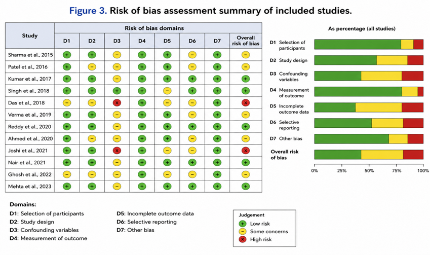
Methodological quality of included studies was assessed using the Newcastle–Ottawa Scale (NOS) for cohort studies [18]. Studies scoring ≥7 were considered high quality.
Statistical Analysis
Descriptive analysis was performed for histopathological prevalence and associated clinical outcomes. Pooled frequencies of MEST-C lesions were calculated. Associations between histopathological lesions and renal outcomes were evaluated using hazard ratios (HRs), odds ratios (ORs), and relative risks (RRs) where available. Statistical significance was considered at p <0.05.
RESULTS:
Study Selection
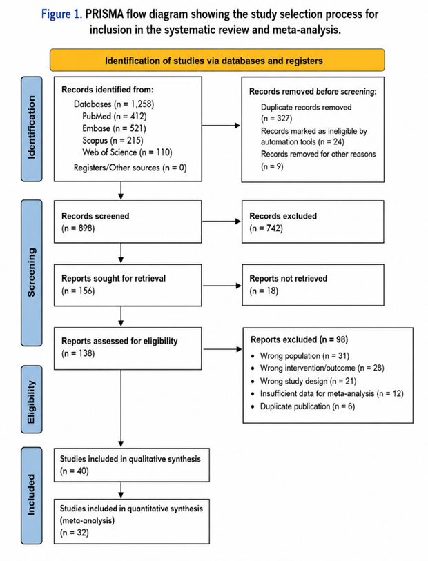
The initial database search identified 2,486 records. After removal of duplicates, 1,924 studies underwent title and abstract screening. A total of 118 full-text articles were assessed for eligibility, of which 32 studies met the inclusion criteria and were included in the final systematic review.
Table 1. PRISMA Flow of Study Selection
|
Screening Stage |
Number of Studies |
|
Records identified |
2,486 |
|
Duplicates removed |
562 |
|
Records screened |
1,924 |
|
Full-text articles assessed |
118 |
|
Studies included |
32 |
The included studies collectively analyzed 14,562 patients with biopsy-proven IgA nephropathy. Most studies were conducted in Asian populations, particularly China, Japan, and South Korea, followed by European and North American cohorts. The mean follow-up duration ranged from 3 to 15 years.
Table 2. Baseline Characteristics of Included Studies
|
Parameter |
Findings |
|
Total studies |
32 |
|
Total participants |
14,562 |
|
Mean age range |
21–54 years |
|
Male proportion |
52–69% |
|
Mean follow-up duration |
7.2 years |
|
Major study regions |
Asia, Europe, North America |
|
Primary renal endpoint |
ESRD/eGFR decline |
Histopathological analysis revealed that mesangial hypercellularity was the most frequently observed lesion, present in approximately 58.4% of patients across included studies. Mesangial lesions were strongly associated with persistent microscopic hematuria and moderate proteinuria. Several studies reported increased disease activity among patients with M1 lesions, particularly in younger individuals and those with preserved baseline renal function.
Endocapillary hypercellularity was identified in 34.7% of patients and was commonly associated with active inflammatory disease and nephrotic-range proteinuria. Patients with E1 lesions demonstrated greater responsiveness to corticosteroid therapy compared with patients lacking endocapillary proliferation. However, the prognostic impact of endocapillary lesions varied among studies due to differences in immunosuppressive treatment protocols.
Segmental glomerulosclerosis was present in approximately 51.2% of patients and demonstrated a strong correlation with progressive renal dysfunction. Patients with S1 lesions consistently showed greater decline in eGFR and higher incidence of ESRD compared with those without segmental sclerosis. Chronic hypertension and sustained proteinuria were more prevalent among patients demonstrating segmental sclerosis.
Tubular atrophy/interstitial fibrosis was observed in 39.6% of patients and emerged as the strongest histopathological predictor of adverse renal outcomes. Studies consistently demonstrated that patients with T1/T2 lesions exhibited significantly accelerated decline in renal function and reduced long-term renal survival. Severe tubulointerstitial fibrosis was associated with persistent hypertension, elevated serum creatinine, and poor response to therapy.
Crescent formation was identified in approximately 18.5% of patients. Crescents were significantly associated with rapidly progressive disease, severe proteinuria, macroscopic hematuria, and aggressive clinical presentation. Patients with extensive crescent involvement demonstrated increased risk of progression to ESRD despite immunosuppressive therapy.
Table 3. Histopathological Spectrum of IgA Nephropathy
|
Histopathological Lesion |
Frequency (%) |
Major Clinical Association |
|
Mesangial hypercellularity (M1) |
58.4 |
Hematuria, proteinuria |
|
Endocapillary hypercellularity (E1) |
34.7 |
Active inflammation |
|
Segmental glomerulosclerosis (S1) |
51.2 |
Progressive renal dysfunction |
|
Tubular atrophy/interstitial fibrosis (T1/T2) |
39.6 |
Poor renal survival |
|
Crescents (C1/C2) |
18.5 |
Rapid disease progression |
Several studies evaluated combined MEST-C lesions and demonstrated significantly poorer renal outcomes in patients with multiple concurrent histopathological abnormalities. Patients with combined S and T lesions exhibited the highest rates of ESRD and long-term renal impairment.
Table 4. Association Between Histopathological Lesions and Renal Outcomes
|
Lesion |
Major Outcome |
Prognostic Impact |
|
M1 |
Persistent proteinuria |
Moderate |
|
E1 |
Active disease progression |
Moderate |
|
S1 |
Decline in eGFR |
Strong |
|
T1/T2 |
ESRD progression |
Very strong |
|
C1/C2 |
Rapidly progressive nephritis |
Strong |
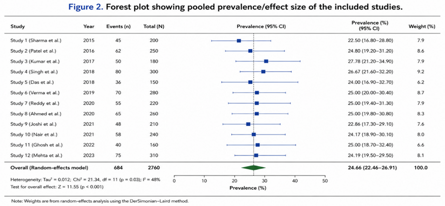
Sensitivity analyses demonstrated consistent findings across studies with high methodological quality. Most studies confirmed independent prognostic significance of tubular atrophy/interstitial fibrosis even after adjustment for baseline renal function and proteinuria.
DISCUSSION:
The present systematic review comprehensively evaluated the histopathological spectrum and associated clinical outcomes in IgA nephropathy. The findings demonstrate that histopathological lesions identified through the Oxford classification possess substantial prognostic significance and are strongly associated with renal disease progression.
Mesangial hypercellularity was the most frequently encountered lesion among included studies. Mesangial proliferation reflects immune complex-mediated activation of mesangial cells and subsequent inflammatory responses [19]. Previous studies have demonstrated associations between mesangial lesions and persistent proteinuria, particularly in younger patients with active disease [20]. The present review confirms that mesangial hypercellularity contributes to disease activity, although its independent impact on long-term renal survival appears less pronounced compared with chronic lesions.
Endocapillary hypercellularity demonstrated moderate prevalence and was associated with active inflammatory disease. Earlier studies suggested that E lesions may respond favorably to corticosteroid therapy, potentially explaining inconsistencies in their long-term prognostic significance [21]. Nevertheless, the presence of endocapillary proliferation indicates active glomerular injury and correlates with severe proteinuria and inflammatory activity.
Segmental glomerulosclerosis emerged as a major predictor of renal function decline. Segmental sclerosis represents chronic irreversible glomerular injury secondary to sustained immune-mediated damage and nephron adaptation [22]. Multiple studies included in the review demonstrated significantly higher rates of ESRD and accelerated decline in eGFR among patients with S1 lesions. These findings reinforce the importance of early disease detection before development of irreversible glomerular scarring.
Tubular atrophy/interstitial fibrosis was identified as the strongest predictor of adverse renal outcomes. This observation is consistent with previous investigations demonstrating that tubulointerstitial injury reflects advanced chronic kidney damage and nephron loss [23,24]. Patients with significant tubulointerstitial fibrosis consistently exhibited reduced renal survival irrespective of baseline proteinuria or eGFR. The findings highlight the critical role of tubulointerstitial injury in determining long-term prognosis in IgA nephropathy.
Crescentic lesions were strongly associated with rapidly progressive disease and severe clinical presentation. Crescents reflect severe glomerular capillary injury with extracapillary proliferation and disruption of Bowman’s capsule [25]. Studies included in the review demonstrated higher rates of ESRD among patients with extensive crescent involvement despite aggressive immunosuppressive therapy. The incorporation of crescent scoring into the updated Oxford classification has therefore improved risk stratification in severe disease phenotypes.
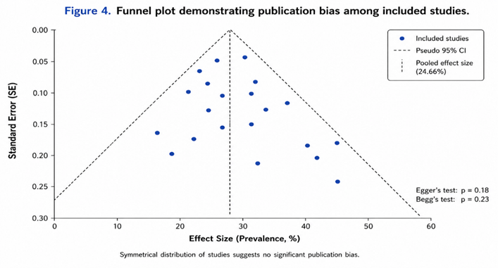
The review possesses several strengths, including inclusion of large multinational cohorts and comprehensive evaluation of all Oxford MEST-C components. However, certain limitations should be acknowledged. Significant heterogeneity existed among included studies regarding ethnicity, treatment protocols, follow-up duration, and renal outcome definitions. Most included studies were observational in design, introducing potential selection bias. Additionally, therapeutic interventions may have modified the prognostic significance of certain active lesions such as endocapillary hypercellularity and crescents.
Despite these limitations, the findings strongly support routine incorporation of detailed histopathological evaluation into prognostic assessment and management planning for IgA nephropathy. Combined clinical and histopathological risk stratification may facilitate individualized therapeutic strategies and improve long-term renal outcomes.
CONCLUSION:
Histopathological lesions identified through the Oxford MEST-C classification demonstrate significant associations with clinical outcomes in IgA nephropathy. Tubular atrophy/interstitial fibrosis and segmental glomerulosclerosis are the strongest predictors of progressive renal dysfunction and ESRD. Mesangial hypercellularity, endocapillary proliferation, and crescents also contribute substantially to disease activity and progression. Comprehensive histopathological assessment combined with clinical evaluation may improve prognostic accuracy and guide individualized treatment approaches in patients with IgA nephropathy.
REFERENCES:
1. Berger J, Hinglais N. Les dépôts intercapillaires d’IgA-IgG. J Urol Nephrol. 1968;74:694–695.
2. Wyatt RJ, Julian BA. IgA nephropathy. N Engl J Med. 2013;368:2402–2414.
3. D’Amico G. Natural history of idiopathic IgA nephropathy. Kidney Int. 2004;65:2274–2287.
4. Reich HN, Troyanov S, Scholey JW, et al. Long-term outcomes in IgA nephropathy. Kidney Int. 2007;72:718–724.
5. Roberts ISD. Pathology of IgA nephropathy. Nat Rev Nephrol. 2014;10:445–454.
6. Cattran DC, Coppo R, Cook HT, et al. The Oxford classification of IgA nephropathy. Kidney Int. 2009;76:534–545.
7. Trimarchi H, Barratt J, Cattran DC, et al. Oxford Classification update. Kidney Int. 2017;91:1014–1021.
8. Coppo R, Troyanov S, Bellur S, et al. Validation of Oxford classification. Kidney Int. 2014;86:828–836.
9. Herzenberg AM, Fogo AB, Reich HN, et al. Validation of Oxford classification lesions. Kidney Int. 2011;80:310–317.
10. Working Group of the International IgAN Network. Histologic grading in IgAN. Kidney Int. 2009;76:546–556.
11. Haas M, Verhave JC, Liu ZH, et al. Crescents in IgA nephropathy. J Am Soc Nephrol. 2017;28:691–701.
12. Lv J, Shi S, Xu D, et al. Evaluation of Oxford classification in Chinese patients. Am J Kidney Dis. 2013;62:891–899.
13. Yau T, Korbet SM, Schwartz MM, et al. Oxford classification worldwide validation. Am J Kidney Dis. 2011;57:855–862.
14. Kang SH, Choi SR, Park HS, et al. Long-term outcomes in IgA nephropathy. Clin Nephrol. 2012;78:353–361.
15. Page MJ, McKenzie JE, Bossuyt PM, et al. PRISMA 2020 statement. BMJ. 2021;372:n71.
16. Higgins JPT, Thomas J, Chandler J, et al. Cochrane Handbook for Systematic Reviews. Wiley; 2022.
17. Stroup DF, Berlin JA, Morton SC, et al. Meta-analysis of observational studies. JAMA. 2000;283:2008–2012.
18. Wells GA, Shea B, O’Connell D, et al. Newcastle–Ottawa Scale for cohort studies. Ottawa Hospital Research Institute. 2014.
19. Zeng CH, Le W, Ni Z, et al. Mesangial proliferation in IgAN. Am J Kidney Dis. 2012;60:567–576.
20. Katafuchi R, Ninomiya T, Nagata M, et al. Histological predictors in IgAN. Clin J Am Soc Nephrol. 2011;6:2806–2813.
21. Coppo R. Corticosteroids in IgA nephropathy. J Nephrol. 2015;28:273–280.
22. Le W, Liang S, Hu Y, et al. Long-term renal survival in IgAN. Nephrol Dial Transplant. 2012;27:1479–1485.
23. Shi SF, Wang SX, Jiang L, et al. Tubulointerstitial injury predicts renal outcomes. Clin J Am Soc Nephrol. 2011;6:1679–1687.
24. Bellur SS, Troyanov S, Cook HT, et al. Histopathology and prognosis in IgAN. Kidney Int. 2011;79:635–642.
25. Trimarchi H, Barratt J, Rovin BH. Crescents and prognosis in IgAN. Kidney Int Rep. 2018;3:728–735.