Clinical and Spirometric Patterns of Pulmonary Function in Patients with Rheumatoid Arthritis
- Mohan B.R. , Professor & HOD, Department of General Medicine, K.C. General Hospital, Bangalore, Karnataka, India.
- N.C. Srinivas Prabhudev , Professor, Department of General Medicine, K.C. General Hospital, Bangalore, Karnataka, India.
- Venkateshiah , Senior Orthopaedic Consultant, K.C. General Hospital, Bangalore, Karnataka, India.
- Kadeeja Shiben , Postgraduate, Department of General Medicine, K.C. General Hospital, Bangalore, Karnataka, India.
Article Information:
Abstract:
Background: Rheumatoid arthritis is a chronic systemic inflammatory disease in which pulmonary involvement may remain clinically silent until functional impairment has already developed. Objective: To assess the clinical profile and spirometric pattern of pulmonary function among subjects with rheumatoid arthritis. Methods: This prospective observational study was conducted in the Department of General Medicine and Orthopedics, KC General Hospital, Bangalore, for a period of 2 years. Sixty subjects aged 18 years or above with rheumatoid arthritis diagnosed according to the 2010 ACR/EULAR classification criteria were included after written informed consent. Clinical examination, complete blood count, erythrocyte sedimentation rate, rheumatoid factor, anti-cyclic citrullinated peptide antibody testing, chest radiography and spirometry were performed. Spirometric patterns were summarized as normal, restrictive, obstructive or mixed. Results: The mean age was 47.22 +/- 11.38 years, with a range of 23-70 years, and 52 subjects (86.7%) were female. Mean rheumatoid arthritis duration was 4.38 +/- 3.43 years, and 46 subjects (76.7%) had disease duration of 0-5 years. Morning stiffness or pain was present in 52 subjects (86.7%), and pulmonary symptoms were recorded in 17 subjects (28.3%). Chest X-ray was suggestive of interstitial lung disease in 2 subjects (3.3%). Spirometry was normal in 50 subjects (83.4%), while 10 subjects (16.7%) had abnormal spirometry: restrictive pattern in 6 (10.0%), mixed pattern in 3 (5.0%), and obstructive pattern in 1 (1.7%). Conclusion: Spirometric abnormality was present in nearly one-sixth of rheumatoid arthritis subjects, with restrictive impairment being the commonest abnormal pattern. Many patients with abnormal pulmonary function had minimal or no respiratory symptoms, supporting the role of spirometry as a practical screening tool in routine rheumatoid arthritis care, particularly in resource-constrained Indian outpatient settings.
Keywords:
Article :
Introduction:
Rheumatoid arthritis (RA) is a chronic immune-mediated inflammatory disease classically marked by symmetric erosive synovitis, yet its clinical significance extends well beyond the joints. It contributes to pain, deformity, work limitation, and long-term systemic morbidity, particularly when diagnosis or disease control is delayed.[1] The 2010 ACR/EULAR classification framework strengthened early identification by combining joint involvement, serology, symptom duration and acute-phase reactants, a useful approach in outpatient medical practice where established deformities should not be the first point of recognition.[2]
Extra-articular disease is one of the major reasons RA remains clinically important for physicians outside rheumatology. Pulmonary, cardiovascular, hematological, neurological, ocular and cutaneous manifestations may accompany the articular disease, and extra-articular involvement has been associated with excess mortality in community cohorts.[3] Among these, lung disease occupies a difficult position: it may be silent, insidious, drug-related, infection-related, inflammatory, or fibrotic, and it often comes to attention only when breathlessness or radiological abnormality appears.[4]
RA-associated interstitial lung disease is the most feared pulmonary phenotype because it can progress despite relatively quiet joint symptoms. Population-based work has shown that interstitial lung disease in RA is associated with substantial morbidity and mortality.[5] Mortality analyses have further underlined that RA-ILD is not merely a radiological label but a clinically consequential extra-articular disease state.[6] In Indian general medicine settings, this matters because patients frequently move between primary care, orthopaedics, rheumatology and medicine clinics, and pulmonary screening may not occur unless symptoms become obvious.
The diagnostic challenge is partly technical. Chest radiography is inexpensive and available, but it lacks sensitivity for early parenchymal involvement. Studies comparing pulmonary function testing and high-resolution computed tomography have shown that functional abnormalities may be identified even when routine radiography is unrevealing.[7] Similarly, fibrosing alveolitis in RA has been better characterized when clinical, radiographic, HRCT and pulmonary function findings are considered together.[8] However, HRCT and diffusion capacity testing are not always feasible as routine screening tools in district and government hospital practice.
Spirometry therefore has a pragmatic role. It is non-invasive, repeatable, relatively low cost, and can be incorporated into screening and follow-up pathways when performed with appropriate quality standards.[9] It cannot replace HRCT or DLCO for diagnosing early ILD, but it can identify restrictive, obstructive and mixed ventilatory patterns that justify closer respiratory evaluation. The present study was undertaken to assess the clinical and spirometric pattern of pulmonary function in subjects with rheumatoid arthritis attending a tertiary care hospital in Bangalore.
Materials and Methods:
Study design and setting
This was a prospective observational study conducted in the Department of General Medicine and Orthopedics, KC General Hospital, Bangalore, Karnataka for a period of 2 years.
Study population and sample size
Male and female patients with rheumatoid arthritis attending the departments were screened. During the study period, 120 patients with rheumatoid arthritis were assessed against the eligibility criteria, and 60 eligible subjects were included in the final analysis.
The calculated sample size was 61 and was rounded to 60. The calculation used the formula N = (Z alpha)2 x p x q/d2, assuming a 20% prevalence of dyspnoea among RA patients, 10% absolute precision, and 95% confidence level.
Inclusion criteria
Patients aged above 18 years with rheumatoid arthritis diagnosed according to the 2010 ACR/EULAR classification criteria and willing to provide written informed consent were included.
Exclusion criteria
Patients with known infective or non-infective respiratory disease, known cardiovascular disease, other connective tissue disorders, or congenital skeletal deformities were excluded.
Clinical and laboratory assessment
Detailed clinical history and physical examination were performed. Clinical variables included age, sex, disease duration, joint involvement, signs of active inflammation, morning stiffness or pain, fever, weight loss, chronic deformity and respiratory symptoms. Hematological and serological investigations included complete blood count, erythrocyte sedimentation rate, rheumatoid factor and anti-cyclic citrullinated peptide antibody testing. Chest X-ray was used to document radiological features suggestive of interstitial lung disease.
Spirometry procedure
Spirometry was performed using an Innotech Respican spirometer. Patients were instructed to avoid heavy exercise for 30 minutes before the test, avoid tight clothing that could restrict breathing, and avoid a large meal within two hours before testing. Forced expiratory volume in one second, forced vital capacity and FEV1/FVC ratio were analyzed. Post-bronchodilator assessment was performed in patients with abnormal pulmonary function tests; improvement of more than 15% was considered compatible with obstructive lung disease.
Statistical analysis
Data were entered in Microsoft Excel and analyzed using SPSS version 20. Continuous variables were summarized as mean and standard deviation, while categorical variables were expressed as frequency and percentage. Bar charts and pie/donut charts were used for visual representation. The analysis was descriptive, and no inferential p-values were added.
Results:
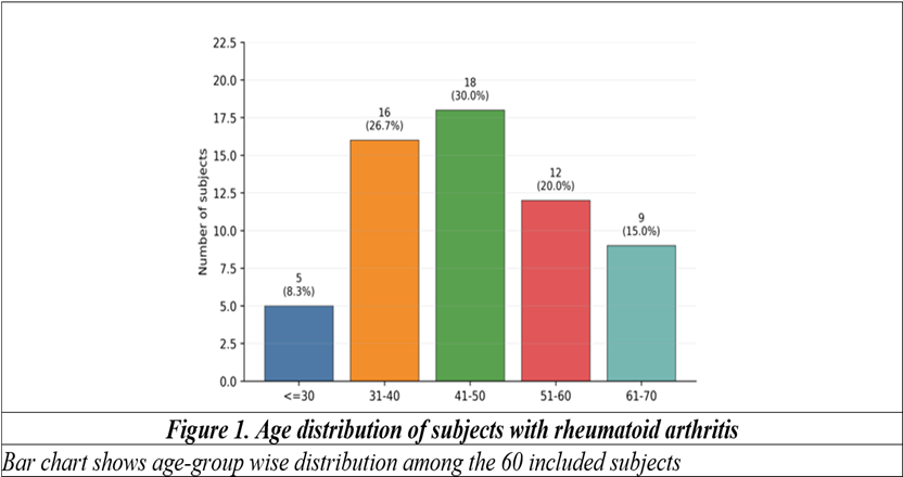
A total of 60 subjects with rheumatoid arthritis were included in the final analysis. The age distribution showed clustering in the fourth and fifth decades. The mean age was 47.22 +/- 11.38 years, ranging from 23 to 70 years. The highest proportion belonged to the 41-50 year age group (18 subjects, 30.0%), followed by 31-40 years (16 subjects, 26.7%) (Table 1, Figure 1).
Table 1. Baseline demographic and disease-duration profile of the study population (N=60)
|
Variable |
Category/Measure |
n (%) or Mean +/- SD |
|
Age group |
<=30 years |
5 (8.3%) |
|
31-40 years |
16 (26.7%) |
|
|
41-50 years |
18 (30.0%) |
|
|
51-60 years |
12 (20.0%) |
|
|
61-70 years |
9 (15.0%) |
|
|
Age |
Mean +/- SD |
47.22 +/- 11.38 years |
|
Range |
23-70 years |
|
|
Sex |
Female |
52 (86.7%) |
|
Male |
8 (13.3%) |
|
|
RA duration |
0-5 years |
46 (76.7%) |
|
6-10 years |
11 (18.3%) |
|
|
>10 years |
3 (5.0%) |
|
|
RA duration |
Mean +/- SD |
4.38 +/- 3.43 years |
|
Range |
1-15 years |
Values are presented as n (%) unless otherwise specified
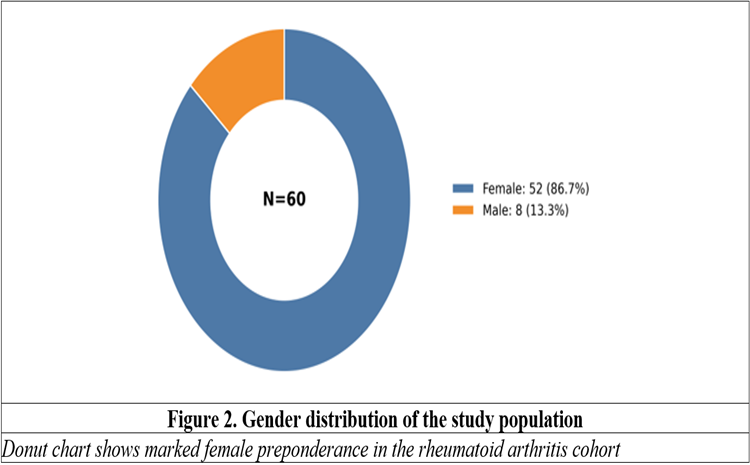
Females formed the major proportion of the cohort, with 52 subjects (86.7%) and a female-to-male ratio of 6.5:1. Among subjects aged above 50 years, the female-to-male ratio was 2.5:1 (Figure 2). The mean duration of RA was 4.38 +/- 3.43 years, and most subjects had a disease duration of 0-5 years.
Articular involvement was extensive in many subjects. The mean number of involved joints was 22.07 +/- 3.63, with a range of 10-32 joints. Forty-two subjects (70.0%) had involvement of more than 20 joints. Upper limb involvement was present in all subjects, lower limb involvement in 15 subjects (25.0%), and axial skeletal involvement in 4 subjects (6.7%). Signs of active inflammation were noted in 9 subjects (15.0%) (Table 2).
Table 2. Articular and general clinical features among subjects with rheumatoid arthritis (N=60)
|
Variable |
Category/Measure |
n (%) or Mean +/- SD |
|
Number of joints involved |
<20 joints |
18 (30.0%) |
|
>20 joints |
42 (70.0%) |
|
|
Mean +/- SD |
22.07 +/- 3.63 |
|
|
Range |
10-32 |
|
|
Pattern of involvement |
Upper limb involvement |
60 (100.0%) |
|
Lower limb involvement |
15 (25.0%) |
|
|
Axial involvement |
4 (6.7%) |
|
|
Signs of active inflammation |
Present |
9 (15.0%) |
|
Absent |
51 (85.0%) |
|
|
Clinical features |
Morning stiffness/pain |
52 (86.7%) |
|
Fever |
8 (13.3%) |
|
|
Weight loss |
5 (8.3%) |
|
|
Chronic deformity |
5 (8.3%) |
|
|
Any pulmonary symptom |
17 (28.3%) |
Clinical features were recorded during routine evaluation.
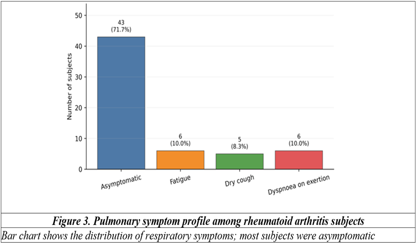
Morning stiffness or pain lasting approximately one hour was reported by 52 subjects (86.7%). Pulmonary symptoms were present in 17 subjects (28.3%), while 43 subjects (71.7%) were asymptomatic from a respiratory perspective. Among respiratory symptoms, fatigue and dyspnoea on exertion were each recorded in 6 subjects (10.0%), while dry cough was recorded in 5 subjects (8.3%) (Table 3, Figure 3).
Laboratory evaluation showed normal haemoglobin in 33 subjects (55.0%). Mild and moderate anaemia were recorded in 13 (21.6%) and 14 (23.4%) subjects, respectively. ESR was elevated above 20 mm/hr in 49 subjects (81.7%). Chest X-ray was normal in 58 subjects (96.7%), while 2 subjects (3.3%) had findings suggestive of interstitial lung disease, described as bilateral lower-zone diffuse shadowing with prominent pulmonary vasculature (Table 3).
Table 3. Pulmonary symptoms, laboratory findings and chest X-ray profile (N=60)
|
Variable |
Category |
n (%) |
|
Pulmonary symptom profile |
Asymptomatic |
43 (71.7%) |
|
Fatigue |
6 (10.0%) |
|
|
Dry cough |
5 (8.3%) |
|
|
Dyspnoea on exertion |
6 (10.0%) |
|
|
Haemoglobin |
Normal (>12 g/dL) |
33 (55.0%) |
|
Mild anaemia |
13 (21.6%) |
|
|
Moderate anaemia |
14 (23.4%) |
|
|
ESR |
<20 mm/hr |
11 (18.3%) |
|
>20 mm/hr |
49 (81.7%) |
|
|
Chest X-ray |
Normal |
58 (96.7%) |
|
Suggestive of ILD |
2 (3.3%) |
ILD: interstitial lung disease; ESR: erythrocyte sedimentation rate.
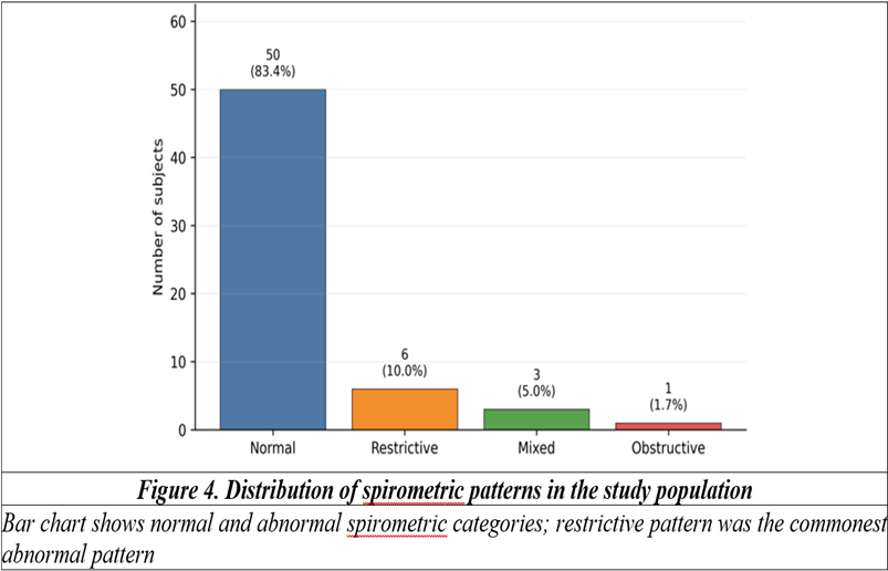
Spirometry was normal in 50 subjects (83.4%). Abnormal spirometry was recorded in 10 subjects (16.7%). Restrictive pattern was the commonest abnormality, seen in 6 subjects (10.0%), followed by mixed pattern in 3 subjects (5.0%) and obstructive pattern in 1 subject (1.7%) (Table 4, Figure 4).
Among subjects with restrictive pattern, 2 were asymptomatic, 3 had dyspnoea on exertion and 1 had dry cough. One of the 6 subjects with restrictive spirometry was male and 5 were female; chest X-ray was suggestive of ILD in one restrictive-pattern subject. Mixed pattern was observed in 3 subjects; one had dyspnoea on exertion, one had dry cough and one had fatigue. Chest X-ray was suggestive of ILD in one subject with mixed spirometry, and all mixed-pattern subjects were female. The single subject with obstructive pattern was male, asymptomatic, and had a normal chest X-ray (Table 4).
Table 4. Spirometric pattern and clinical notes among subjects with rheumatoid arthritis (N=60)
|
Spirometric pattern |
n (%) |
Clinical/radiological notes |
|
Normal |
50 (83.4%) |
No abnormal spirometric pattern recorded |
|
Restrictive |
6 (10.0%) |
2 asymptomatic, 3 with dyspnoea on exertion, 1 with dry cough; 1 male and 5 female; chest X-ray suggestive of ILD in 1 subject |
|
Mixed |
3 (5.0%) |
1 with dyspnoea on exertion, 1 with dry cough, 1 with fatigue; all female; chest X-ray suggestive of ILD in 1 subject |
|
Obstructive |
1 (1.7%) |
Male subject; asymptomatic; chest X-ray normal; post-bronchodilator improvement compatible with obstructive pattern |
Spirometric classification was based on recorded PFT interpretation
Discussion:
This prospective observational study describes pulmonary function patterns among 60 subjects with rheumatoid arthritis attending a government tertiary care hospital in Bangalore. The cohort was predominantly female, with a mean age in the late fourth decade, and most subjects had a disease duration of less than five years. The central clinical finding is straightforward but important: abnormal spirometry was present in 16.7% of subjects, and the restrictive pattern was the commonest abnormality. In a general medicine setting, this is precisely the kind of finding that is easily missed when respiratory evaluation is reserved only for overt breathlessness.
The age profile was broadly similar to previous spirometry-based studies in RA. Kadhum et al. reported a comparable mean age and found that restrictive impairment was the predominant abnormal spirometric pattern among RA patients.[10] Ravikumar et al. also observed a higher frequency of abnormal pulmonary function than the present study, with restrictive impairment being more common than obstructive disease.[11] The lower abnormality rate in the present cohort may be related to its shorter RA duration, exclusion of known respiratory disease, and the limited use of chest X-ray and spirometry rather than HRCT and DLCO.
The female predominance in this study is consistent with the known epidemiology of RA and with regional studies of pulmonary involvement in RA. Banotra et al. reported clinically and radiologically significant pulmonary manifestations in an Indian tertiary-care population, with disease severity, serological status and smoking emerging as relevant clinical considerations.[12] Here, smoking-related interpretation was limited because smoking status was not reported in the final results table; therefore, the obstructive component should be described cautiously rather than attributed to a specific exposure.
A striking aspect of the present findings is the gap between symptoms and functional abnormality. Most subjects were asymptomatic from a respiratory perspective, yet spirometry identified abnormal patterns in 10 subjects. This agrees with the broader literature showing that pulmonary involvement in RA may be clinically subtle, especially in early or minimally symptomatic disease.[13] Fatima et al., in a North Indian cohort, reported a substantially higher proportion of pulmonary symptoms and abnormal PFT findings, likely because their evaluation included HRCT and a broader pulmonary work-up.[14]
Chest X-ray was suggestive of ILD in only 3.3% of subjects, whereas spirometry was abnormal in 16.7%. This difference is clinically relevant. Plain radiography may remain normal in early or mild pulmonary involvement, while spirometry can reflect ventilatory limitation before obvious radiographic changes appear. Similar practical concerns have been raised in Indian and international studies, where chest X-ray alone has been considered insufficient for identifying early RA lung disease.[15]
The predominance of restrictive impairment has biological plausibility. RA-related parenchymal involvement, early interstitial change, pleural restriction, respiratory muscle limitation, and thoracic cage mechanics can all reduce ventilatory reserve. However, spirometry alone cannot diagnose RA-ILD. It should be understood as a screening and triage tool, especially where HRCT and DLCO are not routinely available for every RA patient. In such settings, abnormal spirometry should prompt more detailed respiratory assessment rather than being treated as a final diagnosis.
Anaemia was recorded in 45% of subjects and ESR was elevated in 81.7%, suggesting a considerable inflammatory burden. Anaemia of inflammation is common in chronic immune-mediated disease and is mediated partly by cytokine-driven changes in iron handling and erythropoiesis.[16] In practical terms, fatigue in RA patients may therefore be mixed in origin; it may arise from anaemia, systemic inflammation, respiratory limitation, or deconditioning. This matters when interpreting pulmonary symptoms such as easy fatigability.
The study has limitations. It was a single-centre observational study with a modest sample size of 60 subjects. The analysis was descriptive and did not include inferential comparisons across disease duration, serology, symptom status or treatment exposure. HRCT and DLCO were not performed, so early interstitial lung disease could not be confirmed or excluded with high sensitivity. Drug history, including methotrexate or other DMARD exposure, was not analyzed in the final results. Despite these limitations, the study has a useful outpatient message: spirometry is simple, inexpensive and clinically meaningful when incorporated into routine RA evaluation, particularly for patients with pulmonary symptoms, elevated inflammatory markers, longer disease duration, or persistent clinical concern.
Conclusion:
In this prospective observational study of 60 subjects with rheumatoid arthritis, spirometric abnormality was identified in 16.7% of subjects. Restrictive impairment was the commonest abnormal pattern, followed by mixed and obstructive patterns. Most subjects were asymptomatic from a respiratory perspective, and chest X-ray was normal in the large majority, indicating that spirometry may detect functional pulmonary involvement that is not clinically or radiographically obvious. Routine spirometric screening can therefore serve as a practical, non-invasive and low-cost component of RA care in general medicine settings, while abnormal findings should be followed by more definitive pulmonary assessment wherever feasible.
References:
1. Smolen JS, Aletaha D, McInnes IB. Rheumatoid arthritis. Lancet. 2016;388(10055):2023-2038. doi:10.1016/S0140-6736(16)30173-8.
2. Aletaha D, Neogi T, Silman AJ, Funovits J, Felson DT, Bingham CO 3rd, et al. 2010 rheumatoid arthritis classification criteria: an American College of Rheumatology/European League Against Rheumatism collaborative initiative. Ann Rheum Dis. 2010;69(9):1580-1588. doi:10.1136/ard.2010.138461.
3. Turesson C, O'Fallon WM, Crowson CS, Gabriel SE, Matteson EL. Occurrence of extraarticular disease manifestations associated with excess mortality in a community based cohort of patients with rheumatoid arthritis. J Rheumatol. 2002;29(1):62-67.
4. Shaw M, Collins BF, Ho LA, Raghu G. Rheumatoid arthritis-associated lung disease. Eur Respir Rev. 2015;24(135):1-16. doi:10.1183/09059180.00008014.
5. Bongartz T, Nannini C, Medina-Velasquez YF, Achenbach SJ, Crowson CS, Ryu JH, et al. Incidence and mortality of interstitial lung disease in rheumatoid arthritis. Arthritis Rheum. 2010;62(6):1583-1591. doi:10.1002/art.27405.
6. Olson AL, Swigris JJ, Sprunger DB, Fischer A, Fernandez-Perez ER, Solomon J, et al. Rheumatoid arthritis-interstitial lung disease-associated mortality. Am J Respir Crit Care Med. 2011;183(3):372-378. doi:10.1164/rccm.201004-0622OC.
7. Cortet B, Perez T, Roux N, Flipo RM, Duquesnoy B, Delcambre B, et al. Pulmonary function tests and high resolution computed tomography of the lungs in patients with rheumatoid arthritis. Ann Rheum Dis. 1997;56(10):596-600. doi:10.1136/ard.56.10.596.
8. Dawson JK, Fewins HE, Desmond J, Lynch MP, Graham DR. Fibrosing alveolitis in patients with rheumatoid arthritis as assessed by high resolution computed tomography, chest radiography, and pulmonary function tests. Thorax. 2001;56(8):622-627. doi:10.1136/thorax.56.8.622.
9. Graham BL, Steenbruggen I, Miller MR, Barjaktarevic IZ, Cooper BG, Hall GL, et al. Standardization of spirometry 2019 update: an official American Thoracic Society and European Respiratory Society technical statement. Am J Respir Crit Care Med. 2019;200(8):e70-e88. doi:10.1164/rccm.201908-1590ST.
10. Kadhum HJ, Al Hamza AN, Abdualwahid AB. Pattern of pulmonary function test in rheumatoid arthritis patients. Med J Basrah Univ. 2017;35(2):109-115.
11. Ravikumar P, Das D, Bhattacharjee K. A comparative study of pulmonary involvement in patients with rheumatoid arthritis. J Evol Med Dent Sci. 2017;6(4):296-300. doi:10.14260/Jemds/2017/67.
12. Banotra P, Wangnoo A, Sharma M, Mahajan A. Study of the pulmonary manifestations in patients with rheumatoid arthritis. Int J Res Rev. 2020;7(4):427-436.
13. Zayeni H, Haji-Abbasi A, Foumani SAA, Tohidi M, Masooleh IS, Parsa BG, et al. Pulmonary involvement in rheumatoid arthritis: a cross-sectional study in Iran. Lung India. 2016;33(1):49-52. doi:10.4103/0970-2113.173062.
14. Fatima N, Shameem M, Malik A, Khan PA, Shujatullah F, Ahmed S, et al. A study on the pulmonary manifestations of rheumatoid arthritis from a North Indian town. Open J Respir Dis. 2013;3(3):128-131. doi:10.4236/ojrd.2013.33020.
15. Sen S, Ghosh S, Dhua A. Profile of pulmonary manifestations of rheumatoid arthritis: a cross-sectional study. Int J Healthc Biomed Res. 2020;9(1):4-12. doi:10.36848/IJHBR/2020/12100.51214.
16. Weiss G, Ganz T, Goodnough LT. Anemia of inflammation. Blood. 2019;133(1):40-50. doi:10.1182/blood-2018-06-856500.