Correlation of Hs-CRP and IL-6 With Severity of Acute Coronary Syndrome Undergoing Percutaneous Coronary Intervention : An Observational Study.
- Akash Priyadarshi , Assistant Professor, Department of Medicine, Jawaharlal Nehru Medical College, Aligarh Muslim University, Aligarh, Uttar Pradesh, India.
- Siddharth Gupta , Assistant Professor, Department of Medicine, Jawaharlal Nehru Medical College, Aligarh Muslim University, Aligarh Uttar Pradesh, India.
- MU Rabbani , Professor, Department of Cardiology, Jawaharlal Nehru Medical College, Aligarh Muslim University, Aligarh (Uttar Pradesh, India.
- Anjum Mirza Chughtai. , Professor, Department of Medicine, Jawaharlal Nehru Medical College, Aligarh Muslim University, Aligarh, Uttar Pradesh, India.
Article Information:
Abstract:
Background: Inflammation is pivotal in the initiation and progression of atherosclerosis and in precipitating acute coronary syndrome (ACS) by influencing plaque stability. Serum high-sensitivity C-reactive protein (Hs- CRP) and interleukin-6 (IL-6) are key inflammatory biomarkers, and their levels are strong, independent predictors of plaque rupture and mortality in patients with ACS. Objective: To analyze the correlation between Hs-CRP and IL-6 levels with severity and burden of atherosclerosis in ACS patients. Materials and methods: An observational, cross-sectional, open-label, hospital-based study was carried out at a tertiary care centre between December 2019 and December 2021. Patients aged 18 years or older who were diagnosed with ACS were enrolled after obtaining informed consent. Data on risk factors and clinical history were collected. All participants underwent routine investigations, including lipid profile, HbA1c, Hs-CRP and IL-6, electrocardiography, echocardiography, and coronary angiography (CAG). Patient management was conducted in accordance with current clinical guidelines. Serum hs-CRP and IL-6 levels were measured and correlated with coronary angiographic findings, like, extent of coronary artery stenosis and GENSINI score, and their associations were analyzed for coronary artery disease (CAD) severity. Results: A total of 100 ACS patients were enrolled, including 62 STEMI, 34 NSTEMI, and 4 unstable angina cases. Both biomarkers were significantly higher in STEMI and unstable angina than in NSTEMI (P < 0.05) and showed a positive correlation with each other (r = 0.55). Higher levels were observed in severe CAD compared to moderate and mild CAD, though this difference was not statistically significant (P > 0.05). Conclusions: Raised hs-CRP and IL-6 levels in ACS, particularly in STEMI and severe CAD, reflect increased inflammation, plaque instability, and disease severity. hs-CRP showed a stronger association with angiographic severity, supporting its potential role as a marker of atherosclerotic burden and CAD assessment. Categories: Cardiology, Internal Medicine.
Keywords:
Article :
INTRODUCTION:
Cardiovascular diseases (CVD) are the foremost cause of mortality globally and in India [1]. Coronary artery disease (CAD), a major form of CVD, results from atherosclerotic narrowing of the coronary arteries leading to myocardial oxygen supply-demand imbalance and may manifest as chest pain, arrhythmias, or sudden death. Inflammation is fundamental to the development, progression, and destabilization of atherosclerotic plaques, with acute coronary syndrome (ACS) occurring following plaque rupture or erosion. Data from the PGIMER-ACS registry in North India indicate higher mortality in STE-ACS compared to NSTE-ACS (8.9% vs 4.5%, p<0.001), with an overall in-hospital ACS mortality of 7.6% [2]. High-sensitivity C-reactive protein (Hs-CRP) and interleukin-6 (IL-6), key pro-inflammatory markers-IL-6 being more specific-are proposed indicators of CAD severity and ACS-related mortality [3,4]. The GENSINI score, which quantifies CAD burden, is a recognized predictor of prognosis in ACS, yet limited studies have explored its correlation with hs-CRP and IL-6 levels.
Given that therapies such as statins, aspirin, and tirofiban reduce ACS mortality by modulating inflammatory pathways, this study attempts to enhance understanding of the relationship between inflammatory marker levels and actual angiographic disease severity.
Acute Coronary Syndrome (ACS) represents a spectrum of clinical conditions resulting from acute myocardial ischemia and remains a leading cause of morbidity and mortality worldwide. Despite significant advances in early diagnosis, pharmacotherapy, and revascularization strategies such as percutaneous coronary intervention (PCI), risk stratification and prediction of disease severity continue to pose clinical challenges. Increasing evidence suggests that inflammation plays a pivotal role in the initiation, progression, and destabilization of atherosclerotic plaques, ultimately leading to ACS [5,6].
High-sensitivity C-reactive protein (hs-CRP) and interleukin-6 (IL-6) are well-established inflammatory biomarkers implicated in atherosclerosis and plaque rupture. Hs-CRP, an acute-phase reactant synthesized by the liver in response to inflammatory cytokines, has been shown to correlate with endothelial dysfunction, plaque vulnerability, and adverse cardiovascular outcomes [7,8]. IL-6, a pro-inflammatory cytokine, serves as a key upstream mediator in the inflammatory cascade and stimulates hepatic production of CRP, thereby linking systemic inflammation with cardiovascular pathology [9,10].
Several studies have demonstrated elevated levels of Hs-CRP and IL-6 in patients with ACS, suggesting their potential utility as markers of disease activity and prognosis [11-13]. However, the relationship between these inflammatory markers and the severity of ACS, particularly in patients undergoing PCI, remains an area of ongoing investigation. Understanding this correlation may aid in improved risk stratification, assessment of disease burden, and optimization of therapeutic strategies in the acute setting. The GENSINI score, which quantifies CAD burden, is a recognized predictor of prognosis in ACS, yet limited studies have explored its correlation with hs-CRP and IL-6 levels.
Therefore, the present observational study aims to evaluate the correlation of hs-CRP and IL-6 levels with the severity of ACS in patients undergoing PCI. By elucidating the association between inflammatory biomarkers and clinical severity, this study seeks to contribute to the growing body of evidence supporting the role of inflammation in ACS and its potential implications for patient management and outcomes.
Objectives
The primary objective of the study is to evaluate the association between inflammatory biomarkers, Hs-CRP and IL-6, and the severity of coronary artery disease as assessed by invasive coronary angiography in patients with acute coronary syndrome in terms of number of vessels involved. The secondary objective seeks to determine the correlation of these biomarkers with CAD burden quantified by the GENSINI score and across different ACS subtypes.
MATERIALS AND METHODS:
Study design
The study was a single centre descriptive as well as analytical type of cross-sectional study, concluded within 24 months between December 2019 and December 2021 at Department of Medicine and Department of Cardiology, at a tertiary healthcare centre in accordance with the STROBE guidelines.
Ethical approval
The study was approved by the Institutional Ethics Committee (Regd.), at our tertiary care centre as per the standards of Good Clinical Practice and the Helsinki Declaration vide letter number IECJNMC/585 on 28.12.2019. Written informed consent was obtained from all participants, who had the right to opt out of the study at any time.
Sample size
As per the study conducted by Trideep J. Deori et al. [14] in 2017-2018, the prevalence of coronary artery disease in Uttar Pradesh, India was around 10%. At 95% CI, power of 0.80 and 5% margin of error, the sample size for this observational study came out to be 138. Due to pandemic related recruitment constraints, final enrolment was 100. Consequently the study is underpowered to detect the originally specified primary endpoint effect size, and results should be interpreted as exploratory.
Participants
Patients presenting to the Emergency Department of our hospital with Acute Coronary Syndrome (ACS), diagnosed according to the Fourth Universal Definition of Myocardial Infarction, and who were willing to undergo percutaneous coronary angiography were enrolled in the study. Inclusion criteria comprised patients diagnosed with ACS as per the fourth universal definition of MI, those willing to undergo invasive coronary angiography (CAG), and those who provided informed written consent for active participation. Exclusion criteria included patients with chronic inflammatory diseases; those on long-term statin or NSAID therapy; patients with acute or chronic ongoing infections; autoimmune diseases; a prior history of cerebrovascular accident (CVA) or coronary artery disease (CAD); underlying malignancy; and patients who refused to provide written informed consent.
As depicted in Figure 1, 371 individuals were initially screened and 233 were found to be eligible to participate in the study. Of the 233 individuals, 32 patients (13.7%) declined to provide informed consent, citing personal, logistical, or cultural reasons. Nine patients (3.9%) experienced sudden cardiac arrest and died before enrolment procedures could be completed. 38 patients (16.3%), including 26 STEMI and 12 NSTEMI, were found to have contraindications for coronary angiography, including advanced chronic kidney disease, allergy to contrast media, or unstable hemodynamic status. 54 patients (23.2%), consisting 31 STEMI and 23 NSTEMI, withdrew from the study during the pre-procedure phase, due to personal choice, deterioration in condition and referral to other centre. Following these exclusions and due to difficulty in enrolment because of the ongoing COVID-19 pandemic during the time of study, only a total of 100 participants were enrolled by the end of recruitment deadline after meeting all inclusion criteria, provided informed consent, and successfully underwent CAG, constituting the final study cohort.
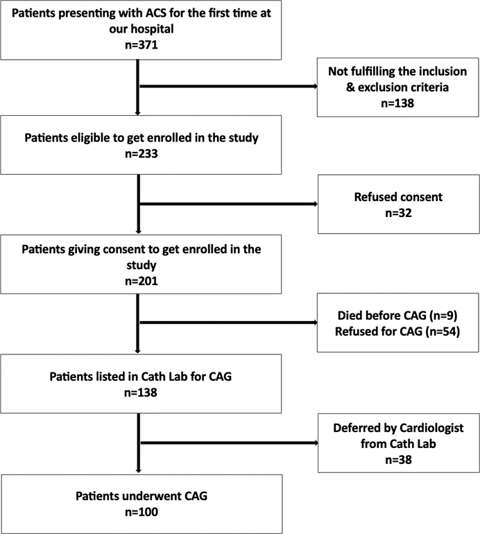
FIGURE 1: Flowchart depicting the selection of study participants. ACS: acute coronary syndrome; CAG: coronary angiography.
Data collection
Risk factors including BMI, lifestyle, socioeconomic status, diet, smoking, hypertension, diabetes, family history, and dyslipidemia were measured using self-administered questionnaire as in Appendix 1. The questionnaire was developed based on existing literature and standard clinical definitions. Content validity was assessed through expert review by three public health specialists. Face validity and feasibility were evaluated through pilot testing among 20 participants. Minor revisions were made prior to final data collection. Blood samples for high-sensitivity Troponin I (hs-Trop I) levels were withdrawn at the time of presentation. Fasting blood samples were analyzed for HbA1C, LDL, HDL, triglycerides, total cholesterol levels and Hs-CRP and IL-6 levels. While all the samples were withdrawn before undergoing PCI, the time to sampling for Hs-CRP and IL-6 ranged from 48 to 72 hours after the symptom onset.
Instruments
Total cholesterol was measured using the cholesterol esterase-oxidase-peroxidase method, and serum triglycerides by a modified enzymatic method. HDL cholesterol was determined after precipitating chylomicrons, VLDL, and LDL with buffered polyethylene glycol; the supernatant was used for analysis. All assays followed the manufacturer’s protocols (Ranbaxy Diagnostic Division, New Delhi, 2019). LDL was calculated using the Friedewald equation [LDL = Total cholesterol - (HDL + VLDL)]. Apo A1 and Apo B were measured turbidimetrically, and Lp(a) by latex turbidimetry, using kits from Euro Diagnostic Division, Peerless Biotech Pvt. Ltd., New Delhi (2019). All analyses were performed on a BioLis 24i® analyzer.
HbA1c were measured by high-performance liquid chromatography (HPLC), and IL-6 and Hs-CRP by electro chemiluminescent assay (ECLIA) and nephelometric assays, respectively, using the Siemens Dimension® RxL Max® Integrated Chemistry System (Siemens Healthcare, Vadodara, India, 2016). Patients were grouped as per their hs-CRP levels into low (<1 mg/L), average (1-3 mg/L) and high risk (>3 mg/L) groups.
Diabetes was defined having a fasting blood glucose (FBS) level ≥126 mg/dl; a 2-hour post prandial blood glucose level ≥200 mg/dl; HbA1C ≥6.5% or already on treatment, either insulin or oral anti-diabetic drug therapy [15]. Systemic hypertension was defined as systolic blood pressure (SBP) ≥130 mmHg and/or diastolic blood pressure (DBP) ≥80 mmHg [16]. Smoking was described as patients who were actively smoking during last six months. Dyslipidemia was labelled with either total cholesterol ≥200 mg/dl; triglyceride ≥150 mg/dl; HDL ≤40 mg/dl (males) and ≤50 mg/dl (females) or LDL ≥100 mg/dl [17]. Family history was clarified as patients having a history of coronary artery disease (CAD) or cerebrovascular accident (CVA) in their first degree blood relatives. Metabolic syndrome was defined as having any three of the following five traits:
waist circumference ≥102 cm (males) and ≥88 cm (females); serum triglyceride levels ≥150 mg/dl; HDL ≤40 mg/dl (males) and ≤50 mg/dl (females); BP ≥130/85 mmHg or on anti-hypertensive drugs; FBG ≥100 mg/dl or on treatment for elevated blood glucose [18].
All participants underwent 2D-Echocardiography and, with informed consent, coronary angiography (CAG). All study patients underwent Coronary catheterization either by trans-femoral or trans-radial routes in the Cath Lab, Department of Cardiology at our tertiary care centre. CAG was performed using Philips Cath Lab (Philips FD10/20, Pune, India). CAG reports were prepared by a cardiologist having experience of more than two decades. Based on CAG findings, patients were classified as having single (SVD), double (DVD), or triple vessel disease (TVD) according to stenosis severity (≥50% in Left Main coronary artery or ≥70% in other major vessels).
The lower limit of detection (LLoD) was 0.3 mg/L for Hs-CRP and 0.0 pg/mL for IL-6. To ensure analytical precision, internal quality controls were run daily. The intra-assay and inter-assay coefficients of variations (CV) were 4.0% and 7.8% for Hs-CRP and 3.2% and 6.0% for IL-6, respectively. Assays were calibrated against international reference materials (CRM 470) and the laboratory participates in an External Quality Assessment (EQA) scheme to ensure long-term accuracy and inter-laboratory traceability. The researcher who prepared the report had an experience of more than two decades in their relevant field.
Degree of stenosis in a coronary artery was defined as a percentage of the luminal obstruction caused by a particular lesion upon direct visualization by the interventional cardiologist doing CAG as: 0% (completely normal vessel lumen); <50% (mild stenosis); 50-70% (moderate stenosis); 70-99% (severe stenosis); and 100% (total occlusion). GENSINI score was used to quantify severity and burden of CAD in the patients [19]. The severity of coronary artery stenosis was subcategorized into mild (<50 points), moderate (50-99 points) and severe lesion (≥100 points) groups as per their GENSINI score.
Bias was handled by using clear inclusion/exclusion criteria, full outcome reporting and standardized data collection while selecting participants. Missing data, if any, was handled by multiple imputation by chain equations.
Study objective
The primary objective of the study was to analyse the correlation between inflammatory biomarkers, Hs-CRP & IL-6, and angiographic parameters in terms of degree of severity (i.e. number of vessels involved) of CAD among ACS patients. The study also seemed correlation between these biomarkers and CAD burden in terms of GENSINI score among different ACS patients, which was the secondary objective.
Statistical Analysis
After collecting the data, it was documented and inscribed in the worksheet of MS Excel software program. The data was then analyzed with the aid of SPSS 23.0 software (IBM Corp.). Descriptive data was elaborated in the form of means ± SDs. Medians and inter-quartile range was used to express the continuous variables whereas categorical variables were reported as frequencies and percentages. The continuously distributed data were compared among two groups by employing an independent sample t-Test, whereas, non- parametric test like Wilcoxon Test was applied for data that was not normally distributed. Group comparison of categorical data was done by Chi-Square Test. Evaluation of a linear correlation for non-normally distributed data was done by using the Spearman’s correlation Coefficient in contrast to the normally distributed data where Pearson’s Correlation Coefficient was used. To isolate the independent predictive value of the biomarkers, we employed multivariable linear/logistic regression models. A p-value <0.05 was considered statistically significant with a confidence interval (CI) of 95%.
RESULTS:
Demographic and clinical characteristics
The mean age was 54.96 ± 11.36 years, with 79 (79%) males and 21 (21%) females as in Table 1. Among the patients, 51 (51%) were smokers, 56 (56%) hypertensive, 29 (29%) diabetic, 95 (95%) dyslipidemic, and 74 (74%) had metabolic syndrome. In the study, 34 (34%) patients reported a positive family history of CAD.
STEMI was the most common presentation (62%), 34 (34%) were NSTEMI with only 4 (4%) had Unstable Angina. 74 (74%) patients presented within 48 hours of chest pain onset. Among 100 participants, 75% (n=75) had elevated LDL (≥70mg/dL; mean115.46±43.4mg/dL). Elevated triglycerides (≥150mg/dL) occurred in 43% [n=43 (mean167.15±78.21mg/dL)]. High total cholesterol (≥160mg/dL) was present in 58% [n=58 (mean181.14±56.64mg/dL)].
TABLE 1: Demographic and clinical characteristics of the study population. (Adjusted p-values are given in Table 3 in appendix)
|
Parameters |
Gender |
p value |
|
|
Male |
Female |
||
|
Age (Years) |
54.19 ± 10.86 |
57.86 ± 12.96 |
0.2441 |
|
Age |
|
|
0.2892 |
|
21-30 Years |
2 (2.5%) |
1 (4.8%) |
|
|
31-40 Years |
6 (7.6%) |
1 (4.8%) |
|
|
41-50 Years |
25 (31.6%) |
3 (14.3%) |
|
|
51-60 Years |
26 (32.9%) |
9 (42.9%) |
|
|
61-70 Years |
13 (16.5%) |
5 (23.8%) |
|
|
71-80 Years |
7 (8.9%) |
1 (4.8%) |
|
|
81-90 Years |
0 (0.0%) |
1 (4.8%) |
|
|
Duration of Chest Pain |
43.65 ± 56.43 |
46.29 ± 61.86 |
0.4383 |
|
Smoking (Present)*** |
48 (60.8%) |
3 (14.3%) |
<0.0014 |
|
Hypertension (Present)*** |
39 (49.4%) |
17 (81.0%) |
0.0104 |
|
Diabetes Mellitus (Present)*** |
17 (21.5%) |
12 (57.1%) |
0.0014 |
|
Family History (Present) |
4 (5.1%) |
1 (4.8%) |
1.0002 |
|
Diagnosis |
|
|
0.1432 |
|
AWMI |
35 (44.3%) |
6 (28.6%) |
|
|
NSTEMI |
23 (29.1%) |
12 (57.1%) |
|
|
IWMI |
17 (21.5%) |
3 (14.3%) |
|
|
Unstable Angina |
4 (5.1%) |
0 (0.0%) |
|
|
Late Presentation (Yes) |
22 (27.8%) |
3 (14.3%) |
0.2024 |
|
BNP (pg/mL) |
3238.13 ± 5271.97 |
6344.86 ± 8961.90 |
0.1593 |
|
Trop-I (ng/L) |
20306.77 ± 17738.10 |
20600.53 ± 16090.66 |
0.8633 |
|
LDL (mg/dL) |
109.22 ± 44.65 |
110.90 ± 38.45 |
0.5823 |
|
HDL (mg/dL) |
36.84 ± 9.58 |
35.67 ± 7.70 |
0.7383 |
|
Total Cholesterol (mg/dL) |
167.19 ± 42.54 |
164.00 ± 34.96 |
0.9933 |
|
TGA (mg/dL) |
163.37 ± 67.33 |
168.10 ± 55.23 |
0.5263 |
|
HbA1C (%)*** |
6.32 ± 1.12 |
7.00 ± 1.43 |
0.0193 |
|
ASCVD Risk (%) |
13.74 ± 12.54 |
13.25 ± 14.97 |
0.5343 |
|
Hs-CRP (mg/L) |
31.62 ± 38.33 |
30.65 ± 20.42 |
0.2363 |
|
IL-6 (pg/mL) |
24.16 ± 32.39 |
26.44 ± 31.84 |
0.8463 |
|
Number of Vessels Involved |
|
|
0.7952 |
|
SVD |
20 (25.3%) |
5 (23.8%) |
|
|
DVD |
18 (22.8%) |
7 (33.3%) |
|
|
TVD |
37 (46.8%) |
8 (38.1%) |
|
|
TVD + LM |
4 (5.1%) |
1 (4.8%) |
|
|
Dominance |
|
|
0.3872 |
|
Right |
65 (82.3%) |
15 (71.4%) |
|
|
Left |
10 (12.7%) |
5 (23.8%) |
|
|
Codominant |
4 (5.1%) |
1 (4.8%) |
|
|
Predominant Vessel Involvement |
|
|
0.4922 |
|
LAD |
33 (41.8%) |
11 (52.4%) |
|
|
LAD + RCA |
1 (1.3%) |
1 (4.8%) |
|
|
LCX |
17 (21.5%) |
2 (9.5%) |
|
|
LCX + LAD |
1 (1.3%) |
0 (0.0%) |
|
|
RCA |
27 (34.2%) |
7 (33.3%) |
|
|
Type of Coronary Artery Stenosis |
|
|
0.3492 |
|
OBS |
50 (63.3%) |
12 (57.1%) |
|
|
CTO |
25 (31.6%) |
6 (28.6%) |
|
|
NOBS |
3 (3.8%) |
3 (14.3%) |
|
|
ATO |
1 (1.3%) |
0 (0.0%) |
|
|
Severity of Stenosis |
|
|
0.1612 |
|
Mild |
3 (3.8%) |
3 (14.3%) |
|
|
Moderate |
4 (5.1%) |
1 (4.8%) |
|
|
Severe |
72 (91.1%) |
17 (81.0%) |
|
|
Metabolic Syndrome (Yes) |
56 (71.8%) |
18 (85.7%) |
0.1934 |
|
Left Main: Condition |
|
|
1.0002 |
|
Normal |
72 (91.1%) |
20 (95.2%) |
|
|
Involved |
7 (8.9%) |
1 (4.8%) |
|
|
Left Main: Type Of Disease |
|
|
0.3752 |
|
Significant |
2 (28.6%) |
1 (100.0%) |
|
|
Non-Significant |
5 (71.4%) |
0 (0.0%) |
|
|
LAD: Condition |
|
|
1.0002 |
|
Normal |
10 (12.7%) |
2 (9.5%) |
|
|
Involved |
69 (87.3%) |
19 (90.5%) |
|
|
LAD: Type of Disease |
|
|
1.0002 |
|
Significant |
60 (87.0%) |
17 (89.5%) |
|
|
Non-Significant |
9 (13.0%) |
2 (10.5%) |
|
|
LCX: Condition |
|
|
0.3964 |
|
Normal |
26 (32.9%) |
9 (42.9%) |
|
|
Involved |
53 (67.1%) |
12 (57.1%) |
|
|
LCX: Type of Disease |
|
|
0.7242 |
|
Significant |
39 (73.6%) |
8 (66.7%) |
|
|
Non-Significant |
14 (26.4%) |
4 (33.3%) |
|
|
RCA: Condition |
|
|
0.9614 |
|
Normal |
23 (29.1%) |
6 (28.6%) |
|
|
Involved |
56 (70.9%) |
15 (71.4%) |
|
|
RCA: Type of Disease |
|
|
0.7452 |
|
Significant |
43 (76.8%) |
11 (73.3%) |
|
|
Non-Significant |
13 (23.2%) |
4 (26.7%) |
|
AWMI: Anterior wall MI; NSTEMI: Non ST-elevation MI; IWMI: Inferior wall MI; BNP: brain natriuretic peptide; Trop-I: Troponin-I; LDL: low density lipoprotein; HDL: high density lipoprotein; TGA: triglycerides; HbA1C: glycated haemoglobin; ASCVD: atherosclerotic cardiovascular disease; Hs-CRP: High-sensitivity C-reactive protein; IL-6: interleukin-6; SVD: single vessel disease; DVD: double vessel disease; TVD: triple vessel disease; TVD+LM: triple vessel disease with left main disease; LAD: left anterior descending; RCA: right coronary artery; LCX: left circumflex; OBS: obstructive; CTO: chronic total occlusion; NOBS: non obstructive; ATO: acute total occlusion.
23% (n=23) of the participants had CAG diagnosis as SVD. 26% (n=26) had DVD. 41% (n=41) had TVD. 6% (n=6) of the participants had non-critical SVD. 4% (n=4) of the participants had TVD along with left main disease.45 (45%) participants had predominant vessel involvement as LAD, 35 (35%) had RCA and 20 (20%) of the participants had LCX. 6% (n=6) of the participants had mild stenosis. Moderate and severe stenosis were present in 6% (n=6) and 63% (n=63) of the participants respectively. Only 25 (25%) patients were found to have total occlusions.
The variables Hs-CRP and IL-6 were not normally distributed among the study population and the median (IQR) of Hs-CRP (mg/L) and IL-6 (pg/mL) were 18.05 (8.12-44.06) and 11.60 (7.43-30.59). 89 (89%) patients out of total study population were categorized as high risk on the basis of their hs-CRP values with only 3 (3%)and 8 (8%) belonging to low and moderate risk category respectively.
Correlation of Hs-CRP and IL-6 levels with type and severity of ACS
Analysis of Hs-CRP levels across ACS types revealed significant differences between groups (χ2=6.626,p=0.036) as shown in Table 2. The USA group exhibited the highest median hs-CRP at 34.35 mg/L (IQR: 25.64-37.16; Range: 0.58-44.54), followed by STEMI at 20.74 mg/L (IQR: 10.32-48.6; Range: 1.8-215.5) and NSTEMI at 11.17 mg/L(IQR: 3.86-29.14; Range: 0.77-75.7). Despite the statistical significance, the strength of association was relatively low (Kendall's Tau = 0.19, small effect size).
TABLE 2: Comparison of Hs-CRP and IL-6 levels among different type of ACS.
|
Inflammatory Biomarker |
Type Of ACS |
Kruskal Wallis Test |
|||
|
STEMI |
NSTEMI |
USA |
χ2 |
p value |
|
|
Hs-CRP (mg/L) Median (IQR) |
37.64 (40.47) |
20.42 (21.61) |
28.46 (19.20) |
6.626 |
0.036 |
|
IL-6 (pg/mL) Median (IQR) |
12.55 (9.38-32.94) |
8.25 (2.53-20.03) |
10.35 (7.72-26.2) |
7.716 |
0.021 |
Hs-CRP: High-sensitivity C-reactive protein; IL-6: interleukin-6; STEMI: ST-elevation MI; NSTEMI: Non ST-elevation MI; USA: unstable angina; IQR: interquartile range.
Significant differences in IL-6 levels were observed across the three ACS groups (χ2=7.716,p=0.021), with the highest median levels found in the STEMI group at 12.55 pg/mL (IQR: 9.38-32.94; Range: 2.27-152.7) (Table 2). The USA group followed with a median of 10.35 pg/mL (IQR: 7.72-26.2; Range: 7.19-66.38), while the NSTEMI group showed the lowest median at 8.25 pg/mL (IQR: 2.53-20.03; Range: 1.5-140). Despite the statistical significance, the association strength remained small (Kendall's Tau = 0.21).
Upon CAG, the mean Hs-CRP and IL-6 values showed a positive linear correlation with the degree of severity of CAD. However, the strength of association was weak and statistically not significant for both hs-CRP (rho=0.16, p=0.183) and IL-6 levels (rho=0.18, p=0.074). Across the different angiographic groups, mean Hs- CRP levels increased with the number of vessels involved: SVD(24.96±24.34 mg/L), DVD (34.03±25.11 mg/L), and TVD, which showed the highest mean and widest range (38.06±46.51 mg/L; range 0.58-215.5).
Conversely, levels were notably lower in the Non-Critical SVD(12.87±11.26 mg/L) and TVD+LM (11.25±11.07 mg/L) groups. Mean IL-6 levels generally increased with the extent of coronary involvement, peaking in the TVD+LM group (29.56±39.21 pg/mL) and TVD group (28.59±36.16 pg/mL). In contrast, the Non-Critical SVD group showed the lowest mean levels (13.12±18.60 pg/mL).
Our study found a weak positive correlation between stenosis extent (%) and Hs-CRP (rho= 0.16, p=0.122) and IL-6 (rho=0.12, p=0.243) levels which was not statistically significant (Figure 2). In our study the CAD burden as estimated by GENSINI score showed a weak positive correlation with Hs-CRP (rho=0.13, p=0.194) and IL-6 levels (rho=0.18, p=0.074) levels which was not statistically significant (Figure 3).
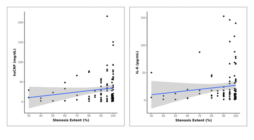
Figure 2: Correlation of Hs-CRP and IL-6 levels withe extent of stenosis. For Hs-CRP and stenosis extent rho=0.16, p=0.183, whereas for IL-6 and stenosis extent rho=0.12, p=0.243.
Hs-CRP: High-sensitivity C-reactive protein; IL-6: interleukin-6.
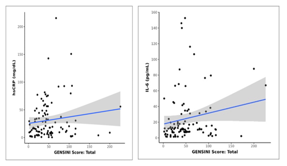
Figure 3: Correlation of Hs-CRP and IL-6 levels with GENSINI score. For Hs-CRP and GENSINI score, rho=0.13, p=0.194, whereas, for IL-6 and GENSINI score, rho=0.18, p=0.074.
Hs-CRP: High-sensitivity C-reactive protein; IL-6: interleukin-6.
DISCUSSION:
This cross-sectional observational study evaluated the relationship between inflammatory biomarkers and angiographic severity of ACS in patients undergoing Percutaneous Coronary Intervention. The study population demonstrated a marked male predominance (79%) with a relatively younger mean age, which is consistent with existing Indian and South Asian data [20]. This gender disparity has been attributed not only to higher exposure of males to traditional cardiovascular risk factors but also to socio-cultural barriers, under-recognition of symptoms in women, and delayed access to healthcare services, all of which continue to influence ACS presentation patterns.
ST-elevation myocardial infarction (STEMI) was the most common clinical presentation, followed by NSTEMI and unstable angina. While several contemporary registries have reported a rising proportion of NSTEMI due to widespread adoption of high-sensitivity cardiac troponin assays, STEMI remains predominant in many low- and middle-income settings, likely reflecting delayed presentation, limited access to early diagnostic facilities, and a higher burden of untreated risk factors [21,22]. The low prevalence of unstable angina in this cohort aligns with current evidence suggesting a declining incidence of unstable angina as a distinct clinical entity in the era of sensitive biomarker-based diagnosis [23].
The high prevalence of metabolic syndrome and dyslipidemia in this study underscores the substantial atherosclerotic burden in ACS patients. These metabolic risk factors are well known to promote endothelial dysfunction, systemic inflammation, and plaque instability, thereby contributing to both the onset and severity of ACS [24]. The coexistence of these conditions likely amplifies inflammatory responses, reflected in elevated hs-CRP and IL-6 levels.
Inflammatory marker analysis revealed significant differences in Hs-CRP and IL-6 levels across ACS subtypes, consistent with current evidence supporting the role of inflammation in plaque rupture and myocardial injury [25]. The STEMI group exhibited the highest mean Hs-CRP and mean IL-6 levels, reinforcing the concept that STEMI represents the most intense inflammatory and thrombotic phenotype of ACS. Elevated inflammatory markers in STEMI have been linked to larger infarct size, greater myocardial necrosis, and worse short- and long-term outcomes [26]. Interestingly, the unstable angina group showed the highest median Hs-CRP, which may reflect active plaque inflammation in the absence of overt myocardial necrosis, as previously described in inflammatory models of ACS [27].
With respect to angiographic severity, higher levels of Hs-CRP and IL-6 were observed in patients with multi-vessel disease, severe coronary stenosis, and higher GENSINI scores. These findings are in agreement with prior studies demonstrating positive correlations between inflammatory biomarkers and the extent and complexity of coronary artery disease [28,29]. However, the lack of statistically significant associations in this cohort suggests that while Hs-CRP and IL-6 are reflective of systemic inflammatory burden, they may not reliably discriminate anatomical disease severity in isolation. This weak to moderate correlation has also been reported in other observational studies, highlighting the multifactorial nature of coronary atherosclerosis, where local plaque characteristics, genetic predisposition, and hemodynamic factors interact alongside systemic inflammation [12,29].
Overall, the findings support the role of Hs-CRP and IL-6 as surrogate markers of inflammatory activity and global atherosclerotic burden rather than precise indicators of angiographic severity. Their greatest utility may lie in risk stratification and prognostication rather than anatomical assessment. Larger prospective studies with serial biomarker measurements and integration of imaging-based plaque characterization may further clarify their role in guiding management strategies in ACS patients undergoing PCI.
Limitations
A primary limitation of this study is the failure to meet the planned enrolment target of 138 participants. With a final sample size of 100, the study lacks the statistical power to definitively comment upon the correlation of Hs-CRP and IL-6 levels and ACS severity. The study had a small sample size and was conducted in a single location, limiting its ability to represent the general population and establish causality. A larger study cohort is required to confirm the predictive values of Hs-CRP and IL-6 and intensity of population. As a cross-sectional study, it lacked follow-up data and a single measurement of Hs-CRP and IL-6 provides less information than a kinetic measurement about their role in CAD.
By only including patients who underwent percutaneous coronary intervention (PCI), the study excluded many patients with acute coronary syndrome (ACS) who were not candidates for angiography. Consequently, some of the findings may not accurately reflect the full range of ACS in the broader population. The temporal variability among patients with regards to time of sampling for Hs-CRP and IL-6, may have masked or exaggerated the observed correlations between biomarkers and clinical outcomes. By focusing on patients undergoing coronary intervention, the study might have excluded those with very advanced multi-vessel disease deemed or those with MINOCA. This limits the generalizability of the findings to the broader, all-comer ACS population. The Unstable Angina (UA) cohort was limited by small sample size-reflecting the increased diagnostic sensitivity of modern troponin assays-predisposing these analyses to Type II errors. Consequently, UA-related comparisons are considered exploratory and hypothesis-generating rather than definitive.
CONCLUSION:
This study suggests that Hs-CRP and IL-6 are vital biomarkers for gauging the inflammatory activity and severity of Acute Coronary Syndrome. While anatomical correlations with angiographic profiles were not always statistically significant, the systemic inflammatory markers were significantly higher in STEMI compared to NSTEMI/UA, highlighting the fact that they have an important role in acute plaque instability. A non-significant trend toward higher biomarker levels in multi-vessel disease are reported as exploratory observations that require larger, powered cohorts to confirm. Trends suggesting higher inflammatory peaks in women were not significant after adjusting for age and diabetes.
Notably, Hs-CRP showed a stronger correlation with GENSINI Scores than IL-6, suggesting it may serve as a more precise surrogate for total atherosclerotic burden. Both markers increased alongside the degree of luminal stenosis and multi-vessel involvement, reflect inflammatory activity in Ischemic Heart Disease.
In this cross-sectional observational study, we identified a positive association between inflammatory biomarkers and acute myocardial injury. While these markers provide insight into the biological activity during an index ACS event, their utility for prospective screening or long-term prognosis remains unproven. Further longitudinal studies are required to determine if this inflammatory 'snapshot' translates into future clinical outcomes.
REFERENCES:
1. Di Cesare M, Perel P, Taylor S, et al.: The Heart of the World. Glob Heart [Internet]. [cited . 2026, 7:19. 10.5334/gh.1288
2. Sharma YP, Santosh Vemuri K, Bootla D, et al.: Epidemiological profile, management and outcomes of patients with acute coronary syndrome: Single centre experience from a tertiary care hospital in North India. Indian Heart J [Internet. 20212026, 7:174-9.
3. Interleukin-6 and High-sensitivity C-reactive Protein for the Prediction of Outcomes in Non-ST-segment Elevation Acute Coronary Syndromes. Revista Española de Cardiología (English Edition) [Internet. 201312026, 7:185-92.
4. Silva D, Pais de Lacerda A: High-sensitivity C-reactive protein as a biomarker of risk in coronary artery disease. Rev Port Cardiol [Internet. 201212026, 7:733-45.
5. Ajoolabady A, Pratico D, Lin L, et al.: Inflammation in atherosclerosis: pathophysiology and mechanisms. Cell Death Dis [Internet. 2024, 11:817.
6. Brie DM, Mornoș C, Adam O, Tîrziu A, Popescu R, Brie AD: Inflammatory Mechanisms in Acute Coronary Syndromes: From Pathophysiology to Therapeutic Targets. Cells [Internet. 20262026, 7:72-2073. 10.3390/cells15010072
7. Kamath DY, Xavier D, Sigamani A, Pais P: High sensitivity C-reactive protein (hsCRP) & cardiovascular disease: An Indian perspective. Indian J Med Res [Internet. 20152026, 7:261-8.
9. Su JH, Luo MY, Liang N, et al.: Interleukin- 6: Novel Target for Cardio-Cerebrovascular Diseases . Front Pharmacol [Internet. 2021, 24:745061.
10. Alfaddagh A, Martin SS, Leucker TM, et al.: Inflammation and cardiovascular disease: From mechanisms to therapeutics. Am J Prev Cardiol [Internet. 2020, 21:100130.
11. Singh A, Bind MK, Sinha N, Singh A, Rai A, Kumar D: The Role of Inflammatory Markers in Coronary Artery Disease Severity: Insights from a High vs. Low Inflammation Group. European Journal of Cardiovascular Medicine [Internet. 2024202026, 7:1368-73.
12. Rafiqi K, Hoeks CB, Løfgren B, Mortensen MB, Bruun JM: Diagnostic Impact of Hs-CRP and IL-6 for Acute Coronary Syndrome in Patients Admitted to the ED with Chest Pain: Added Value to the HEART Score?. Open Access Emerg Med [Internet. 2023212026, 7:333-42.
13. Yang C, Deng Z, Li J, Ren Z, Liu F: Meta-analysis of the relationship between interleukin-6 levels and the prognosis and severity of acute coronary syndrome. Clinics [Internet. 202112026, 7:2690.
14. Deori TJ, Agarwal M, Masood J, Sharma S, Ansari A: Estimation of cardiovascular risk in a rural population of Lucknow district using WHO/ISH risk prediction charts. J Family Med Prim Care [Internet. 2020302026, 7:4853-60.
15. American Diabetes Association Professional Practice Committee. 2: Diagnosis and Classification of Diabetes: Standards of Care in Diabetes—2024. Diabetes Care [Internet. 2023, 11:20-42.
16. 2023 ESH Hypertension Guideline Update: Bringing Us Closer Together Across the Pond - American College of Cardiology [Internet]. [cited. (20267). https://www.acc.org/Latest-in- Cardiology/Articles/2024/02/05/11/43/2023-ESH-Hypertension-Guideline-Update.
17. Pappan N, Awosika AO, Rehman A: Dyslipidemia. In. StatPearls [Internet, Treasure Island (FL): StatPearls Publishing; 202520267.
18. Lam DW, LeRoith D: Metabolic Syndrome. In: Feingold KR, Adler RA, Ahmed SF, Anawalt B, Blackman MR, Chrousos G, et al., editors. Endotext [Internet]. South Dartmouth (MA): MDText.com, Inc. 200020267,
19. Charach L, Blatt A, Jonas M, et al.: Using the Gensini score to estimate severity of STEMI, NSTEMI, unstable angina, and anginal syndrome. Medicine (Baltimore. 2021, 15:27331. 10.1097/MD.0000000000027331.
21. High-Sensitivity Cardiac Troponin for the Rapid Diagnosis of Acute Coronary Syndrome in the Emergency Department: A Clinical and Cost-Effectiveness Evaluation. CADTH Technol Overv [Internet. 201312026, 8:3203.
24. Młynarska E, Bojdo K, Frankenstein H, et al.: Endothelial Dysfunction as the Common Pathway Linking Obesity, Hypertension and Atherosclerosis. Int J Mol Sci [Internet. 2025, 16:10096.
25. Held C, White HD, Stewart RAH, et al.: Inflammatory Biomarkers Interleukin‐6 and C‐Reactive Protein and Outcomes in Stable Coronary Heart Disease: Experiences From the STABILITY (Stabilization of Atherosclerotic Plaque by Initiation of Darapladib Therapy) Trial. J Am Heart Assoc [Internet. 2017, 24:005077.
26. Gadhavi DN, Ghantiya UV, Gondaliya TK: Correlation of Inflammatory Biomarkers (IL-6, CRP, and TNF-α) With Severity and Outcomes in Patients with ST-Elevation Myocardial Infarction (STEMI): A Prospective Cohort Study. Journal of Heart Valve Disease [Internet. 2025, 10:15-9.
28. Bouzidi N, Gamra H: Relationship between serum interleukin-6 levels and severity of coronary artery disease undergoing percutaneous coronary intervention. BMC Cardiovasc Disord [Internet. 2023282026, 8:586.
29. Sharma A: Association of Hs-CRP Levels in Patients with Acute Coronary Syndromes and it’s Correlation with Angiographic Severity of Coronary Artery Stenosis. Indian J Cardiovasc Dis Women [Internet. 2023222026, 8:37-42.