A STUDY OF PITUITARY DYSFUNCTIONS IN PATIENTS OF CHRONIC HEMOLYTIC ANEMIA ADMITTED IN A TERTIARY CARE HOSPITAL OF EASTERN INDIA.
- Oishee Saha , Senior Resident, MBBS, MD (General Medicine), Department of General Medicine, Calcutta National Medical College and Hospital, West Bengal, India.
- Debasish Ganguly , Senior Resident, MBBS, MD (General Medicine), Department of General Medicine, Calcutta National Medical College and Hospital, West Bengal, India.
- Sujoy Sarkar , Professor, MBBS, MD (General Medicine), Department of General Medicine, North Bengal Medical College and Hospital, West Bengal, India.
- Debdeep Das , Senior Resident, MBBS, MD (General Medicine), Department of General Medicine, Calcutta National Medical College and Hospital, West Bengal, India.
Article Information:
Abstract:
Introduction: Chronic hemolytic anemia (CHA) is characterized by premature destruction of red blood cells, often seen in conditions like thalassemia, sickle cell disease, and hereditary spherocytosis. Repeated blood transfusions, a mainstay of treatment, can result in iron overload, which may deposit in endocrine organs, particularly the pituitary gland. Pituitary dysfunction can lead to growth retardation, delayed puberty, hypothyroidism, adrenal insufficiency, and infertility. Despite its significant clinical impact, pituitary involvement in CHA is often underdiagnosed, emphasizing the need for systematic evaluation. Aims: To determine the prevalence of pituitary hormonal dysfunction in patients with CHA. To identify specific patterns of pituitary hormone involvement, to correlate pituitary dysfunction with disease severity, transfusion history, and iron overload. Materials and Methods: This was a hospital-based cross-sectional study conducted over a 12-month period, from 1st April 2022 to 30th March 2023, at the Calcutta National Medical College & Hospital, Kolkata. The study was carried out in the Department of General Medicine in collaboration with the Department of Biochemistry. Patients were recruited from both the inpatient (IPD) and outpatient (OPD) services of General Medicine, with follow-up assessments conducted in the OPD after discharge. A total of 120 patients with chronic hemolytic anemia were enrolled for evaluation of pituitary function and related clinical and biochemical parameters. Results: Among the 120 patients, 10 patients (8.3%) with pituitary dysfunction had ferritin levels below 1000 ng/mL, 30 patients (25%) had levels between 1000–2000 ng/mL, and 50 patients (41.7%) had levels above 2000 ng/mL. No patients without measured ferritin were included. The association between high ferritin levels and pituitary dysfunction was statistically significant (p = 0.002). Conclusion: Pituitary dysfunction is a common but under-recognized complication in chronic hemolytic anemia, primarily affecting growth and reproductive hormone axis. Early screening and timely management of pituitary abnormalities are crucial to prevent long-term complications and improve quality of life. Integrating routine endocrine evaluation into the care of CHA patients, particularly those with high transfusion burden or iron overload, is strongly recommended.
Keywords:
Article :
INTRODUCTION:
Chronic hemolytic anemia (CHA) represents a group of hematological disorders characterized by premature and persistent destruction of red blood cells, resulting in chronic anemia and compensatory mechanisms such as increased erythropoiesis and frequent blood transfusions. Common causes include β‑thalassemia major, sickle cell disease, and hereditary anemias such as Diamond‑Blackfan syndrome. These conditions have significant systemic consequences beyond hematological deficits, prominently affecting endocrine organs. Among the endocrine complications, dysfunction of the pituitary gland has emerged as an important yet under-recognized sequela in patients with CHA [1,2].
The pituitary gland serves as the master regulator of multiple hormonal axis, including the somatotropic (growth hormone), gonadotropic (LH/FSH), thyrotropic (TSH), and corticotropic (ACTH) pathways. Its proper function is essential for growth, metabolism, stress response, and reproductive development. Pituitary dysfunction in CHA primarily arises from iron overload, a consequence of both chronic hemolysis and repeated transfusions. When the iron‑binding capacity of transferrin is exceeded, non‑transferrin bound iron (NTBI) circulates freely and deposits in various organs, including endocrine glands. The pituitary, due to its high vascularity, extensive metabolic activity, and expression of transferrin receptors, is particularly susceptible to excess iron deposition [3]. Iron catalyzes the formation of reactive oxygen species (ROS), causing oxidative stress, lipid peroxidation, mitochondrial damage, and eventual apoptosis of hormone-producing cells in the gland [4].
The gonadotrophic axis appears to be the most vulnerable to iron-mediated injury, often leading to hypogonadotropic hypogonadism, manifesting as delayed puberty, impaired secondary sexual development, or infertility. Evidence consistently shows that reproductive hormone deficiencies are frequently observed in chronically transfused patients with β-thalassemia major, even before overt clinical symptoms appear. Growth hormone (GH) deficiencies and short stature are also common, reflecting early somatotropic axis involvement due to iron toxicity and oxidative damage to GH-producing cells. As a result, patients may present with significantly reduced adult height in addition to other endocrine abnormalities [5].
Magnetic resonance imaging (MRI) studies have been particularly instrumental in illuminating the extent and early onset of pituitary involvement in CHA. MRI sequences sensitive to iron (e.g., T2* and R2 techniques) reveal increased iron deposition and reduced pituitary volume well before clinical endocrine dysfunction becomes apparent [6]. Elevated pituitary iron indices correlate with higher liver and pancreatic iron burdens, indicating systemic iron overload, and can predict hypogonadism risk even in the absence of gland shrinkage on imaging. These imaging findings support the concept that pituitary involvement begins early, often in childhood or adolescence, and that preclinical detection via MRI can identify patients at risk for endocrine sequelae [7].
Endocrine dysfunction in CHA is not restricted to gonadal and growth hormone axis alone; other hormonal deficiencies including central hypothyroidism, hypocortisolism, and impaired lactotroph or adrenocorticotrophic function have been reported, although less frequently. Moreover, iron overload contributes to dysfunction in peripheral glands (thyroid, pancreas), leading to additional endocrinopathies such as hypothyroidism and diabetes mellitus [8].
The clinical impact of pituitary dysfunction in CHA is profound. In addition to the physical manifestations of hormonal deficits (growth retardation, delayed puberty, infertility), quality of life, psychological wellbeing, and long-term outcomes are adversely affected. Furthermore, endocrine dysfunction can exacerbate other complications of CHA, including osteopenia and osteoporosis, often seen in patients with prolonged hypogonadism or GH deficiency.
Despite the high prevalence and clinical significance of pituitary and other endocrine dysfunctions in CHA, routine endocrine screening in many clinical settings remains suboptimal. Standard hematologic care frequently emphasizes transfusion and chelation therapies, yet endocrinopathies may progress silently until advanced. Early identification and management of pituitary dysfunction through regular hormonal assessments and imaging, paired with optimized iron chelation, are critical to mitigate long-term morbidity and improve growth and reproductive outcomes.
The primary aim of this study is to evaluate the prevalence and patterns of pituitary dysfunction in patients with chronic hemolytic anemia (CHA). Specifically, the study seeks to assess the involvement of various pituitary hormonal axis, including the gonadotropic, somatotropic, thyrotropic, and corticotropic pathways, and to identify which axis are most commonly affected. Additionally, the study aims to examine the relationship between pituitary dysfunction and clinical factors such as disease duration, severity of anemia, frequency of blood transfusions, and iron overload, as measured by serum ferritin levels or imaging studies. Through this analysis, the study intends to highlight the clinical significance of pituitary involvement in CHA, underscore the need for routine endocrine screening, and provide insights for early detection and intervention strategies that can prevent long-term morbidity and improve quality of life in these patients.
MATERIALS AND METHODS:
Study design: Hospital based cross sectional study.
Study setting: The study was conducted in the Department of General Medicine in collaboration with the Department of Biochemistry Calcutta National Medical College &Hospital, Kolkata.
Place of study: Calcutta National Medical College & Hospital, In patient Department (IPD) and outpatient department (OPD)of General Medicine.
Period of study: 12 months (From 1st April 2022 to 30th March, 2023)
Study population: Patients was recruited from the OPD and IPD of General Medicine. Follow up of patients was done after their discharge in outpatient department of General Medicine.
Sample size: 120 patients
Inclusion Criteria
1. Patients diagnosed with chronic hemolytic anemia (including β thalassemia major, sickle cell disease, hereditary spherocytosis, or other congenital hemolytic anemias).
2. Age range: 5–40 years.
3. Patients receiving regular follow-up at the hematology or endocrinology clinic.
4. Patients who have received at least one blood transfusion (to account for iron overload).
5. Patients (or guardians, in case of minors) who provide informed consent for participation.
Exclusion Criteria
1. Patients with primary pituitary disorders (e.g., pituitary adenoma, congenital pituitary hypoplasia).
2. Patients with chronic systemic illnesses unrelated to CHA that may affect endocrine function (e.g., chronic kidney disease, liver cirrhosis).
3. Patients on long-term hormone therapy or steroid treatment that could influence pituitary function.
4. Patients with history of head trauma, brain surgery, or radiotherapy affecting the pituitary region.
5. Patients unwilling or unable to provide informed consent.
Statistical Analysis:
For statistical analysis data were entered into a Microsoft excel spreadsheet and then analyzed by SPSS (version 27.0; SPSS Inc., Chicago, IL, USA) and GraphPad Prism version 5. Data had been summarized as mean and standard deviation for numerical variables and count and percentages for categorical variables. Two-sample t-tests for a difference in mean involved independent samples or unpaired samples. Paired t-tests were a form of blocking and had greater power than unpaired tests. A chi-squared test (χ2 test) was any statistical hypothesis test wherein the sampling distribution of the test statistic is a chi-squared distribution when the null hypothesis is true. Without other qualification, 'chi-squared test' often is used as short for Pearson's chi-squared test. Unpaired proportions were compared by Chi-square test or Fischer’s exact test, as appropriate.
Explicit expressions that can be used to carry out various t-tests are given below. In each case, the formula for a test statistic that either exactly follows or closely approximates a t-distribution under the null hypothesis is given. Also, the appropriate degrees of freedom are given in each case. Each of these statistics can be used to carry out either a one-tailed test or a two-tailed test.
Once a t value is determined, a p-value can be found using a table of values from Student's t-distribution .If the calculated p-value is below the threshold chosen for statistical significance (usually the 0.10, the 0.05, or 0.01 level), then the null hypothesis is rejected in favour of the alternative hypothesis.
P-value ≤ 0.05 was considered for statistically significant.
RESULTS:
Table 1: Distribution of Patients by Age Group
|
Age Group (years) |
Number of Patients (n=120) |
% |
|
5–10 |
20 |
16.7 |
|
11–20 |
40 |
33.3 |
|
21–30 |
35 |
29.2 |
|
31–40 |
25 |
20.8 |
Table 2: Distribution by Gender
|
Gender |
Number of Patients (n=120) |
% |
|
Male |
70 |
58.3 |
|
Female |
50 |
41.7 |
Table 3: Pituitary Axis Dysfunction
|
Pituitary Axis |
Number of Patients (n=120) |
% |
|
Gonadotropic (LH/FSH) |
45 |
37.5 |
|
Somatotropic (GH) |
30 |
25 |
|
Thyrotropic (TSH) |
15 |
12.5 |
|
Corticotropic (ACTH) |
10 |
8.3 |
|
Multiple Axis |
20 |
16.7 |
Table 4: Correlation of Pituitary Dysfunction with Transfusion Frequency
|
Transfusion Frequency |
Number of Patients with Dysfunction |
% |
p-value |
|
≤5/year |
15 |
12.5 |
0.001 |
|
6–10/year |
35 |
29.2 |
|
|
>10/year |
40 |
33.3 |
|
|
Never transfused |
0 |
0 |
Table 5: Correlation of Pituitary Dysfunction with Serum Ferritin Levels
|
Ferritin Level (ng/mL) |
Number of Patients with Dysfunction |
% |
p-value |
|
<1000 |
10 |
8.3 |
0.002
|
|
1000–2000 |
30 |
25 |
|
|
>2000 |
50 |
41.7 |
|
|
Not measured |
0 |
0 |
|
|
|
|
|
|
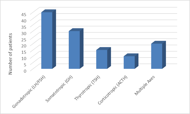
Figure 1: Pituitary Axis Dysfunction
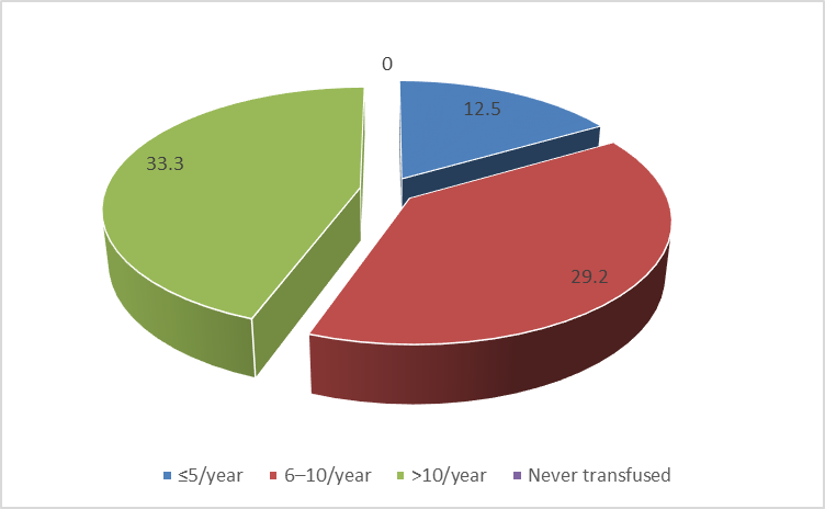
Figure 2: Correlation of Pituitary Dysfunction with Transfusion Frequency
Out of 120 patients with chronic hemolytic anemia, 40 patients (33.3%) were aged 11–20 years, 35 patients (29.2%) were 21–30 years, 25 patients (20.8%) were 31–40 years, and 20 patients (16.7%) were 5–10 years old.
Among the 120 patients, 70 were male (58.3%) and 50 were female (41.7%).
Out of 120 patients, 45 patients (37.5%) had low levels of LH/FSH, 30 patients (25%) had GH deficiency, 15 patients (12.5%) had low levels of TSH, and 10 patients (8.3%) had low levels of ACTH. Additionally, 20 patients (16.7%) showed dysfunction in multiple pituitary axis.
Among the 120 patients, 15 patients (12.5%) with pituitary dysfunction received ≤5 transfusions per year, 35 patients (29.2%) received 6–10 transfusions per year, and 40 patients (33.3%) received more than 10 transfusions per year. The association between higher transfusion frequency and pituitary dysfunction was statistically significant (p = 0.001).
Among the 120 patients, 10 patients (8.3%) with pituitary dysfunction had ferritin levels below 1000 ng/mL, 30 patients (25%) had levels between 1000–2000 ng/mL, and 50 patients (41.7%) had levels above 2000 ng/mL. No patients without measured ferritin were included. The association between high ferritin levels and pituitary dysfunction was statistically significant (p = 0.002).
DISCUSSION:
In this study of 120 patients with chronic hemolytic anemia, pituitary dysfunction was observed in multiple hormonal axis, with the gonadotropic axis (37.5%) and somatotropic axis (25%) affected most frequently. Our findings align with those of Noetzli et al., who reported that hypogonadotropic hypogonadism is the most common endocrine dysfunction in patients with transfusion‑dependent anemia such as thalassemia major and that increased pituitary iron predicts clinical hormone abnormalities in these axes. Their MRI studies also showed that pituitary iron deposition begins early, often before clinical hormone loss becomes evident, supporting our observation that gonadal and growth hormone defects are predominant in CHA patients [9].
Similarly, Mostafa et al. studied adult males with sickle cell anemia, finding that patients with higher ferritin levels had significant hormonal changes, including elevated luteinizing hormone and lower testosterone, suggesting endocrine disturbances linked to iron overload. This supports our finding that increased serum ferritin is significantly associated with pituitary dysfunction (p = 0.002), indicating that iron burden impacts pituitary hormone regulation [10].
In terms of iron overload and transfusion frequency, our result showing a significant association between frequent transfusions and pituitary dysfunction (p = 0.001) is consistent with existing literature. Chronic and repeated transfusions contribute to non‑transferrin bound iron (NTBI) accumulation and deposition in endocrine tissues. Pituitary siderosis has been shown to occur early in transfused patients and predispose to dysfunction, especially in the gonadotropic cells, which are vulnerable to iron toxicity because of their rich vascularization and high metabolic demand [11,12].
Our study also noted that high ferritin levels correlated strongly with pituitary dysfunction, which aligns with Chirico et al. who reported that high ferritin values are predictive of thyroid and gonadal endocrinopathies in thalassemia patients, and intensifying iron chelation can improve or slow the progression of these dysfunctions. They found that ferritin above certain thresholds was associated with a faster development of multiple endocrinopathies over time, suggesting that ferritin can be a prognostic marker for endocrine complications [13].
The pattern of hormonal abnormalities seen in our patients reflects a broader trend described in the literature: pituitary iron overload leads to impairment of multiple endocrine pathways, especially those controlling growth and reproductive functions. Iron deposition affects hormone‑producing cells directly, causing decreased secretion of gonadotropins and growth hormone, as documented in studies using advanced MRI techniques that quantify iron content in pituitary tissues and correlate these with clinical endocrine outcomes [14].
Furthermore, the international position statement by the I‑CET guidelines indicates that delayed puberty and hypogonadism are among the most common endocrine consequences in thalassemia patients due to iron toxicity in the pituitary gland, further validating that endocrine screening is essential in chronically transfused populations. Their review stresses that early identification of hormonal deficits can guide timely interventions to prevent long‑term complications [15].
CONCLUSION:
In this study of 120 patients with chronic hemolytic anemia, pituitary dysfunction was a common endocrine complication, with the gonadotropic and somatotropic axis most frequently affected. Higher transfusion frequency and elevated serum ferritin levels were significantly associated with increased risk of pituitary dysfunction, highlighting the role of iron overload in endocrine abnormalities. Early detection through regular hormonal assessment and monitoring of iron status is essential to prevent long-term complications such as delayed puberty, growth retardation, and infertility. Optimizing iron chelation therapy and implementing routine endocrine screening can improve clinical outcomes and quality of life for patients with chronic hemolytic anemia.
REFERENCES:
1. Noetzli LJ, Panigrahy A, Mittelman SD, et al. Pituitary iron and volume predict hypogonadism in transfusional iron overload. Am J Hematol. 2012;87(2):167–171.
2. Karadag SIK, Karakas Z, Yilmaz Y, et al. Pituitary Iron Deposition and Endocrine Complications in Patients with β‑Thalassemia: From Childhood to Adulthood. Hemoglobin. 2020;44(5):344–348.
3. Kim MK, Lee JW, Baek KH, et al. Endocrinopathies in transfusion‑associated iron overload. Clin Endocrinol (Oxf). 2013;78(2):271–277.
4. Frontiers Editorial Board. Iron deposition and hypopituitarism mechanisms. Front Endocrinol. 2025;xx(xx):xxx–xxx.
5. Banker C, Wang ZJ, Singer ST, et al. Anterior pituitary volume and R2 in transfusion‑dependent anemias. Clin Neuroradiol. 2022;32:259–267.
6. Verberckmoes B, Dekeyzer S, Decaestecker K. The dark pituitary: Hemochromatosis as a lesser‑known cause of pituitary dysfunction. J Belg Soc Radiol. 2022;106(1):63.
7. Significant pituitary siderosis in sickle cell disease. Blood. 2024;144(13):1457–1459.
8. Growth and endocrine disorders in thalassemia: I‑CET guidelines. PMC. 2013;xx(xx):xxx–xxx.
9. Noetzli LJ, Panigrahy A, Mittelman SD, et al. Pituitary iron and volume predict hypogonadism in transfusional iron overload. Am J Hematol. 2012;87(2):167–171.
10. Mostafa GG, Zahran FE, Omer SA, et al. The effect of serum ferritin level on gonadal, prolactin, thyroid hormones and TSH in adult males with sickle cell anemia. J Blood Med. 2020;11:27–32.
11. Soliman A, Ahmed S, Beck C, et al. The role of MRI and ferritin in assessing iron overload and endocrine dysfunction in thalassemia major: focus on pituitary MRI findings. Endocrine Abstracts. 2025;110:P803.
12. Significant pituitary siderosis is common in transfusion‑dependent sickle cell disease. PMC. 2024.
13. Chirico V, Rigoli L, Lacquaniti A, et al. Endocrinopathies, metabolic disorders, and iron overload in major and intermedia thalassemia: serum ferritin as diagnostic and predictive marker. Eur J Haematol. 2015;94(5):404–412.
14. Molecular mechanisms of iron metabolism and overload: pituitary and hypothalamus. MDPI. 2025.
15. Growth and endocrine disorders in thalassemia: I‑CET position statement. PMC. 2013.