EFFECTIVENESS OF 3D VIRTUAL DIGITAL CADAVER IN MEDICAL TRAINING.
- Rajarshi Roy , MBBS, MD (Anatomy), Department of Anatomy, JMN Medical College, Chakdaha, Nadia, West Bengal.
- Anushree Das , MBBS, MD (Anatomy), MBA (Healthcare Management), Department of Anatomy, JIMSH, Budge budge, West Bengal.
Article Information:
Abstract:
Introduction: The integration of virtual reality (VR) and simulation-based learning has transformed medical education by providing immersive, interactive, and risk-free environments. Among these innovations, 3D virtual digital cadavers offer unique opportunities to enhance anatomical understanding, skill acquisition, and learner engagement. Aims: This study aimed to evaluate the impact of digital cadaver-based learning on knowledge acquisition, retention, and student satisfaction among Phase I MBBS students, compared with traditional cadaveric teaching. Materials and methods: An interventional study was conducted at JMN Medical College, West Bengal, with 132 Phase I MBBS students randomized into two groups: Group A (digital cadaver + traditional teaching) and Group B (traditional teaching only). Assessments included pre- and post-test MCQs, Objective Structured Practical Examinations (OSPE), a retention test after two weeks, and student feedback via a Likert scale. Statistical analysis was performed using SPSS v27.0, with p ≤ 0.05 considered significant. Result: Pre-test and post-test scores showed no significant differences between groups (p > 0.05), indicating comparable baseline knowledge and immediate learning outcomes. However, retention scores were significantly higher in Group A (17.2 ± 2.4) compared to Group B (14.8 ± 2.6; p < 0.001). Student feedback highlighted improved engagement, confidence, and satisfaction with the blended approach. Conclusion: Digital cadaver-based learning, when integrated with traditional teaching, enhances long-term retention of anatomical knowledge and improves learner engagement. While challenges such as cost and technological barriers remain, hybrid curricula incorporating VR and simulation represent a promising direction for modern medical education.
Keywords:
Article :
INTRODUCTION:
The integration of virtual reality (VR) and simulation-based learning has significantly transformed modern medical education by providing immersive, interactive, and risk-free environments for skill development. Traditional anatomy teaching methods, particularly cadaveric dissection, have long been considered the cornerstone of medical training.1 However, advances in digital technology have introduced three-dimensional (3D) virtual digital cadavers as innovative educational tools that enhance anatomical visualization, learner engagement, and clinical understanding.2,3 3D virtual cadaver systems allow medical students to explore human anatomy in a detailed and repeatable manner without the limitations associated with conventional cadaveric dissection, such as limited availability, ethical concerns, specimen degradation, and high maintenance requirements.
Studies have shown that virtual anatomy platforms improve spatial understanding, knowledge retention, procedural confidence, and academic performance, especially in complex subjects like musculoskeletal and neurological anatomy.4,5,6 Many learners also report increased satisfaction and engagement when digital dissection is combined with traditional cadaver-based teaching. Simulation-based learning further strengthens medical training by enabling repetitive practice, immediate feedback,7 objective assessment, and exposure to diverse clinical scenarios without compromising patient safety. VR-assisted training has demonstrated improvements in surgical precision, operative speed, psychomotor skills, and clinical decision-making across multiple medical specialties. Despite these advantages, challenges such as high initial costs,8 technological limitations, and variability in skill transfer to real clinical settings continue to affect widespread implementation.
Nevertheless, with rapid technological advancement and increasing accessibility,9 3D virtual digital cadavers and VR-based simulation are emerging as valuable adjuncts to conventional medical education and are likely to play a major role in future medical training systems. Aims this study aimed to evaluate the impact of digital cadaver-based learning on knowledge acquisition, retention, and student satisfaction among Phase I MBBS students, compared with traditional cadaveric teaching.10.
MATERIALS AND METHODS:
Study design: Interventional study
Study setting: JMN Medical College, Chakdaha, Nadia, WB
Study period: 6 weeks
Study population: Phase I MBBS students
Sample size: To calculate the sample size for the study comparing two groups with significance level of 0.05, power of the study 80% & standard deviation 10, the sample size for each group comes as 63. We made two groups of students comprising 66 in each groups.
Sampling method: Purposive sampling.
Inclusion criteria: The students who appeared in 1st internal assessment examination and scored pass marks 50% and had 75% attendance and were willing to be the part of the study were included.
Exclusion criteria: The students who appeared in 1st internal assessment examination and scored less than pass marks 50% and had below 75% attendance and were not willing to be the part of the study were not included.
Ethical considerations:
Clearance from the college scientific and ethical committees
Conflict of interest: None, no sponsorship or financial liabilities obtained from manufacturer of Digital cadaver used in the study.
Data collection & analysis methods:
Group Allocation
Participants were randomly assigned into two groups:
· Group A (Intervention Group): Received training using Digital cadaver in addition to conventional cadaveric teaching and practical sessions.
· Group B (Control Group): Received conventional cadaveric teaching and practical sessions.
· Randomization was achieved using a computer-generated sequence.
Intervention
· Group A (n=66): To Undergo structured digital cadaver-based training using Anatomage virtual dissection table. Each session lasted for 40-45 min and was repeated over several sessions depending upon the topic. In addition to standard lectures, video demonstrations, and hands-on sessions with faculty supervision on actual cadavers & viscera of the same topics.
· Group B (n=66): To attend only standard lectures, video demonstrations, and hands-on sessions with faculty supervision on actual cadavers & viscera of the same topics.
Assessment Tools
1. Pre- and Post-Test Knowledge Assessment: To be conducted using a validated multiple-choice questionnaire (MCQ) designed to test theoretical knowledge.
2. Skill Performance Assessment: Objective Structured Practical Examination (OSPE) stations and the same validated multiple-choice questionnaire (MCQ) designed to test theoretical knowledge used for theoretical knowledge improvement, evaluated by blinded faculty assessors.
3. Retention Test: Reassessment after 2 weeks to evaluate knowledge retention.
4. Feedback and Satisfaction: Collected using a 5-point Likert scale questionnaire regarding the learning experience, engagement, and perceived usefulness.
Outcome Measures
· Improvement in knowledge scores (pre- vs post-intervention) in both groups.
· Retention of knowledge at follow-up.
· Participant satisfaction and engagement.
Study Variable:
Independent Variable
• 3D Virtual Digital Cadaver-based learning method
• Traditional cadaveric teaching method
Dependent Variables
• Knowledge acquisition (pre-test and post-test scores)
• Knowledge retention
• Student satisfaction
• Learner engagement
• Confidence level
• Perceived usefulness/effectiveness
Demographic Variables
• Age
• Gender
• Academic year/Phase I MBBS status
Outcome Variables
• Academic performance
• Retention test scores
• Student perception toward learning methods
Recommendations: Institutions should adopt hybrid teaching models, invest in faculty training, and explore scalable deployment strategies. Future research should focus on multicentric validation, longitudinal outcomes, and cost-effectiveness analyses to support widespread adoption.
Statistical Analysis: For statistical analysis, data were initially entered into a Microsoft Excel spreadsheet and then analysed using SPSS (version 27.0; SPSS Inc., Chicago, IL, USA) and GraphPad Prism (version 5). Numerical variables were summarized using means and standard deviations, while Data were entered into Excel and analyzed using SPSS and GraphPad Prism. Numerical variables were summarized using means and standard deviations, while categorical variables were described with counts and percentages. Two-sample t-tests were used to compare independent groups, while paired t-tests accounted for correlations in paired data. Chi-square tests (including Fisher’s exact test for small sample sizes) were used for categorical data comparisons. P-values ≤ 0.05 were considered statistically significant.
RESULTS:
Table 1: Comparison of Mean Scores (Out of 25)
|
Test |
Group A Mean ± SD |
Group B Mean ± SD |
t-value |
p-value |
|
Pre-test |
10.2 ± 2.1 |
10.0 ± 2.0 |
0.56 |
0.58 |
|
Post-test |
18.5 ± 2.3 |
18.1 ± 2.5 |
0.96 |
0.34 |
|
Retention Test |
17.2 ± 2.4 |
14.8 ± 2.6 |
5.51 |
<0.001 |
Table 2. Student Satisfaction Scores on a 5-Point Likert Scale:
|
Domain |
Group A (Digital Cadaver + Traditional) |
Group B (Traditional Only) |
|
Satisfaction |
4.3 ± 0.5 |
3.6 ± 0.6 |
|
Engagement |
4.5 ± 0.4 |
3.7 ± 0.5 |
|
Confidence |
4.4 ± 0.5 |
3.5 ± 0.6 |
|
Usefulness |
4.6 ± 0.4 |
3.8 ± 0.5 |
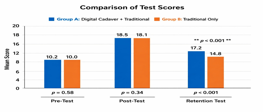
Figure 1. Comparison of Test Scores between Digital Cadaver and Traditional Teaching Groups.
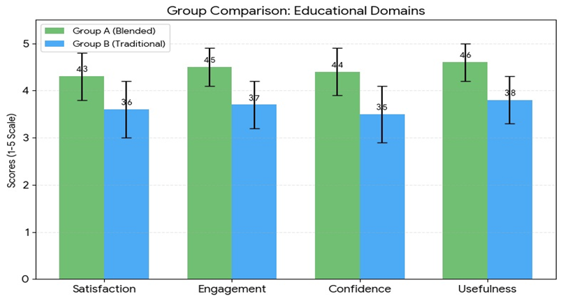
Figure 2. Comparative Student Satisfaction Scores on a 5‑Point Likert Scale.
A total of 132 Phase I MBBS students participated in the study and were equally divided into Group A (Digital Cadaver + Traditional Teaching) and Group B (Traditional Teaching Only). Pre-test scores showed no significant difference between Group A (10.2 ± 2.1) and Group B (10.0 ± 2.0) with a p-value of 0.58, indicating comparable baseline knowledge among students. Similarly, post-test scores were comparable between Group A (18.5 ± 2.3) and Group B (18.1 ± 2.5) with a p-value of 0.34, suggesting that both teaching approaches were equally effective for immediate knowledge acquisition. However, retention test scores conducted after two weeks revealed significantly higher scores in Group A (17.2 ± 2.4) compared with Group B (14.8 ± 2.6), with a highly significant p-value of <0.001, demonstrating superior long-term knowledge retention with the blended digital cadaver approach. Feedback analysis using a 5-point Likert scale showed that Group A consistently achieved higher ratings across satisfaction, engagement, confidence, and perceived usefulness domains, with mean scores ranging from 4.3 to 4.6, whereas Group B scored between 3.5 and 3.8. Students exposed to digital cadaver-based learning reported better visualization of anatomical structures, improved interactivity, easier repetition of concepts, and greater confidence in identifying complex anatomical regions, indicating that the blended teaching model enhanced overall learner experience and educational effectiveness compared with traditional teaching alone.
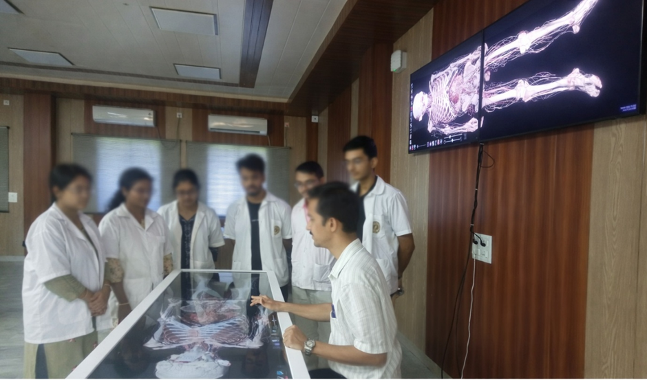
Figure 3. Digital Anatomy Classroom Using a Virtual Cadaver System (Faces Blurred).
Students and an instructor engage in an interactive anatomy session using a digital cadaver table and large wall‑mounted display. The setup demonstrates the integration of virtual dissection technology into medical education, enabling detailed visualization of anatomical structures without physical specimens. The environment reflects a modern, technology‑enhanced learning space that supports hybrid teaching approaches combining digital and traditional methods. All human faces are intentionally blurred to preserve participant anonymity.
DISCUSSION:
The findings of the present study demonstrate that the integration of 3D virtual digital cadavers with traditional anatomy teaching significantly improves long-term knowledge retention, learner engagement, satisfaction, confidence, and perceived usefulness among Phase I MBBS students. Although immediate post-test performance was comparable between both groups, students exposed to blended digital cadaver-based learning achieved significantly higher retention scores after two weeks, suggesting better memory consolidation and deeper understanding of anatomical concepts. Similar findings were reported by Koucheki et al. [11],
who demonstrated that immersive virtual reality and cadaveric bone teaching were equally effective in skeletal anatomy education, supporting the educational value of VR-assisted anatomy learning. Tiznado-Matzner et al. [12] also observed that 3D-scanned cadaveric models enhanced student perception, spatial understanding, and interactive learning experiences in anatomy education. Likewise, Yoon [13] reported that 3D virtual cadaver practice significantly improved learning motivation, academic achievement, and self-efficacy among first-year nursing students, which aligns with the higher confidence and satisfaction scores observed in the present study. Chen et al. [14] further demonstrated that virtual reality-based 3D anatomical models enhanced student engagement and understanding of complex anatomical structures compared with traditional approaches alone. Michalik et al. [15] highlighted the growing importance of high-fidelity 3D cadaveric models as effective alternatives and adjuncts in medical education and clinical practice, emphasizing their value in improving accessibility and visualization. In addition, Lim et al. [16] found that innovative 3D educational models were comparable to cadaveric materials in teaching anatomy, reinforcing the effectiveness of technology-enhanced anatomy education. Collectively, these studies support the present findings and suggest that blended digital cadaver-based learning provides a valuable supplement to conventional cadaveric teaching by improving educational outcomes, learner engagement, and long-term retention in medical training.
CONCLUSION:
The present study demonstrates that the integration of 3D virtual digital cadaver learning with traditional cadaveric teaching is an effective approach in medical education. Although immediate learning outcomes were comparable between groups, students exposed to the blended digital cadaver method showed significantly better long-term knowledge retention. Additionally, higher levels of satisfaction, engagement, confidence, and perceived usefulness were observed among students using digital cadaver-based learning. These findings suggest that 3D virtual digital cadavers serve as valuable adjuncts to conventional anatomy teaching by enhancing learner experience and retention. Incorporating digital cadaver technology into undergraduate medical training may improve the overall effectiveness of anatomy education.
REFERENCES:
1. Alasmari WA. Medical Students’ Feedback of Applying the Virtual Dissection Table (Anatomage) in Learning Anatomy: A Cross-sectional Descriptive Study. AMEP. 2021 Nov 11;12(1):1303–1307.
2. Chen S, Zhu J, Cheng C, Pan Z, Liu L, Du J, et al. Can virtual reality improve traditional anatomy education programmes? A mixed-methods study on the use of a 3D skull model. BMC Med Educ. 2020 Oct 31;20(1):395.
3. Alharbi Y, Al-Mansour M, Al-Saffar R, Garman A, Al-Radadi A. Three-dimensional Virtual Reality as an Innovative Teaching and Learning Tool for Human Anatomy Courses in Medical Education: A Mixed Methods Study. Cureus. 2020 Feb 24;12(2):e7085.
4. Neyem A, Cadile M, Burgos-Martínez SA, Farfán Cabello E, Inzunza O, Alvarado MS, et al. Enhancing medical anatomy education with the integration of virtual reality into traditional lab settings. Clinical Anatomy. 2024 Sept 8;38(3):249–268.
5. Elbeshbeishy R, Salama R, Goud BM, Babiker R, Jhancy M, Hamed N, et al. Unveiling the perceptions of medical and allied health students towards cadaveric dissection and virtual resources in anatomy education: a cross sectional study. BMC Med Educ. 2025 June 3;25(1):829.
6. Ghosh SK. Cadaveric dissection as an educational tool for anatomical sciences in the 21st century. Anatomical Sciences Ed. 2016 Aug 30;10(3):286–299.
7. Chatha WA. From Scalpel to Simulation: Reviewing the Future of Cadaveric Dissection in the Upcoming Era of Virtual and Augmented Reality and Artificial Intelligence. Cureus. 2024 Oct 16;16(10):e71578.
8. Boillat T, Prithishkumar IJ, Suresh D, Naidoo N. Integrating virtual reality to enhance remote teaching of anatomy during unprecedented times. Anat Cell Biol. 2024 Dec 3;58(1):112–121.
9. Arora A, Agrawal A, Dutta AD, Kumar Y. Role of cadaveric dissection in enhancing clinical skills and anatomical knowledge in dental education. JADE. 2025 Dec 1;11:106–112.
10. Čavka M, Petanjek Z, Zanze M, Beader B, Mathew T, Brassett C, et al. Balancing tradition and innovation: rethinking the dichotomy in anatomy teaching. Croat Med J. 2025 Feb 1;66(1):62–70.
11. Koucheki R, Lex JR, Morozova A, Ferri D, Hauer TM, Mirzaie S, Ferguson PC, Ballyk B. Immersive virtual reality and cadaveric bone are equally effective in skeletal anatomy education: a randomized crossover noninferiority trial. Journal of surgical education. 2023 Jul 1;80(7):1028-38.
12. Tiznado-Matzner G, Bucarey-Arriagada S, Pérez RL, TIZNADO-MATZNER G, BUCAREY-ARRIAGADA S, LIZAMA P. Three-dimensional virtual models of 3d-scanned real cadaveric samples used as a complementary educational resource for the study of human anatomy: Undergraduate student’s perception of this new technology. Int. j. morphol. 2020 Dec 1;38(6):1686-92.
13. Yoon H. Effects of 3D virtual cadaver practice on learning motivation, academic achievement and self-efficacy among first-year nursing students. Nurse education in practice. 2025 Mar 1;84:104302.
14. Chen S, Zhu J, Cheng C, Pan Z, Liu L, Du J, Shen X, Shen Z, Zhu H, Liu J, Yang H. Can virtual reality improve traditional anatomy education programmes? A mixed-methods study on the use of a 3D skull model. BMC medical education. 2020 Oct 31;20(1):395.
15. Michalik W, Szczepanik M, Biel K, Mordarski M, Bak K, Fryzlewicz K, Jaszewski K, Maciaszek S, Pierzchała M, Arshad A, Rams D. High-fidelity 3D models of human cadavers and their organs with the use of handheld scanner–Alternative method in medical education and clinical practice. Frontiers in Medicine. 2025 Sep 24;12:1644808.
16. Lim KH, Loo ZY, Goldie SJ, Adams JW, McMenamin PG. Use of 3D printed models in medical education: A randomized control trial comparing 3D prints versus cadaveric materials for learning external cardiac anatomy. Anatomical sciences education. 2016 May 6;9(3):213-21.