Study Of Diaphragmatic Excursion And Thickening Fraction In Healthy Individuals And Patients With Impending Respiratory Failure For Early Management Of Respiratory Failure.
- Udaykumar , Associate professor, Dept. of Emergency Medicine, MRMC, KALABURGI.
- Shwetha Badad , Assistant Professor, Dept.of Periodontics, H.K.E.S S Nijalingappa Dental College and Research Kalaburgi.
- Mahadev Diggi , Associate Professor, Dept.of Emergency Medicine, MRMC Kalaburgi.
Article Information:
Abstract:
Background: The diaphragm is the central muscle in respiration and plays a crucial role in the process of effective ventilation and gas exchange. Diaphragmatic dysfunction is increasingly recognised as a key contributor to respiratory distress and deterioration into respiratory failure. Objectives: To evaluate the use and effectiveness of ultrasonographic assessment of the diaphragm as a diagnostic and predictive tool in patients with impending respiratory failure and to compare diaphragmatic parameters in patients with respiratory distress with those of healthy individuals. Methods: This hospital-based observational comparative cohort study was conducted in the Emergency Department and Intensive Care Units of of MRMC, Kalaburgi. Duration of study was from August 2025 to April 2026. A total of 166 participants were enrolled: 83 patients with impending respiratory failure (Group A) and 83 age- and sex-matched healthy volunteers (Group B). Diaphragmatic excursion was measured using M-mode ultrasonography with a curvilinear probe, while diaphragmatic thickness and thickening fraction were assessed at the zone of apposition using a high-frequency linear probe. Rapid Shallow Breathing Index (RSBI) was calculated concurrently. Clinical outcomes, including need for assisted ventilatory support, and ICU mortality, were recorded. Statistical analysis was done using SPSS version 22, and was presented as intergroup comparisons, correlation analysis, ROC curve analysis, and multivariable logistic regression. Results: The two groups were comparable in age, sex, and BMI (p > 0.05). Patients with impending respiratory failure had significantly higher respiratory rates and lower oxygen saturation compared to healthy controls (p < 0.001). Ultrasonographic parameters demonstrated marked diaphragmatic dysfunction in patients, with significantly reduced DE during quiet and deep breathing (1.1 ± 0.4 cm vs 1.8 ± 0.5 cm; 2.8 ± 1.0 cm vs 4.6 ± 1.2 cm, respectively; p < and lower DTF (28.7 ± 12.5% vs 52.3 ± 14.8%; p < 0.001). RSBI was significantly higher in patients (p < 0.001). DTF and DE showed strong negative correlations with RSBI (r= −0.72 and −0.65, respectively) and positive correlations with SpO₂ (p < 0.001). ROC analysis revealed excellent predictive performance for need of mechanical ventilation, with DTF showing the highest accuracy (AUC 0.90). On multivariable logistic regression, DTF ≤30%, DE ≤3 cm, and RSBI ≥105 emerged as independent predictors of mechanical ventilation requirement. Conclusion: Diaphragm ultrasonography is a reliable, non-invasive and repeatable bedside tool that could effectively differentiate patients with impending respiratory failure from healthy individuals.
Keywords:
Article :
INTRODUCTION:
The diaphragm is the principal muscle of inspiration and plays a crucial role in maintaining effective respiration and adequate gas exchange. It contributes nearly 60–70% of the tidal volume during quiet breathing and is essential for sustaining normal pulmonary mechanics. [1].
Traditionally, diaphragmatic function has been assessed through invasive or radiation-based techniques such as fluoroscopy, phrenic nerve stimulation, and trans-diaphragmatic pressure measurement [2]. However, these modalities are cumbersome, require patient transport, and are not feasible for repeated bedside monitoring. Ultrasonography, on the other hand, provides a rapid, reliable, non-invasive, radiation-free, and reproducible method for the dynamic assessment of diaphragmatic structure and function [3].
Ultrasound assessment of the diaphragm allows evaluation of diaphragmatic excursion (DE), diaphragmatic thickness (DT), and diaphragmatic thickening fraction (DTF), which reflect contractility and strength [4]. When performed at the bedside, especially in emergency and critical care settings, ultrasound can detect subclinical diaphragmatic dysfunction even before overt respiratory failure manifests [5]. Diaphragm ultrasound can be performed using a high-frequency linear or curvilinear probe in the zone of apposition between the 7th and 9th intercostal spaces, yielding quantitative indices that correlate well with pulmonary function and weaning success [6].
Moreover, the thickening fraction — the percentage change in diaphragmatic thickness between inspiration and expiration — has been identified as a robust marker of diaphragmatic contractility, and is been shown often as a more sensitive than excursion alone [7].
Although there is growing evidence from recent studies on the use of diaphragm ultrasound in the intensive care practice and mechanically ventilated patients, there isn’t adequate data giving comparison of the various ultrasonographic parameters between healthy individuals and in patients with impending respiratory failure — a subset of patients in respiratory distress who might decompensate, but are not yet started on assisted ventilation [8]. In the emergency department, this “grey zone” population, consisting of acutely ill patients who might worsen anytime, present unique diagnostic and therapeutic challenges.
Hence, this study aims to evaluate and compare the ultrasonographic parameters of the diaphragm—excursion and thickening fraction—between healthy controls and patients with impending respiratory failure, thereby determining its potential for use and further, effectiveness as an early predictor of respiratory compromise. The findings can be used to measure and further establish ultrasound-based cut-off values for clinical decision-making in the emergency setting, where early assisted or mechanical ventilation can significantly change patient outcomes.
MATERIALS AND METHODS:
This hospital-based observational comparative cohort study was conducted in the Emergency Department and Intensive Care Units (ICCU/CCU/SICU) of MRMC, Kalaburgi. Duration of study was from August 2025 to April 2026. The study compares ultrasonographic parameters of the diaphragm between:
• Patients (Group A): Adult patients (18–60 years) presenting to the Emergency Room or admitted to ICCU/CCU/SICU with impending respiratory failure, fulfilling the inclusion criteria.
• Healthy Volunteers (Group B): Adults (18–60 years) with no known respiratory, neuromuscular, or major systemic disease, who consent to participate as controls
• Study period: From March 2024 to December 2025, as per the approved synopsis.
Inclusion Criteria
Group A – Patients with Impending Respiratory Failure
1) Age between 18 and 60 years.
2) Normal performance status prior to current illness.
3) Conscious and oriented at the time of assessment.
4) Presenting to the ER or admitted in ICCU/CCU/SICU with impending respiratory failure, defined clinically by:
• Tachypnoea, increasing work of breathing, or use of accessory muscles
• Worsening hypoxia despite oxygen therapy
• Rising PaCO₂ or features of CO₂ retention, but not yet intubated
Group B – Healthy Volunteers
5) Age between 18 and 60 years.
6) No history of chronic respiratory disease, neuromuscular disorder, or significant systemic illness.
7) Willingness to participate and provide written informed consent.
Exclusion Criteria
1) Obesity: BMI ≥ 30 kg/m².
2) Pregnant and lactating women.
3) Patients who are already tracheostomised.
4) Patients who are already intubated.
5) Patients with pre-presentation Clinical Frailty Score ≥ 6.
Sample Size Calculation
The total sample size for the study was fixed at 166 subjects (83 per group), based on a comparison of mean diaphragmatic thickening fraction (DTF) between healthy individuals and patients with impending respiratory failure.
The comparison involves two independent means (DTF in Group A vs Group B).
Method of Data Collection
Recruitment Procedure
• Patients (Group A): Consecutive eligible patients presenting with respiratory distress to the ER or admitted to ICCU/CCU/SICU with respiratory distress and/or impending respiratory failure will be screened using the inclusion and exclusion criteria. Those fulfilling the laid out criteria and providing consent were enrolled.
• Healthy Volunteers (Group B): Age- and sex-matched healthy volunteers (staff, attendants, students, or community members) were recruited after screening and consent.
Ultra sonographic Assessment of the Diaphragm
All ultrasound examinations were performed at the bedside by a trained investigator using a portable ultrasound machine.
Measurement of Diaphragmatic Thickness and Thickening Fraction (DTF)
1) A high-frequency linear probe was placed on the zone of apposition of the diaphragm, between the 7th and 9th intercostal spaces along the anterior axillary line.
2) The diaphragm could be identified as a three-layered structure:
• Two echogenic lines (pleural and peritoneal)
• A hypoechoic muscle layer between them.
Thickness was measured at:
• End-expiration → Te
• End-inspiration → Ti
DTF will be calculated as:
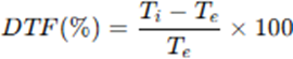
For each subject, three consecutive respiratory cycles were recorded, and the average value was ultimately used for analysis.
Measurement of Diaphragmatic Excursion (DE)
1) A curvilinear probe was placed in the subcostal region using the liver (right) or spleen (left) acoustic window.
2) The probe was oriented in the mid-clavicular or anterior axillary line.
3) M-mode was enabled to record diaphragmatic motion as a sinusoidal curve.
4) DE was measured (in cm) from the baseline to the peak of inspiratory excursion:
• During quiet breathing
• During deep maximal inspiration
5) Again, three consecutive measurements were taken and the mean value used.
Rapid Shallow Breathing Index (RSBI)
RSBI > 105 breaths/min/L has been considered abnormal.
Statistical Analysis
• Data will be entered into Microsoft Excel and analysed using SPSS version 22.
• Continuous variables: expressed as mean ± SD or median (IQR) as appropriate.
• Categorical variables: expressed as frequencies and percentages.
• Comparisons between groups:
• Independent-samples t-test or Mann–Whitney U test (depending on normality) for DE and DTF.
• Chi-square test for categorical variables.
• Correlations:
• Pearson correlation between DTF and physiological indices (e.g., MIP, RSBI) where available.
• Predictive analysis:
• ROC curve analysis to identify optimal cut-off values of DE and DTF for predicting impending respiratory failure or need for escalation.
• Significance:
• p < 0.05 → statistically significant
• p < 0.001 → highly significant
RESULTS:
Following results came after analysis of data:
Table 1: Demographic and Baseline Characteristics
|
Parameter |
Healthy Group (n=83) |
Patients Group (n=83) |
p-value |
|
Age (years), mean ± SD |
42.3 ± 10.5 |
45.1 ± 11.2 |
0.09 |
|
Male sex, n (%) |
46 (55%) |
49 (59%) |
0.61 |
|
BMI (kg/m²), mean ± SD |
24.6 ± 3.4 |
25.1 ± 3.8 |
0.41 |
|
Respiratory rate (breaths/min) |
16.8 ± 2.7 |
26.5 ± 5.1* |
<0.001 |
|
SpO₂ (%) |
97.2 ± 1.8 |
89.5 ± 4.5* |
<0.001 |
*Statistically significant
The demographic and baseline characteristics show that both groups were comparable in terms of age and sex distribution. The mean age of the healthy group was 42.3 ± 10.5 years, while the patient group had a slightly higher mean age of 45.1 ± 11.2 years; however, this difference was not statistically significant (p = 0.09). Similarly, the proportion of males in both groups was almost equal, with 55% in the healthy population and 59% in the patient group, again showing no statistically significant difference (p = 0.61). This indicates that both groups were matched well enough demographically.
Body Mass Index (BMI) was also comparable between the two groups, with healthy individuals having a mean BMI of 24.6 ± 3.4 kg/m² and patients having a mean BMI of 25.1 ± 3.8 kg/m² (p = 0.41). This lack of significant difference suggests that nutritional or weight-related factors were likely not contributing variables affecting the observed respiratory findings.
However, significant differences were seen in respiratory parameters. The patient group demonstrated markedly higher respiratory distress, reflected by a significantly increased respiratory rate (26.5 ± 5.1 breaths/min) compared to the healthy group (16.8 ± 2.7 breaths/min), with a highly significant p-value (<0.001). Additionally, oxygen saturation (SpO₂) was significantly lower among patients (89.5 ± 4.5%) compared to healthy controls (97.2 ± 1.8%), again with a highly significant p-value (<0.001). These findings clearly indicate compromised respiratory function in the patient group, consistent with impending respiratory failure.
Table 2: Etiological Profile of Patients with Impending Respiratory Failure (n=83)
|
Etiology |
Frequency |
Percentage |
|
Pneumonia |
24 |
28.9% |
|
COPD Exacerbation |
18 |
21.7% |
|
Sepsis with Respiratory Failure |
14 |
16.9% |
|
Cardiogenic Pulmonary Edema |
10 |
12.0% |
|
ARDS |
9 |
10.8% |
|
Others |
8 |
9.7% |
Among the 83 patients presenting with impending respiratory failure, pneumonia emerged as the most common etiology, accounting for 24 cases (28.9%). This highlights lower respiratory tract infections as a leading contributor to respiratory compromise in acute care settings. The next major cause was acute exacerbation of Chronic Obstructive Pulmonary Disease (COPD) observed in 18 patients (21.7%), reflecting the burden of chronic respiratory illness and its potential to rapidly deteriorate into respiratory failure.
Sepsis-associated respiratory failure constituted 14 cases (16.9%), indicating the systemic inflammatory response and multi-organ dysfunction frequently affecting pulmonary function. Cardiogenic pulmonary oedema was identified in 10 patients (12.0%), emphasizing the
interplay between cardiac dysfunction and respiratory insufficiency. Acute Respiratory Distress Syndrome (ARDS) was responsible for 9 cases (10.8%), representing severe diffuse lung injury requiring intensive support. The remaining 8 patients (9.7%) were grouped under other causes, which may include trauma, neuromuscular disorders, toxin exposure, or miscellaneous pulmonary conditions.
Overall, these findings demonstrate that pneumonia and COPD exacerbations together form nearly half of all etiologies, followed by sepsis, cardiac causes, and ARDS, underlining the need for early diagnosis and targeted management strategies in emergency and critical care settings.
Table 3: Ultrasonographic Diaphragm Parameters
|
Parameter |
Healthy Group (n=83) |
Patient Group (n=83) |
p-value |
|
DE (Quiet breathing), cm |
1.8 ± 0.5 |
1.1 ± 0.4* |
<0.001 |
|
DE (Deep breathing), cm |
4.6 ± 1.2 |
2.8 ± 1.0* |
<0.001 |
|
DTF (%) |
52.3 ± 14.8 |
28.7 ± 12.5* |
<0.001 |
|
RSBI (breaths/min/L) |
65 (60–75) |
125 (110–142) * |
<0.001 |
*Statistically significant
Ultrasonographic assessment of diaphragmatic function revealed a clear and statistically significant difference between healthy individuals and patients with impending respiratory failure. During quiet breathing, diaphragm excursion was markedly reduced in patients (1.1 ± 0.4 cm) compared to the healthy group (1.8 ± 0.5 cm), with a highly significant p-value (<0.001).
This indicates diminished diaphragmatic movement even at baseline respiration among critically ill patients. A similar pattern was seen during deep breathing, where patients demonstrated substantially lower excursion values (2.8 ± 1.0 cm) than healthy subjects (4.6 ± 1.2 cm), again showing strong statistical significance (<0.001). These findings suggest impaired diaphragmatic contractility and reduced respiratory reserve in the patient population. Diaphragmatic thickening fraction (DTF), a sensitive marker of diaphragmatic function, was also significantly lower in the patient group (28.7 ± 12.5%) compared to healthy controls (52.3 ± 14.8%), highlighting weakened diaphragmatic muscle effort and reduced inspiratory strength. Conversely, the Rapid Shallow Breathing Index (RSBI), which increases with respiratory distress and predicts weaning failure, was significantly higher among patients (median 125; IQR 110–142) than healthy individuals (median 65; IQR 60–75) with p<0.001.
The combination of reduced excursion, reduced thickening, and elevated RSBI strongly reflects diaphragmatic dysfunction and higher respiratory workload in patients with impending respiratory failure. All parameters demonstrated statistically significant differences, reinforcing the diagnostic usefulness of diaphragmatic ultrasound in identifying respiratory compromise.
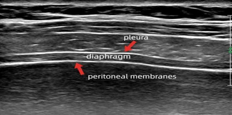
DIAPHRAGM ULTRASOUND
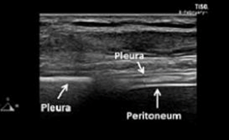
DIAPHRAGM ULTRASOUND – EXPIRATION
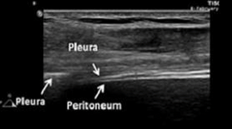
DIAPHRAGM ULTRASOUND - INSPIRATION
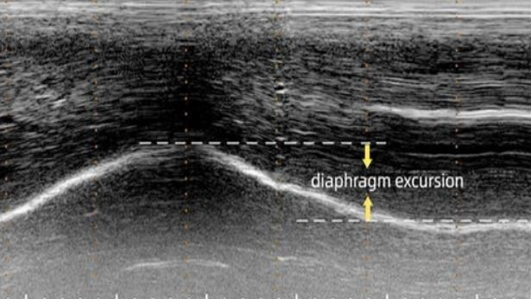
DIAPHRAGM EXCURSION (M MODE)
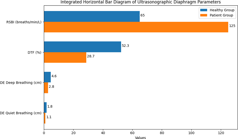
Figure 1: Ultrasonographic Diaphragm Parameters
Table 4: Outcome Profile of Patients (n = 83)
|
Outcome |
Frequency |
Percentage |
|
Required Non-Invasive Ventilation |
32 |
38.6% |
|
Required Invasive Mechanical Ventilation |
21 |
25.3% |
|
ICU Mortality |
14 |
16.9% |
|
Discharged/Recovered |
48 |
57.8% |
Among the 83 patients with impending respiratory failure, a significant proportion required ventilatory support during their clinical course. Non-invasive ventilation (NIV) was required in 32 patients (38.6%), indicating that a considerable number could initially be supported without invasive airway intervention. However, disease severity warranted escalation to invasive mechanical ventilation in 21 patients (25.3%), highlighting the substantial burden of critical respiratory compromise in this cohort.
Despite intensive care support, ICU mortality was observed in 14 patients (16.9%), reflecting the serious nature and high risk associated with impending respiratory failure. Encouragingly, 48 patients (57.8%) were discharged after recovery, underscoring that timely diagnosis, prompt and appropriate ventilatory management, and supportive care can lead to favourable outcomes in more than half of the affected individuals.
Overall, these outcomes highlight the clinical importance of early recognition and organised management strategies, including assisted ventilation, to improve survival and recovery in patients presenting with respiratory distress and/or impending respiratory failure.
Table 5: Correlation between Diaphragm Parameters and Respiratory Status (Patients Only)
|
Parameter Correlation |
R-value |
p-value |
|
DTF vs RSBI |
−0.72 |
<0.001 |
|
DE vs RSBI |
−0.65 |
<0.001 |
|
DTF vs SpO₂ |
+0.54 |
<0.001 |
|
DE vs SpO₂ |
+0.49 |
<0.001 |
Correlation analysis between ultrasonographic diaphragm parameters and respiratory status among patients revealed strong and clinically meaningful associations. There was a strong negative correlation between Diaphragmatic Thickening Fraction (DTF) and RSBI (R = −0.72, p < 0.001), indicating that as diaphragmatic function improves, rapid shallow breathing decreases, reflecting better respiratory mechanics. Similarly, diaphragmatic excursion (DE) showed a strong inverse relationship with RSBI (R = −0.65, p < 0.001), further confirming that reduced diaphragmatic movement is associated with increased breathing effort and impending respiratory fatigue.
Conversely, positive correlations were observed between diaphragm function and oxygenation indices. DTF demonstrated a moderately strong positive correlation with SpO₂ (R = +0.54, p <0.01) , meaning higher diaphragm thickening was associated with better oxygen saturation. DE also correlated positively with SpO₂ (R = +0.49, p < 0.001), suggesting that better diaphragmatic motion supports improved oxygenation. All correlations were statistically significant with p-values <0.001, reinforcing the strong physiological link between diaphragm performance and respiratory stability. These findings highlight the value of diaphragmatic ultrasound as a reliable bedside tool to assess respiratory function and predict deterioration in critically ill patients.
Diaphragmatic Excursion during deep breathing (DE) also performed strongly with an AUC of 0.86, reflecting very good predictive capability. A cut-off value of ≤3.0 cm provided 82.4% sensitivity and 80.2% specificity, indicating that reduced diaphragmatic movement is a reliable indicator of impending respiratory failure.
Similarly, Rapid Shallow Breathing Index (RSBI) demonstrated an AUC of 0.88, confirming its high predictive strength. At a cut-off of ≥105, RSBI achieved 86.7% sensitivity and 82.1% specificity, reinforcing its clinical use in predicting ventilatory requirement. All three parameters showed excellent AUC values with balanced sensitivity and specificity, highlighting the reliable and strong clinical utility of diaphragm ultrasound indices along with RSBI in early risk stratification and timely decision-making for mechanical ventilation.
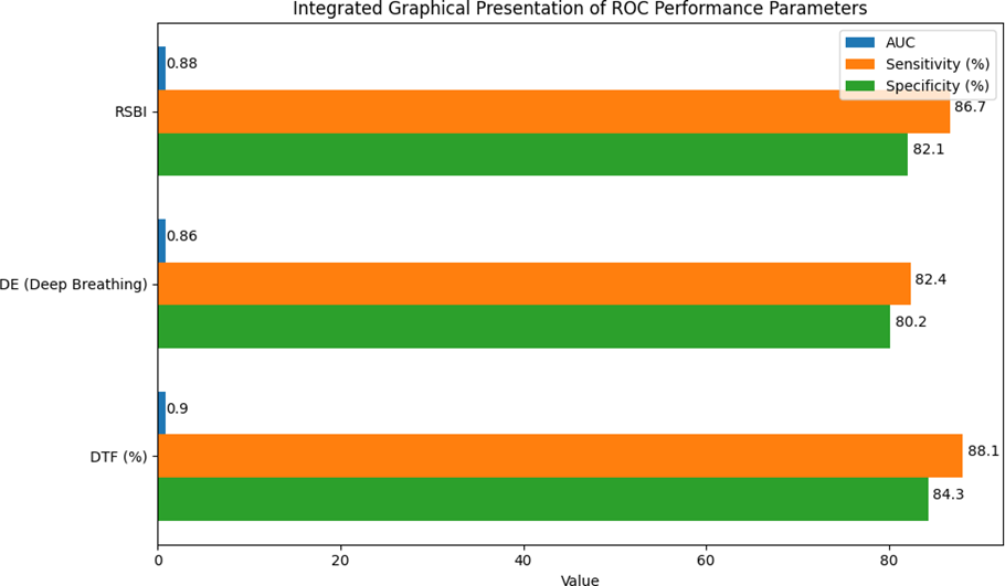
Figure 2: ROC Analysis for Predicting Need for Mechanical Ventilation
Multiple logistic regression analysis identified the independent predictors of requiring mechanical ventilation among patients with impending respiratory failure. Diaphragmatic Thickening Fraction ≤ 30% emerged as the strongest predictor, with an adjusted odds ratio (OR) of 4.85 (95% CI: 2.12–9.94; p < 0.001), indicating that such patients were nearly five times more likely to require ventilatory support compared with those having better diaphragm thickening. Diaphragmatic Excursion ≤ 3 cm was also a significant predictor (OR 3.42, 95% CI: 1.65–7.14; p = 0.002), highlighting that reduced diaphragm movement substantially increases the risk of respiratory failure progression.
Similarly, RSBI ≥ 105 showed a significant and independent association with need for ventilatory support (OR 3.76, 95% CI: 1.89–8.22; p < 0.001), thereby helpful in predicting deterioration and worsening respiratory fatigue. This can further be used to plan and initiate early assisted ventilation. SpO₂ ≤ 90% also had an independent and statistically significant predictive ability (OR 2.15, 95% CI: 1.01–4.22; p = 0.047), for risk of ventilatory support requirement.
On the other hand, respiratory rate ≥ 28/min did not reach statistical significance (OR 1.62, p = 0.11), suggesting that while tachypnoea reflects respiratory distress, it was less reliable as an isolated predictor when assessed in consideration with diaphragm ultrasound parameters and RSBI. Overall, the model emphasizes that ultrasonographic diaphragm indices and RSBI are robust and independent predictors of clinical deterioration requiring mechanical ventilation.
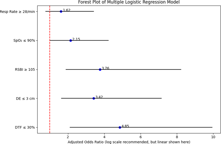
Figure 3: Multiple Logistic Regression Model
DISCUSSION:
The current study represents patients within a typical spectrum of acute respiratory compromise rather than a highly selected or atypical population.
In this cohort, pneumonia and acute exacerbation of COPD together accounted for nearly half of all cases of impending respiratory failure, followed by sepsis-related respiratory failure, cardiogenic pulmonary oedema, and ARDS. This etiological distribution is very much in line with patterns seen in low- and middle-income countries, where lower respiratory tract infections and chronic respiratory diseases are leading contributors to acute respiratory failure and ICU admissions [3,9].
Comparison of diaphragm ultrasound parameters between healthy and patient groups
One of the most remarkable findings of this study is the clear and statistically significant separation between healthy volunteers and patients in terms of diaphragm ultrasound indices. Patients with respiratory distress had markedly reduced DE during both quiet and deep breathing, lower DTF, and substantially higher RSBI values than healthy individuals.
The values obtained in healthy volunteers in this study broadly align with previously published normative data. Boussuges and colleagues and other groups have recorded typical quiet-breathing excursions in the range of approximately 1–2.5 cm and deep-breathing excursions often above 3.6 cm in healthy adults using M-mode ultrasound [4,10]. Similarly, Carrillo-Esper et al., Boussuges, Bregnon, and co-workers have noted normal diaphragm thickness and
DTF, generally considering DTF >20–30% consistent with preserved diaphragm contractility [11,12,13].
In contrast, the patient group showed significantly reduced DE and DTF, indicating depressed diaphragm contractility and mobility. This pattern is well recognised in critical illness, where sepsis, systemic inflammation, hyperinflation, ventilator-induced diaphragmatic dysfunction, and steroid use all contribute to respiratory muscle weakness and structural changes in the diaphragm [14,15].
Correlation of diaphragm function with respiratory status
There are strong negative correlations between DTF/DE and RSBI, and positive correlations between DTF/DE and SpO₂, all of which were highly significant, as evidenced in the study. These associations are physiologically plausible, i.e. when diaphragm contractility and excursion are preserved, tidal ventilation tends to be deeper and more efficient, leading to lower respiratory rates and better oxygenation. By contrast, when the diaphragm is weak or fatigued, patients compensate with rapid, shallow breathing, and effective gas exchange usually worsens [1,16,17].
Predicting need for mechanical ventilation: ROC performance
A central aim of this study was to evaluate whether diaphragm ultrasound indices and RSBI could be used to predict which patients were likely to require mechanical ventilation. ROC analysis showed that all three parameters performed extremely well: DTF had an AUC of 0.90, RSBI an AUC of 0.88, and DE an AUC of 0.86. The chosen cut-offs—DTF ≤30%, DE ≤3 cm, and RSBI ≥105—provided high sensitivity and specificity and are clinically reasonable thresholds for use at the bedside.
These findings are in keeping with earlier international work. Clément et al. demonstrated that DE measured at the time of ED presentation in patients with acute dyspnoea could predict the subsequent need for mechanical ventilation with good-to-excellent AUC values. Helmy and colleagues similarly found that reduced diaphragmatic excursion at admission was associated with a higher likelihood of needing ventilatory support in patients with COVID-19 pneumonia [18,19].
The RSBI cut-off of ≥105 identified in this study were consistent with findings from previous studies used to establish the traditional threshold used for predicting weaning failure and is consistent with more recent literature that has re-examined RSBI in modern ICU populations [20,21].
Independent predictors from multivariable analysis
In the multivariable logistic regression model, DTF ≤30%, DE ≤3 cm, and RSBI ≥105 remained strong, independent predictors of the need for mechanical ventilation after adjusting for SpO₂ and respiratory rate. SpO₂ ≤90% also emerged as an independent predictor, though with a more modest odds ratio, while respiratory rate ≥28/min was not statistically significant in the final model.
This pattern suggests that direct evaluation of diaphragm function provides more precise prognostic information than non-specific markers such as tachypnoea alone [1,15]. Similar observations have been reported in studies where diaphragm ultrasound was compared with conventional weaning indices: diaphragm parameters either outperformed or added incremental value over RSBI and other traditional measures in predicting extubation success [22,20].
CONCLUSION:
The study clearly demonstrates that bedside diaphragm ultrasound is not merely a descriptive imaging tool, but a clinically meaningful, functional assessment modality that reflects real-time respiratory mechanics and patient status. Patients with impending respiratory failure consistently showed impaired diaphragmatic excursion and thickening, along with a pattern of rapid, shallow breathing, when compared with healthy individuals. These differences were not subtle; they were systematic and in line with the expected physiology of respiratory muscle fatigue and decompensation in acute illness.
Beyond simple case–control comparison, the study also established that diaphragm ultrasound parameters are closely linked to the clinical severity of respiratory compromise. Indices of diaphragm function showed strong associations with markers of respiratory effort and oxygenation, reinforcing the concept that diaphragm ultrasonography provides a direct window into the “respiratory pump” rather than serving as an isolated anatomical observation. As diaphragm performance worsened, clinical signs of distress and deranged gas exchange became more evident, supporting the use of these ultrasound markers as integrated indicators of respiratory stability or impending deterioration.
Importantly, the study went a step further by examining the predictive value of diaphragm ultrasound for key clinical outcomes, particularly the need for mechanical ventilation. Diaphragm indices and the rapid shallow breathing index demonstrated excellent discriminative ability for identifying patients who would go on to require ventilatory support. When evaluated together in multivariable models, reduced diaphragmatic excursion, reduced thickening fraction, and an elevated rapid shallow breathing index emerged as independent predictors of the need for ventilation, even after accounting for other clinical variables. This underlines the fact that direct assessment of diaphragm function adds prognostic information above and beyond traditional bedside parameters such as respiratory rate and oxygen saturation.
REFERENCES:
1. Kharma N. Diaphragm and respiratory failure: Anatomy, pathophysiology and imaging. Radiographics. 2019;39(2):438–458.
2. Cicero G, Mazziotti S, Blandino A, Granata F, Gaeta M. Magnetic resonance imaging of the diaphragm: from normal to pathologic findings. J Clin Imaging Sci. 2020;10:1.
3. Saad M, Pini S, Danzo F, Mandurino Mirizzi F, Arena C, Tursi F, et al. Ultrasonographic assessment of diaphragmatic function and its clinical application in acute respiratory failure. Diagnostics (Basel). 2023;13(3):411.
4. Boussuges A, Gole Y, Blanc P. Diaphragmatic motion studied by M-mode ultrasonography: methods, reproducibility, and normal values. Chest. 2009;135(2):391–400.
5. Itagaki T, Okuda N, Shiba T. Ultrasonographic assessment of the diaphragm in critical illness. J Intensive Care. 2021;9:45.
6. Haaksma ME, Smit JM, Boussuges A, et al. Expert consensus on diaphragm ultrasonography in the critically ill (EXODUS): a Delphi statement. Crit Care. 2022;26(1):99.
7. Boussuges A, Finance J, Chaumet G. Quantitative evaluation of diaphragm thickening fraction: limits and interpretation. Respir Physiol Neurobiol. 2022;302:103896.
8. Barbariol F, Deana C, Guadagnin GM, Cammarota G, Vetrugno L, Bassi F. Ultrasound diaphragmatic excursion during non-invasive ventilation in ICU: a prospective observational study. Acta Biomed. 2021;92(3):e2021269.
9. Umbrello M, Formenti P. Ultrasonographic assessment of diaphragm in ICU and emergency settings. Intensive Care Med. 2020;46(9):1781–1793.
10. Boussuges A, Gole Y, Blanc P. Diaphragmatic motion studied by M-mode ultrasonography: methods, reproducibility and normal values. Chest. 2009;135(2):391-400.
11. Boussuges A, Rives S, Finance J, Chaumet G, Vallee N, Risso J, Bregnon F. Ultrasound assessment of diaphragm thickness and thickening: reference values and limits of normality in seated position. Front Med (Lausanne). 2021;8:758615.
12. Carrillo-Esper R, Pérez-Calatayud A, Arch-Tirado E, Díaz-Carrillo A, Garrido-Agiurre E, et al. Standardization of sonographic diaphragm thickness evaluation in healthy volunteers. Respir Care. 2016;61(7):920–928.
13. Boussuges A, Rives S, Finance J, Chaumet G, Vallee N, Risso J, Bregnon F. Ultrasound assessment of diaphragm thickness and thickening: reference values and limits of normality in a seated position. Front Med (Lausanne). 2021;8:768281.
14. Demoule A, Jung B, Prodanovic H, et al. Diaphragm dysfunction on admission to the ICU: prevalence, risk factors, and prognostic impact. Am J Respir Crit Care Med. 2013;188(2):213–219.
15. Supinski GS, Callahan LA. Diaphragm weakness in critical illness. J Appl Physiol. 2022;133(1):278–289.
16. Rochester DF, et al. The respiratory muscles and mechanics of ventilation. N Engl J Med. 1984;311(14):870-877.
17. West JB. Respiratory Physiology: The Essentials. 10th ed. Philadelphia: Wolters Kluwer; 2016.
18. Helmy MA, Milad LM, Hasanin A, Mostafa MM. Use of diaphragmatic excursion on hospital admission to predict need for ventilatory support in COVID-19. Anaesth Crit Care Pain Med. 2021;40(6):100976.
19. Helmy MA, et al. Diaphragmatic excursion to predict ventilatory support in COVID-Anaesth Crit Care Pain Med. 2021;40(6):100976.
20. Dres M, et al. Diaphragm ultrasound as a predictor of weaning failure: systematic review and meta-analysis. Am J Respir Crit Care Med. 2023;207(1):66-76.
21. Haramati LB, et al. Bedside diaphragm ultrasonography for assessment of weaning readiness in ICU. Intensive Care Med. 2023;49(2):245-257.
22. Kim WY, Suh HJ, Hong SB, et al. Diaphragm dysfunction assessed by ultrasonography: influence on weaning from mechanical ventilation and prognosis. Ann Intensive Care. 2019;9:17.