Acetabular measurements of hip bone and its clinical implications: A cross-sectional study on human adult dry hipbones.
- S.Ahmed Peer , Associate Professor, Department of Anatomy, Government Medical College, Anantapuramu-515001.
- K.Tirumala Chaitanya , Assistant Professor, Department of Orthopaedics, Government Medical College, Anantapuramu-515001.
- S. Umamaheswara Rao , Professor, Department of Anatomy, Government Medical College, Anantapuramu-515001.
Article Information:
Abstract:
Background: The acetabulum is a key component of the hip joint that influences stability, load transmission, and range of motion. The acetabulum dimensions vary across ethnic groups and show sexual differences. The data exist for some Indian and Asian populations, specific normative values for the South Indian population are limited. Our study aimed to measure important acetabular parameters in dry hip bones of South Indian adults, establish sex-specific and side-specific normative values, and highlight their clinical relevance. Materials and Methods: A cross-sectional osteometric study was conducted on 100 unpaired adult dry hip bones (50 -Right and 50-Left). The bones were selected based on standard age estimation criteria (suture closure and dental wear) and examined after excluding damaged or pathological specimens. Measurements of acetabular depth, transverse diameter, acetabular angle, and center-edge angle were taken using a digital vernier caliper (0.01 mm accuracy) following established protocols. Data were analysed and expressed as mean ± standard deviation with p < 0.05 considered statistically significant. Results: The overall mean acetabular depth was 21.2 ± 2.3 mm, transverse diameter 50.5 ± 3.4 mm, acetabular angle 19.2 ± 2.4°, and center-edge angle 40.7 ± 4.2°. Males showed significantly larger linear dimensions than females: acetabular depth was 22.5 ± 2.1 mm in males versus 19.8 ± 1.9 mm in females (p < 0.001), and transverse diameter was 52.3 ± 3.2 mm versus 48.7 ± 2.8 mm (p < 0.001). The center-edge angle was also higher in males (42.5 ± 4.1° vs 38.9 ± 3.7°, p < 0.001), while the acetabular angle was greater in females (18.2 ± 2.4° vs 20.1 ± 2.1°, p = 0.002). Conclusion: Our study acknowledges acetabular dimensions is essential for designing effective prosthetic components in total hip arthroplasty and the parameters aid in precise preoperative assessment, ensuring optimal implant fit and stability. In acetabular reconstruction in hip joint surgeries, the parameters of our study are necessary for the creation of acetabular prosthesis and surgical procedures.
Keywords:
Article :
INTRODUCTION:
The hip joint is a multiaxial synovial ball-and-socket joint formed by the articulation of the femoral head with the acetabulum. It plays a vital role in weight-bearing, locomotion, and maintenance of erect posture. The acetabulum, a deep cup-shaped cavity formed by the confluence of the ilium, ischium, and pubis, contributes to joint stability through its depth, rim orientation, and associated soft-tissue structures, while permitting a wide range of motion(Figure 1). Morphometric parameters such as acetabular depth, transverse diameter, acetabular angle, and center-edge angle significantly influence hip biomechanics, susceptibility to fractures, and outcomes of surgical interventions[1].
Within India, differences have been observed between North and South Indian populations, with several reports indicating relatively smaller acetabular diameters and depths in South Indians, possibly influenced by genetic, nutritional, and environmental factors[2,3].Osteoarthritis of the hip and knee also remains a significant concern in the region, with higher prevalence reported among older adults. Shallower acetabula have been linked to increased risk of anterior dislocation and DDH, with a noted female predominance attributed to hormonal influences and biomechanical stresses associated with childbirth. Existing South Indian osteometric studies have reported mean acetabular depths in the range of approximately 21–23 mm, generally smaller than values documented in North Indian populations, although many of these studies had modest sample sizes and inconsistent sex-specific analysis [4,5,6].
Accurate knowledge of local acetabular norms is essential for optimal cup positioning, selection of appropriate implant sizes, planning of periacetabular osteotomies in DDH, and radiological interpretation using parameters such as the center-edge angle[7,8].Routine examination of osteological measurements and establishing such a normative database is expected to help bridge the existing ethnic data gap and support more precise surgical planning in high-volume trauma and arthroplasty centres in South India.
MATERIALS AND METHODS:
This cross-sectional observational study was conducted on dry adult hip bones from the Department of Anatomy, Forensic Medicine and the Department of orthopaedics, Government Medical College, Anantapuramu. A total of 100 unpaired adult dry hip bones (50 right and 50 left) were utilized in the current study and approved by the Institutional Ethics Committee of Government medical college, Anantapuramu (IEC NO.12233). The bones with an estimated age range of 25–70 years. Sex determination was performed using standard osteological criteria, including the width of the greater sciatic notch, presence of the preauricular sulcus, and robusticity of the ischiopubic ramus. These criteria have been previously validated with high accuracy in the departmental collections [9,10]. Bones were excluded if they showed acetabular rim fractures or erosions, osteophytes, cysts, or any pathological changes, incomplete fusion of pelvic elements, excessive surface wear that obscured landmarks, or if collection records indicated non-South Indian origin. All measurements were performed using a calibrated digital verniercaliper (Mitutoyo, Japan; precision 0.01 mm) and a goniometer (accuracy 0.1°). Measurements were taken under uniform lighting with the help of 2× magnification loupes. Four acetabular parameters were measured three times on each bone:
1. Transverse Diameter (TD): The maximum horizontal distance between the anterior and posterior acetabular rims measured at the mid-depth level (Figure 2).
2. Acetabular Depth (AD): The perpendicular distance from the deepest point of the acetabular floor (cotyloid notch area) to a plane connecting the anterior and posterior rims (Figure 3).
3. Acetabular width (AW): The distance between the two ends of the lunate-shaped articular part of the acetabulum(Figure 4).
4. Acetabular Angle (AA): The angle formed between the tangents drawn to the anterior and posterior acetabular rims, measured relative to the horizontal plane.
5. Center-Edge Angle (CEA): The angle formed between the vertical axis of the acetabulum and a line drawn from the center of the acetabulum to the lateral rim, estimated using bony landmarks as a skeletal proxy.
All measurements were photo documented using a Canon DSLR camera with standardized 11,12,13,14]
Statistical Analysis
Results were expressed as mean ± standard deviation (SD), along with median and interquartile range (IQR) where appropriate. Side-wise (right vs left) comparisons were performed using paired t-tests. Effect sizes were calculated using Cohen’s d. A p-value of less than 0.05 (two-tailed) was considered statistically significant[15,16].
RESULTS:
A total of 100 unpaired adult dry hip bones were included in the present study after application of the inclusion and exclusion criteria. Of these, 50 bones belonged to the right side and 50 to the left side. Based on osteological criteria, 50 bones were categorized as male and 50 as female. All studied variables showed acceptable distribution for statistical comparison. The overall mean acetabular depth in the study sample was 21.2 ± 2.3 mm[Table 1]. The overall mean transverse acetabular diameter was 50.5 ± 3.4 mm.
The overall mean acetabular angle was 19.2 ± 2.4°, and the overall mean center-edge angle was 40.7 ± 4.2°. These values provide baseline osteometric data for the South Indian population represented in the present study. When sex-wise comparison was performed, male hip bones demonstrated significantly greater linear dimensions than female hip bones. The mean acetabular depth in male bones was 22.5 ± 2.1 mm, whereas in female bones it was 19.8 ± 1.9 mm.
The mean acetabular notch width in male bones was 20.3 ± 1.9mm, whereas in female bones it was 18.9 ± 1.8mm[Table 2]. Similarly, the mean transverse diameter was 52.3 ± 3.2 mm in males and 48.7 ± 2.8 mm in females. The mean center-edge angle was also higher in males than in females. In contrast, the mean acetabular angle was greater in female bones, suggesting relatively wider acetabular orientation in females. These findings indicate definite sexual dimorphism in acetabular morphology.
Table 1: Sex-wise comparison of acetabular parameters
|
Parameter |
Males (n=50) |
Females (n=50) |
Mean difference |
p-value |
|
Acetabular depth (mm) |
22.5 ± 2.1 |
19.8 ± 1.9 |
2.7 |
<0.001 |
|
Transverse diameter (mm) |
52.3 ± 3.2 |
48.7 ± 2.8 |
3.6 |
<0.001 |
|
Width of acetabular notch |
20.3 ± 1.9 |
18.9 ± 1.8 |
2.0 |
<0.001 |
|
Acetabular angle (°) |
18.2 ± 2.4 |
20.1 ± 2.1 |
-1.9 |
0.002 |
|
Center-edge angle (°) |
42.5 ± 4.1 |
38.9 ± 3.7 |
3.6 |
<0.001 |
Table 2: Side-wise comparison of acetabular parameters
|
Parameter |
Right (n=50) |
Left (n=50) |
Mean difference |
p-value |
|
Acetabular depth (mm) |
21.3 ± 2.4 |
21.0 ± 2.2 |
0.3 |
0.44 |
|
Transverse diameter (mm) |
50.6 ± 3.5 |
50.4 ± 3.3 |
0.2 |
0.73 |
|
Width of acetabular notch |
22.4 ± 2.1 |
21.6 ± 2.0 |
0.2 |
0.46 |
|
Acetabular angle (°) |
19.1 ± 2.3 |
19.2 ± 2.5 |
-0.1 |
0.84 |
|
Center-edge angle (°) |
40.9 ± 4.0 |
40.5 ± 4.4 |
0.4 |
0.58 |
Table 3: Correlation among acetabular parameters
|
Parameter |
Depth |
Diameter |
width |
Angle |
Center-edge angle |
|
Acetabular depth |
1.00 |
0.71 |
0.86 |
-0.52 |
0.62 |
|
Transverse diameter |
0.71 |
0.86 |
-0.52 |
0.62 |
1.00 |
|
Width of acetabular notch |
0.86 |
-0.52 |
0.60 |
1.00 |
0.71 |
|
Acetabular angle |
-0.52 |
-0.38 |
0.81 |
1.00 |
-0.48 |
|
Center-edge angle |
0.62 |
0.55 |
0.60 |
-0.48 |
1.00 |
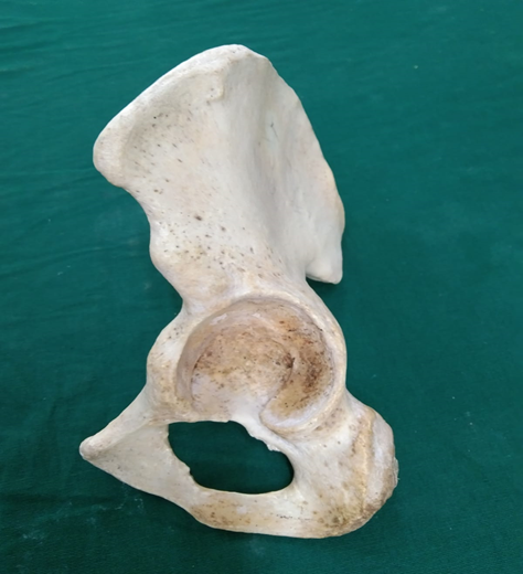
Figure 1: Human dry hip bone with acetabulum
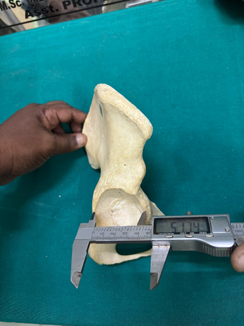
Figure 2: Transverse Diameter - maximum horizontal distance between the anterior and posterior acetabular rims measured at the mid-depth level.
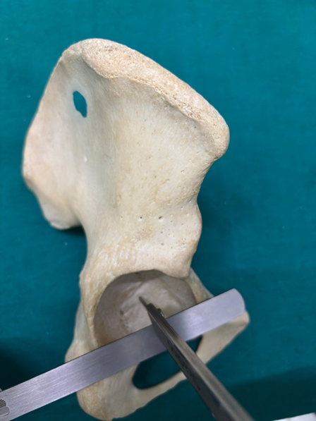
Figure 3: Depth of Acetabulum - perpendicular distance from the deepest point of the acetabular floor (cotyloid notch area) to a plane connecting the anterior and posterior rims.
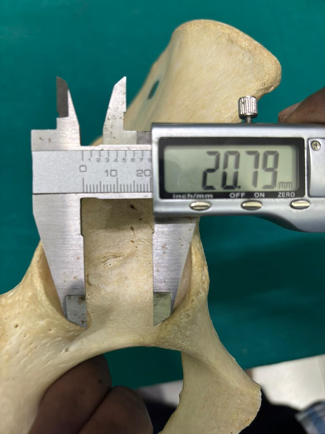
Figure 4: Width of Acetabulum- The distance between the two ends of the lunate-shaped articular part of the acetabulum
DISCUSSION:
The present study provides baseline morphometric data on the acetabulum in the South Indian population and demonstrates clear sexual dimorphism. Male hip bones exhibited significantly greater acetabular depth, transverse diameter, and center-edge angle compared to female bones, while the acetabular angle was larger in females. These findings highlight the need for population-specific and sex-specific reference values in orthopedic and anatomical practice.The overall mean acetabular depth in this study (21.2 ± 2.3 mm) and the sex-specific values (22.5 ± 2.1 mm in males and 19.8 ± 1.9 mm in females) are comparable to previous South Indian osteometric studies.
Deepa et al. reported similar smaller dimensions in South Indian hip bones [2]. Another recent study on 71 dry hip bones from a South Indian population also documented mean acetabular depths of approximately 22.5–23.3 mm, concluding that acetabular dimensions in South Indians are generally smaller than in other populations [17]. These values are typically lower than those reported in North Indian and Western/Caucasian populations [18,19], likely due to differences in skeletal build, ethnicity, nutrition, and genetics. Such variations suggest that hip prosthetic designs based solely on Western anthropometric data may not be ideal for Indian patients, particularly females with smaller and shallower acetabula.The significantly smaller acetabular depth and transverse diameter observed in female bones carry important clinical implications. Shallower acetabula provide reduced femoral head coverage, which may predispose to joint instability, developmental dysplasia of the hip (DDH), and altered load distribution [20]. The lower center-edge angle in females further supports relatively poorer bony coverage. These morphological features align with clinical observations of higher rates of hip instability and dysplastic changes in females.The mean transverse acetabular diameter in the present study was lower than values commonly reported in North Indian studies [21] but consistent with other South Indian data [2,17].
This regional difference is directly relevant to total hip arthroplasty (THA). Accurate knowledge of local acetabular dimensions helps in selecting appropriate implant sizes, achieving better cup fit and alignment, and reducing complications such as dislocation, edge loading, and early loosening. Significantly greater acetabular angle, indicating a relatively wider acetabular orientation was noted in females in our study. When combined with reduced depth and center-edge angle, this pattern may represent a morphological predisposition to borderline dysplasia. These findings are valuable for radiological screening and preoperative planning in patients presenting with hip pain. No statistically significant differences were found between right and left sides for any parameter, indicating bilateral symmetry. This observation is consistent with earlier morphometric studies on the acetabulum [22]. Correlation analysis showed that acetabular depth was positively associated with transverse diameter and center-edge angle, and negatively associated with acetabular angle [Table 3]. These interrelationships suggest that acetabular morphology functions as an integrated structure influencing overall joint stability and femoral head coverage. The clinical relevance of these findings is considerable. Orthopedic surgeons can utilise these normative values for better implant selection in THA, stable fixation of acetabular fractures, and improved radiological assessment of hip disorders. The data may also assist in forensic anthropology for sex estimation from skeletal remains.
CONCLUSION:
Our study acknowledges acetabular dimensions is essential for designing effective prosthetic components in total hip arthroplasty and the parameters aid in precise preoperative assessment, ensuring optimal implant fit and stability. In acetabular reconstruction in hip joint surgeries, the parameters of our study are necessary for the creation of acetabular prosthesis and surgical procedures.
REFERENCES:
1. Moore KL, Dalley AF, Agur AMR. Clinically Oriented Anatomy. 8th ed. Philadelphia: Wolters Kluwer; 2018.
2. Deepa R, Shastri D, Suganya K. Morphometric analysis of acetabulum in South Indian population. Int J Anat Res. 2016;4(3):2785-90.
3. Keerthana MBS, Sangeetha S. Anatomical observation and morphometric study of acetabulum in South Indian population. Biosci Biotechnol Res Asia. 2020;17(3):569-74.
4. Putzer D, Hasler M, List C, et al. Differences in acetabular morphology related to sex and side in Korean population. Hip Int. 2022;32(5):112070002210918.
5. Gupta S, Kotasthane VD, Kotasthane DS. Acetabular morphometry in North Indian population. J Anat Soc India. 2018;67(1):22-7.
6. Kunnoor NS, et al. Acetabular morphometry and prevalence of hip dysplasia in Indian population. Indian J Orthop. 2011;45(4):324-8.
7. Rajan D, et al. Profound morphometric analysis of acetabulum in South Indian population. Biomed Pharmacol J. 2024;17(1):245-52.
8. Indurjeeth K, et al. Variations in acetabular anatomy with reference to total hip replacement. Clin Anat. 2008;21(5):567-71.
9. von Elm E, Altman DG, Egger M, et al. The Strengthening the Reporting of Observational Studies in Epidemiology (STROBE) statement: guidelines for reporting observational studies. Lancet. 2007;370(9596):1453-7.
10. Indian Council of Medical Research. National Ethical Guidelines for Biomedical and Health Research Involving Human Participants. New Delhi: ICMR; 2017.
11. Buikstra JE, Ubelaker DH. Standards for Data Collection from Human Skeletal Remains. Fayetteville: Arkansas Archeological Survey; 1994.
12. Murty OP. Sex determination from skeletal remains. J Indian Acad Forensic Med. 2006;28(2):38-42.
13. Faul F, Erdfelder E, Lang AG, Buchner A. G*Power 3: a flexible statistical power analysis program for the social, behavioral, and biomedical sciences. Behav Res Methods. 2007;39(2):175-91.
14. Shrout PE, Fleiss JL. Intraclass correlations: uses in assessing rater reliability. Psychol Bull. 1979;86(2):420-8.
15. Ponseti IV. Morphology of the acetabulum in congenital dislocation of the hip. J Bone Joint Surg Am. 1978;60(5):586-99.
16. Wiberg G. Studies on dysplastic acetabula and congenital subluxation of the hip joint. Acta Chir Scand. 1939;83 Suppl 58:53-70.
17. Rajila RHS, Abitha R, Logithkumar S, Gnanasundaram V. Profound morphometric analysis of acetabulum in South Indian population (acetabular dimensions). Biomed Pharmacol J. 2024;17(1):123-130.
18. Vaidya VK, et al. Morphometric study of the acetabulum among North Indian population: A cross-sectional radiological study. J Clin Diagn Res. 2025 (In press).
19. Umer M, Sepah YJ, Asif S, Azam I, Jawad MU. Acetabular morphometry and prevalence of hip dysplasia in the South Asian population. J Pak Med Assoc. 2010;60(10):822-825.
20. Yugesh K, Kumar SS. Morphometric analysis of acetabulum and its clinical correlation in South Indian population. Int J Appl Res. 2016;2(6):1011-1014.
21. Pullanna B, Bindhu S, Avadhani R, Jacob M. Morphometry of the adult human dry hip bone in South Indian population. Int J Anat Res. 2019;7(4):6178-6182.