ENDOSCOPIC SURGERY FOR PERSISTENT WOUND COMPLICATIONS AFTER ACHILLES TENDON SUTURE REPAIR
- KAVIN RAJA S , Postgraduate Resident Professor, DEPARTMENT OF ORTHOPAEDICS, SREE MOOKAMBIKA INSTITUTE OF MEDICAL SCIENCES, KULASEKHARAM, KANYAKUMARI - 629161, TAMILNADU, INDIA.
- . K. C. MATHEW , Professor and Head of the DEPARTMENT OF ORTHOPAEDICS, SREE MOOKAMBIKA INSTITUTE OF MEDICAL SCIENCES, KULASEKHARAM, KANYAKUMARI - 629161, TAMILNADU, INDIA.
- GOWTHAM RAJ G , Postgraduate Resident Professor, DEPARTMENT OF ORTHOPAEDICS, SREE MOOKAMBIKA INSTITUTE OF MEDICAL SCIENCES, KULASEKHARAM, KANYAKUMARI - 629161, TAMILNADU, INDIA.
- ANTO JEFRIN VISHAL A , Senior Resident DEPARTMENT OF ORTHOPAEDICS, SREE MOOKAMBIKA INSTITUTE OF MEDICAL SCIENCES, KULASEKHARAM, KANYAKUMARI - 629161, TAMILNADU, INDIA.
- SUGIN GLEN BAISIL J , Postgraduate Resident Professor, DEPARTMENT OF ORTHOPAEDICS, SREE MOOKAMBIKA INSTITUTE OF MEDICAL SCIENCES, KULASEKHARAM, KANYAKUMARI - 629161, TAMILNADU, INDIA.
- ZAKIR HUSSAIN MOHAMED , Postgraduate Resident Professor, DEPARTMENT OF ORTHOPAEDICS, SREE MOOKAMBIKA INSTITUTE OF MEDICAL SCIENCES, KULASEKHARAM, KANYAKUMARI - 629161, TAMILNADU, INDIA.
- . ANNAMALAI T T , Postgraduate Resident Professor, DEPARTMENT OF ORTHOPAEDICS, SREE MOOKAMBIKA INSTITUTE OF MEDICAL SCIENCES, KULASEKHARAM, KANYAKUMARI - 629161, TAMILNADU, INDIA.
Article Information:
Abstract:
Background: Delayed wound healing following Achilles tendon repair remains a challenging postoperative complication associated with tissue ischemia, persistent inflammation, and foreign body reactions to nonabsorbable sutures. Conventional open debridement may lead to prolonged wound care, delayed rehabilitation, and increased soft tissue morbidity. This case series evaluates the clinical outcomes of endoscopic management in patients with persistent postoperative wound complications after Achilles tendon repair. Case Presentation: Three male patients presented with persistent non-healing wounds and localized inflammatory changes following Achilles tendon reconstruction. Conservative wound management failed to achieve satisfactory recovery. All patients underwent minimally invasive endoscopic debridement with removal of retained nonabsorbable suture material and inflamed tissue. Postoperative recovery was favorable, with satisfactory wound healing, reduction in pain, and improvement in functional outcomes during follow-up evaluation. No recurrent deep infection or major wound complication was observed after treatment. Conclusion: Endoscopic debridement appears to be a safe and effective minimally invasive approach for selected patients with delayed wound healing after Achilles tendon repair. The technique may reduce additional soft tissue trauma while facilitating satisfactory wound recovery and functional improvement.
Keywords:
Article :
INTRODUCTION:
Acute Achilles tendon rupture is a common orthopedic injury frequently managed with surgical tendon repair to restore functional stability and reduce the risk of rerupture. Despite favorable surgical outcomes, postoperative complications remain clinically significant, particularly delayed wound healing and persistent inflammatory reactions. The Achilles tendon has a relatively limited vascular supply, especially within its mid-portion, and is covered by a thin soft tissue envelope, making the region vulnerable to impaired healing and tissue breakdown following surgery (1,2). Previous studies have reported postoperative complication rates approaching 28%, including skin necrosis, wound dehiscence, deep infection, and delayed tissue recovery (1,3).
Delayed wound healing after Achilles tendon repair may occur secondary to tissue ischemia, chronic inflammation, infection, or adverse reactions to surgical materials. Among these factors, foreign body reactions associated with nonabsorbable sutures have been increasingly recognized as an important cause of persistent wound drainage, sinus formation, and chronic inflammatory changes (4,5). Although nonabsorbable sutures provide durable mechanical support during tendon reconstruction, retained suture material may occasionally provoke granulomatous inflammation and interfere with normal tissue healing.
Conventional management of persistent postoperative wound complications often involves open surgical debridement combined with prolonged wound care and antibiotic therapy. However, extensive soft tissue exposure during open procedures may further delay recovery and increase postoperative morbidity (6,7). In recent years, minimally invasive endoscopic techniques have emerged as an alternative approach for managing selected Achilles tendon complications. Endoscopic debridement allows direct visualization and removal of inflamed tissue and retained suture material while minimizing additional soft tissue trauma (1,8).
The present case series describes the clinical presentation, surgical management, and postoperative outcomes of three patients with delayed wound healing following Achilles tendon repair who were successfully treated using an endoscopic approach.
CASE PRESENTATIONS:
Case 1
The first patient was a 42-year-old man who ruptured his Achilles tendon four months earlier and had surgery in 2022. Before the rupture, he received local hormone injections for Achilles tendon periarthritis. The injury was about 15mm from the calcaneal prominence, creating a large defect of around 4 cm. The remaining scar tissue was weak, so a gastrocnemius aponeurotic flap was used to repair the tendon. Multiple no. 1 and no. 4 silk sutures fixed the flap and closed the gap between it and the remaining scar tissue.
Two weeks after the initial surgery, wound complications developed with localized skin breakdown, purulent discharge, and progressive soft tissue discoloration around the posterior ankle region. Clinical examination during follow-up demonstrated persistent inflammatory changes and areas of ischemic skin involvement despite repeated local wound care and dressing management over a period of three months. Due to failure of conservative treatment and ongoing delayed wound healing, endoscopic surgical intervention was planned. (Figure 1)
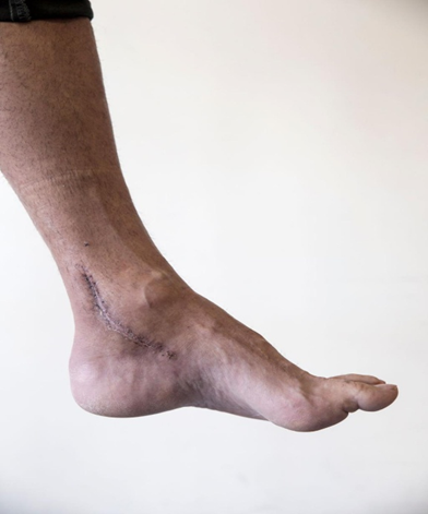
Figure 1. Preoperative clinical appearance demonstrating delayed wound healing, localized inflammation, and soft tissue breakdown following Achilles tendon repair.
Under spinal anesthesia, the patient was positioned prone and minimally invasive endoscopic debridement was performed using portals established adjacent to the wound defect. Preoperative magnetic resonance imaging assisted in identifying areas of tendon degeneration and retained suture material. Intraoperatively, inflamed granulation tissue and devitalized tissue surrounding the Achilles tendon were identified and carefully debrided while preserving viable tendon fibers. Multiple retained nonabsorbable sutures associated with chronic foreign body reaction were removed successfully. Following extensive irrigation, the wound was closed primarily and a temporary drainage strip was placed for short-term postoperative drainage. (Figure 2)
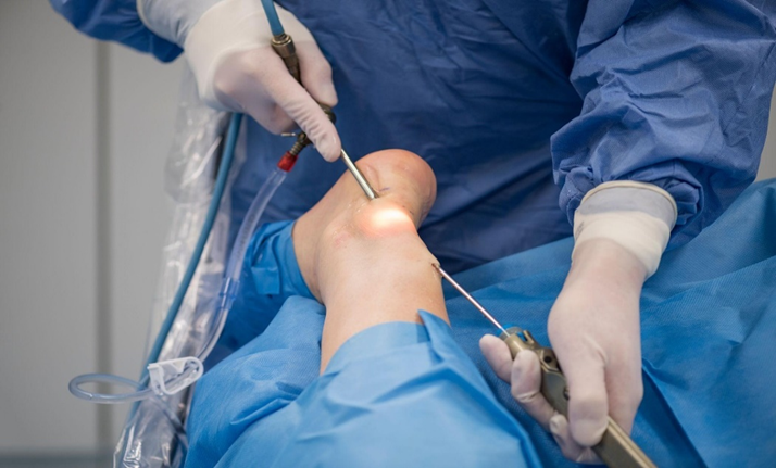
Figure 2. Endoscopic surgical management of delayed wound healing after Achilles tendon repair showing minimally invasive portal placement and debridement of inflamed tissue with removal of retained suture material.
Postoperatively, the patient received intravenous second-generation cephalosporin therapy followed by a short course of oral antibiotics. Limb immobilization was maintained initially using a short leg plaster cast, after which progressive rehabilitation with gradual ankle mobilization and protected weight-bearing was initiated. The patient demonstrated satisfactory wound healing and progressive functional recovery during follow-up, with no recurrent wound breakdown or evidence of deep infection observed at the final evaluation in 2025.
Case 2
The present patient was a 58-year-old man who presented with fluid leakage and a foreign body reaction at the incision site starting 1.5 months after Achilles tendon suture repair. He sought treatment in 2023. We performed the same Endoscopic surgery approach as the first patient to address the delayed wound healing. Instead of using a drainage strip, a sterile dressing and cotton compression dressing were applied. After surgery, the affected leg was set in plaster to keep it still. To ensure no infection, intravenous antibiotics were given once after the operation. Initially, this patient did well after surgery.
However, a year later, he noticed a bulge under the skin in the Achilles tendon area, along with redness and tenderness. He returned to the department for treatment 1 month later. We considered these symptoms to be caused by a persistent foreign body reaction due to a piece of suture that was missed or unreachable in the previous surgery. Therefore, under local anesthesia, we performed a 1-cm incision, found the thread, and removed it. No tourniquet was used in this minor operation, and no postoperative antibiotics were given.
MRI results showed a significant improvement in the Achilles tendon area during follow-up compared with preoperatively. (Figure 3) Pain at the Achilles tendon was assessed using visual analogue scale (VAS) scores, where 0 indicates no pain. The function and movement of the Achilles tendon were assessed using scores on the American Orthopedic Foot and Ankle Society (AOFAS) Ankle-Hindfoot Scale and the Achilles tendon total rupture score (ATRS). At the last follow-up in 2026 (3 years after the original endoscopic operation), the patient's VAS had improved from 6 preoperatively to 0. His AOFAS score changed from 45 to 100, and his ATRS moved from 19 to 92, which indicated that the function and movement of the patient's affected limb had improved satisfactorily.
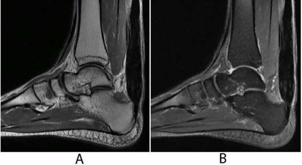
Figure 3. Preoperative and postoperative magnetic resonance imaging of the Achilles tendon region. (A) Preoperative MRI demonstrating inflammatory changes and soft tissue involvement surrounding the Achilles tendon. (B) Follow-up postoperative MRI showing interval improvement following endoscopic debridement and removal of retained suture material.
Case 3
The third patient was a 53-year-old man who presented with persistent pain and delayed wound healing following Achilles tendon suture repair performed two months earlier in 2024. The patient reported continued discomfort around the surgical site along with incomplete healing of the incision despite regular dressing care. Clinical examination revealed localized tenderness and mild inflammatory changes around the Achilles tendon region without evidence of extensive purulent discharge or systemic infection.
Considering the persistent symptoms and poor wound progression, endoscopic surgery was planned. Under spinal anesthesia and tourniquet control, arthroscopic portals were established around the affected region. Inflammatory granulation tissue and degenerative tendon tissue were identified intraoperatively. Careful debridement was performed while preserving the intact tendon fibers. Retained nonabsorbable suture material surrounded by inflamed tissue was identified and removed completely. The wound was thoroughly irrigated, and primary closure was performed.
Postoperatively, the patient received a prophylactic intravenous antibiotic dose and the affected limb was immobilized with a short leg plaster cast. Wound healing progressed satisfactorily, and no secondary wound complications were observed during the early postoperative period. Gradual ankle mobilization exercises were initiated after immobilization, followed by progressive weight-bearing rehabilitation.
Two months after the procedure, the patient sustained a minor fall which resulted in transient pain over the Achilles tendon region during weight-bearing. However, clinical examination confirmed that the skin remained intact without wound breakdown or evidence of re-rupture. Conservative management with activity modification and continued rehabilitation was advised.
At the most recent follow-up in 2026, the patient demonstrated satisfactory clinical recovery with significant reduction in pain and improved ankle function. Functional outcome scores showed improvement compared with preoperative assessment, and the patient was able to return to routine daily activities without major limitations.
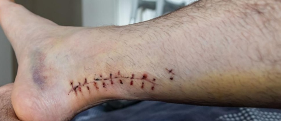
Figure 4. Clinical follow-up demonstrating satisfactory wound healing and recovery of the posterior ankle region following endoscopic management of delayed wound healing after Achilles tendon repair.
DISCUSSION:
Factors Contributing to Delayed Wound Healing After Achilles Tendon Repair
Delayed wound healing following Achilles tendon repair is a recognized postoperative complication resulting from a combination of anatomical, patient-related, and surgical factors. The Achilles tendon possesses a relatively poor vascular supply, particularly within its mid-portion, and is covered by a thin soft tissue envelope. These characteristics reduce tissue perfusion and make postoperative healing more vulnerable to ischemia and tissue breakdown (2,8). In addition, the posterior ankle region is frequently exposed to moisture and friction from footwear, which may further compromise wound healing and increase susceptibility to local contamination (1,9).
Several patient-related risk factors have also been associated with impaired healing. Advanced age, obesity, smoking, diabetes mellitus, cardiovascular disease, and arrhythmias have all been shown to increase postoperative wound complications after Achilles tendon surgery (5,9). Corticosteroid exposure may additionally impair collagen synthesis and suppress inflammatory mechanisms essential for tissue repair (10). In the present report, one patient had a prior history of local steroid injections before tendon rupture, which may have contributed to tissue fragility and delayed recovery.
Surgical factors are equally important in the development of postoperative wound complications. Prolonged operative duration, tissue handling, and the use of nonabsorbable sutures can provoke persistent local inflammation (11,12). Although nonabsorbable sutures provide strong mechanical support during tendon reconstruction, they may occasionally trigger adverse foreign body reactions, granuloma formation, and chronic sinus formation (12). In all three of our patients, retained nonabsorbable sutures and surrounding inflammatory tissue were identified as the major contributors to persistent wound symptoms and delayed healing.
Distinguishing Delayed Healing from Deep Infection
An important aspect in managing postoperative Achilles tendon complications is differentiating delayed wound healing from deep surgical infection. Deep infections are generally associated with purulent discharge, positive bacterial cultures, extensive tissue necrosis, and systemic inflammatory signs (1,7). Such cases often require aggressive debridement, prolonged antibiotic therapy, and occasionally reconstructive soft tissue procedures or negative pressure wound therapy (13,14).
In our patients, persistent drainage, inflammatory changes, and localized tenderness were present; however, there was no evidence of systemic infection or extensive purulence. The clinical presentation was more consistent with chronic inflammatory reaction secondary to retained suture material. Following endoscopic debridement and removal of the offending sutures, all three patients demonstrated satisfactory wound healing without recurrent infection during follow-up.
The third patient experienced transient pain after a minor fall during the rehabilitation period; however, no wound disruption or rerupture was identified clinically, and the skin remained intact. Conservative management and continued rehabilitation resulted in satisfactory recovery. These findings support previous reports suggesting that foreign body reactions should be considered in patients with prolonged wound complications after Achilles tendon repair (1,12).
Role of Endoscopic Surgery in Management
Endoscopic surgery offers several advantages in the treatment of delayed wound healing after Achilles tendon repair. Compared with conventional open debridement, the endoscopic approach minimizes additional soft tissue trauma and allows direct visualization of inflamed tissue and retained suture material through smaller portals (1,8). This technique facilitates accurate removal of necrotic tissue while preserving surrounding tendon integrity.
Another important advantage is the reduction in postoperative wound care requirements. Traditional open procedures often require repeated dressing changes, prolonged wound irrigation, and extended hospital care (6,7). In contrast, the minimally invasive approach used in our cases allowed early wound closure and simplified postoperative management. All three patients required only limited antibiotic administration and achieved satisfactory wound healing without repeated open wound care.
Functional recovery following endoscopic management was also favorable. Improvement in pain and ankle function was observed clinically during follow-up. Similar outcomes have been described in previous studies, where minimally invasive procedures enabled earlier mobilization and improved patient comfort while reducing complications associated with extensive open surgery (1,5,8).
Nevertheless, surgeons should recognize that complete removal of all retained suture fragments may not always be achievable during a single procedure. As observed in our second patient, a small residual foreign body reaction developed later and required minor secondary removal under local anesthesia. Despite this limitation, the overall clinical outcome remained satisfactory in all three patients.
CONCLUSION:
The present cases highlight the importance of considering retained nonabsorbable sutures and chronic inflammatory reactions in patients presenting with persistent wound complications after Achilles tendon repair. Early recognition and timely intervention may help prevent progression to deeper tissue involvement and prolonged disability. Endoscopic debridement represents a safe and effective minimally invasive alternative to conventional open surgery, offering reduced wound morbidity, simplified postoperative care, and satisfactory functional outcomes.
Informed Consent
Written informed consent was obtained from all patients for publication of the clinical details and accompanying images included in this case series.
REFERENCES:
1. Yang YP, Chen HY, Cui GQ, et al. Endoscopic Surgery for Delayed Wound Healing After Achilles Tendon Suture Repair: A Report of Three Cases. Orthopaedic Surgery. 2021;13(4):1126-1131.
2. Dayton P. Anatomic, vascular, and mechanical overview of the Achilles tendon. Clin Podiatr Med Surg. 2017;34:107-113.
3. Pajala A, Kangas J, Ohtonen P, Leppilahti J. Rerupture and deep infection following treatment of total Achilles tendon rupture. J Bone Joint Surg Am. 2002;84:2016-2021.
4. Maffulli N, Gougoulias N. Letter regarding: endoscopic flexor Hallucis Longus transfer for chronic Noninsertional Achilles tendon rupture. Foot Ankle Int. 2019;40:121.
5. Grassi A, Amendola A, Samuelsson K, et al. Minimally invasive versus open repair for acute Achilles tendon rupture: meta-analysis showing reduced complications. J Bone Joint Surg Am. 2018;100:1969-1981.
6. Schipper O, Cohen B. The acute injury of the Achilles: surgical options (open treatment, and, minimally invasive surgery). Foot Ankle Clin. 2017;22:689-714.
7. Mosser P, Kelm J, Anagnostakos K. Negative pressure wound therapy in the management of late deep infections after open reconstruction. J Foot Ankle Surg. 2015;54:2-6.
8. Patel MS, Kadakia AR. Minimally invasive treatments of acute Achilles tendon ruptures. Foot Ankle Clin. 2019;24:399-424.
9. Dombrowski M, Murawski CD, Yasui Y, et al. Medical comorbidities increase the rate of surgical site infection in primary Achilles tendon repair. Knee Surg Sports Traumatol Arthrosc. 2019;27:2840-2851.
10. Wang AS, Armstrong EJ, Armstrong AW. Corticosteroids and wound healing: clinical considerations in the perioperative period. Am J Surg. 2013;206:410-417.
11. Huang J, Wang C, Ma X, et al. Rehabilitation regimen after surgical treatment of acute Achilles tendon ruptures: a systematic review. Am J Sports Med. 2015;43:1008-1016.
12. Kara A, Celik H, Seker A, et al. Granuloma formation secondary to Achilles tendon repair with nonabsorbable suture. Int J Surg Case Rep. 2014;5:720-722.
13. Fourniols E, Lazennec JY, Rousseau MA. Salvage technique for postoperative infection and necrosis of the Achilles tendon. Orthop Traumatol Surg Res. 2012;98:915-920.
14. Saku I, Kanda S, Saito T, et al. Wound management with negative pressure wound therapy in postoperative infection. Int J Surg Case Rep. 2017;37:106-108.
15. Akoh CC, Phisitkul P. Minimally Invasive and Endoscopic Approach for the Treatment of Noninsertional Achilles Tendinopathy. Foot Ankle Clin. 2019;24:495-504.
16. Brumann M, Baumbach SF, Mutschler W, Polzer H. Accelerated rehabilitation following Achilles tendon repair after acute rupture. Injury. 2014;45:1782-1790.