FUNCTIONAL OUTCOME IN OLECRANON FRACTURE MANAGED BY LOCKING COMPRESSION PLATE
- K. C. MATHEW , Professor and Head of the department, DEPARTMENT OF ORTHOPAEDICS, SREE MOOKAMBIKA INSTITUTE OF MEDICAL SCIENCES, KULASEKHARAM, KANYAKUMARI -629161, TAMILNADU, INDIA.
- . ANNAMALAI T T , Postgraduate Resident, DEPARTMENT OF ORTHOPAEDICS, SREE MOOKAMBIKA INSTITUTE OF MEDICAL SCIENCES, KULASEKHARAM, KANYAKUMARI -629161, TAMILNADU, INDIA.
- KIRAN SREEKANDAN , Senior Resident DEPARTMENT OF ORTHOPAEDICS, SREE MOOKAMBIKA INSTITUTE OF MEDICAL SCIENCES, KULASEKHARAM, KANYAKUMARI -629161, TAMILNADU, INDIA.
- ROHIN G , Postgraduate Resident, DEPARTMENT OF ORTHOPAEDICS, SREE MOOKAMBIKA INSTITUTE OF MEDICAL SCIENCES, KULASEKHARAM, KANYAKUMARI -629161, TAMILNADU, INDIA.
- ZAKIR HUSSAIN MOHAMED , Postgraduate Resident, DEPARTMENT OF ORTHOPAEDICS, SREE MOOKAMBIKA INSTITUTE OF MEDICAL SCIENCES, KULASEKHARAM, KANYAKUMARI -629161, TAMILNADU, INDIA.
- KAVIN RAJA S , Postgraduate Resident, DEPARTMENT OF ORTHOPAEDICS, SREE MOOKAMBIKA INSTITUTE OF MEDICAL SCIENCES, KULASEKHARAM, KANYAKUMARI -629161, TAMILNADU, INDIA.
- SUGIN GLEN BAISIL J , Postgraduate Resident, DEPARTMENT OF ORTHOPAEDICS, SREE MOOKAMBIKA INSTITUTE OF MEDICAL SCIENCES, KULASEKHARAM, KANYAKUMARI -629161, TAMILNADU, INDIA.
Article Information:
Abstract:
Introduction: Olecranon fractures account for approximately 10% of adult upper-extremity fractures. While Tension Band Wiring (TBW) is a traditional treatment, it is frequently associated with hardware migration and symptomatic prominence. This study evaluates the clinical and functional outcomes of using Locking Compression Plates (LCP) for the internal fixation of olecranon fractures. Materials and Methods: A prospective study was conducted at a tertiary care centre from November 2023 to October 2025, involving 32 patients (21 males, 11 females) with a mean age of 42.96± 13.24 years. Inclusion criteria focused on patients over 18 with displaced fractures managed via Open Reduction and Internal Fixation (ORIF) using a 3.5 mm LCP. Functional outcomes were assessed six months postoperatively using the Mayo Elbow Performance Score (MEPS). Results: Road traffic accidents (59.4%) were the most common mechanism of injury, and Mayo Class IIA was the most frequent fracture pattern (56.25%). At the 6-month follow-up, 19 patients (59.3%) achieved "Excellent" functional outcomes, 11 (34.3%) were "Good," and 2 (6.25%) were "Fair." No "Poor" outcomes or significant secondary displacements were recorded. The LCP maintained superior stable compression compared to historical data for TBW, allowing for early mobilization. Conclusion: Plate osteosynthesis using LCP for olecranon fractures provides excellent fragment stability and superior functional restoration. It significantly reduces common complications associated with traditional wiring, such as hardware prominence and reoperation rates, making it a highly effective fixation strategy for displaced and comminuted olecranon fractures.
Keywords:
Article :
INTRODUCTION:
The elbow functions as a complex hinge assembly facilitating flexion, extension, and forearm rotation through supination and pronation. Traumatic injuries resulting in periarticular fractures often lead to significant functional impairment and a restricted range of motion (ROM). Among these, olecranon fractures represent approximately 10% of all adult upper-extremity fractures (1)These injuries exhibit a bimodal age distribution and encompass a broad clinical spectrum, ranging from stable, non-displaced lesions to high-energy fracture-dislocations (2)
The etiology of olecranon process fractures typically involves high-energy road traffic accidents, falls from height, or direct physical assault. These injuries result from both direct and indirect traumatic mechanisms (3). Direct trauma, such as a forceful posterior impact or a fall onto a flexed elbow, frequently culminates in comminuted fracture patterns, the severity of which correlates with the kinetic energy involved.
Conservative management is generally reserved for stable, non-displaced fractures involves immobilization for 4 to 6 weeks, followed by a gradual mobilization protocol. While some evidence suggests favorable outcomes for minimally displaced fractures, the vast majority of cases are displaced and necessitate surgical intervention. Open reduction and internal fixation (ORIF) are indicated to restore articular congruity and reconstruct the anatomical alignment of the joint surface.(4) The primary objective of operative stabilization is to achieve sufficient rigid fixation to permit early active range of motion, thereby mitigating the risk of post-traumatic stiffness.
Tension band wiring (TBW) remains a staple in orthopedic practice due to its cost-effectiveness and technical simplicity(5). However, its clinical utility is often hampered by a high incidence of symptomatic hardware prominence—specifically related to K-wire migration—and a reduced mechanical capacity to stabilize comminuted or unstable fracture patterns (6)
In cases of complex olecranon morphology, dorsal plating is the preferred surgical intervention (7). While providing superior rigid internal fixation this approach is frequently associated with specific morbidity, including compromised wound healing and persistent soft tissue irritation. These complications often necessitate a secondary surgical procedure for hardware removal following osseous union [13].
While extensive literature has characterized the functional outcomes of tension band wiring (TBW) in elbow trauma, there remains a need for further high-quality data regarding alternative stabilization methods. Given the known complications associated with TBW, such as hardware prominence and secondary displacement, this study was designed to evaluate the clinical and functional efficacy of plate fixation in the management of olecranon fractures.
MATERIALS AND METHODS:
This prospective study was conducted in the Department of Orthopaedics, Sree Mookambika Institute of Medical Sciences, Kanyakumari from November 2023 to October 2025. All patient diagnosed with olecranon fracture and those who are willing to participate this study were managed with LCP
Inclusion and Exclusion Criteria
Inclusion criteria: 1. Diagnosed with olecranon fracture; 2. Age > 18 years; 3. Good joint function before elbow injury; 4. Complete follow-up data.
Exclusion criteria: 1. Patients with fractures of other parts of the elbow joint (distal humerus, radial head, coronoid process); 2. Patients with severe complications and inoperable; 3. Nondisplaced fracture fragments or fragment displacement < 2 mm; 4. Time from injury to operation > 2 weeks; 5. Open fractures.
Surgical Technique
Under general or regional anaesthesia, patients were positioned supine with the affected limb draped across the chest. A standard posterior approach was utilized to achieve exposure. Direct visualization guided the reduction, prioritizing the restoration of the articular surface using the distal ulnar fragment and humeral trochlea as anatomical benchmarks.
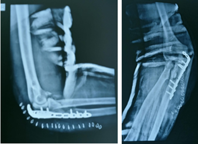
Impacted articular fragments were elevated to ensure congruity with the trochlear surface. Following temporary stabilization with Kirschner wires, the proximal fragment was reduced with the elbow in extension to counteract the pull of the triceps brachii tendon. Definitive internal fixation was achieved using a 3.5 mm locking compression plate (LCP) contoured to the proximal dorsal ulna. Upon fluoroscopic confirmation of anatomical alignment, the surgical site was closed in a layered fashion.
Postoperative Protocol and Rehabilitation
A standardized rehabilitation regimen was initiated for all patients. Prophylactic antibiotics were administered for 24 hours postoperatively, and drainage tubes were removed on the second postoperative day.
For the initial 72 hours, the elbow was immobilized in an extension brace. Starting on postoperative day four, passive range of motion (ROM) exercises were introduced, emphasizing maximal flexion and extension within pain tolerance. Between postoperative weeks two and four, patients transitioned to active functional exercises. Full mobilization and activities of daily living (ADLs)—excluding resisted or gravity-loaded tasks—commenced after the fourth week.
Clinical Assessment
Functional outcomes were quantified using the Mayo Elbow Performance Score (MEPS), which evaluates pain intensity, joint stability, and the ability to perform daily activity. Functional outcome was assessed 6 months post operatively. Clinical outcomes were stratified as follows:
· Excellent: 90–100 points
· Good: 75–89 points
· Fair: 60–74 points
· Poor: <60 points
Table 1: Mayo classification of olecranon fracture
|
Type |
Classification |
|
Type I |
Undisplaced fractures |
|
Type IA |
Non-comminuted |
|
Type IB |
Comminuted |
|
Type II |
Fractures with ≥3 mm displacement, intact collateral ligaments, and preserved forearm–humerus relationship |
|
Type IIA |
Non-comminuted |
|
Type IIB |
Comminuted |
|
Type III |
Fractures with disruption of the relationship between the forearm and the humerus, constituting a fracture-dislocation |
|
Type IIIA |
Non-comminuted |
|
Type IIIB |
Comminuted |
Table 2: Mayo elbow performance score
|
Function |
Definition |
Points |
Score Classification |
|
Pain |
None |
45 |
Excellent > 90 |
|
Mild |
30 |
||
|
Moderate |
15 |
||
|
Severe |
0 |
||
|
Motion |
Arc > 100° |
20 |
Good, 75–89 |
|
Arc 50–100° |
15 |
||
|
Arc < 50° |
5 |
||
|
Stability |
Stable |
10 |
Fair, 60–74 |
|
Moderate instability |
5 |
||
|
Gross instability |
0 |
||
|
Function |
Comb hair |
5 |
Poor < 60 |
|
Feed |
5 |
||
|
Hygiene |
5 |
||
|
Shirt |
5 |
||
|
Shoe |
5 |
||
|
Total |
100 |
Table 3: Patient demographics and basic information
|
Variables |
Outcome in plating group |
|
Age |
42.96±13.24 |
|
Gender |
|
|
Male |
21 |
|
Female |
11 |
|
Cause of trauma |
|
|
Fall |
13 |
|
Traffic accident |
19 |
|
MAYO CLASSIFICATION |
|
|
I A |
0 |
|
I B |
0 |
|
II A |
18 |
|
II B |
12 |
|
III A |
2 |
|
III B |
0 |
Table 4: FUNCTIONAL OUTCOME OF LCP PLATING USING MEPS SCORE
|
EXCELLENT |
19 (59.3%) |
|
GOOD |
11 (34.3%) |
|
FAIR |
2 (6.25%) |
|
POOR |
0 (0%) |
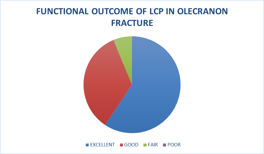
Figure 1: Functional outcome of LCP in olecranon fracture
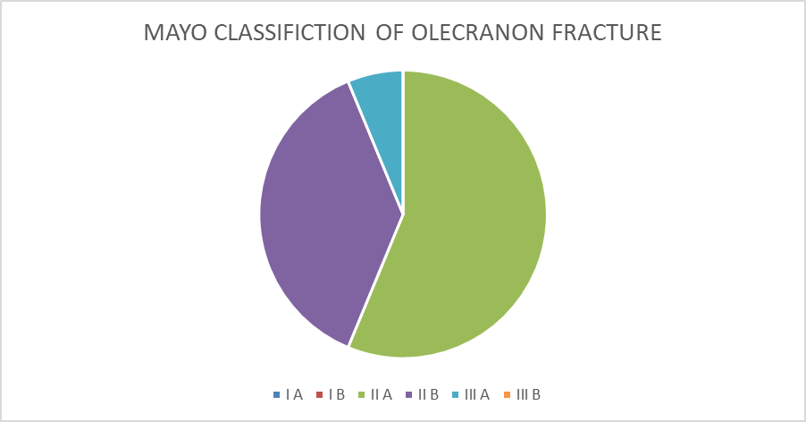
Fig 2 : Olecranon Fracture pattern of study individuals
RESULTS:
This study includes 32 patient who are diagnosed with olecranon fracture and those patients who have met with inclusion criteria and willing to take part in the study. The mean age group of patients who took place in this study was 42.96±13.24. The study cohort comprised 21 males (65.6%) and 11 females (34.4%). Male to female ratio of olecranon fracture in this study is 1.9 :1 This data clearly shows that males are more susceptible to olecranon fractures due to their higher involvement in high-impact trauma and road traffic accidents Road Traffic Accidents (RTAs) were the most frequent etiology for olecranon fracture followed by fall. Table 3 also shows the fracture pattern of olecranon fracture using Mayo classification. Most frequently Mayo class IIA fracture (56.25%) pattern identified in this study followed by Mayo class II B fracture (37.5%).
Table 4 indicates functional outcome of olecranon fracture stabilized with LCP using Mayo elbow performance score 6 months post operatively. This study indicates 19 patients had excellent outcome, 11 had good outcome and 2 had fair outcome.
DISCUSSION:
Olecranon fractures are predominantly intra-articular disruptions characterized by a distinct clinical presentation. Standard orthogonal radiography typically suffices for definitive diagnosis and morphological characterization. While these injuries are nearly always intra-articular, a notable exception is the avulsion fracture of the olecranon tip, which compromises the elbow’s extensor mechanism. Biomechanically, the distracted force of the triceps brachii muscle typically results in proximal fragment displacement, necessitating surgical restoration.(8) Surgical strategy and long-term prognosis are dictated by fracture severity, morphological patterns, concomitant elbow trauma, and ligamentous stability (9,10). The Mayo Classification serves as a vital clinical guide, categorizing injuries based on displacement, degree of comminution, and humeroulnar stability (11)The overarching goal of intervention is the anatomical restoration of the articular surface to facilitate early postoperative mobilization and mitigate joint stiffness.
Olecranon fractures represent approximately 10% of adult upper-extremity fractures and 20% of proximal forearm injuries. Various internal fixation techniques are employed, including tension band wiring (TBW), locking compression plates (LCP), one-third tubular plates , and reconstruction plates (12,13). Tension Band Wiring (TBW): Historically considered the "gold standard" for simple, transverse fractures TBW is frequently limited by hardware-related morbidity. The smooth architecture of standard Kirschner wires (K-wires) lacks sufficient rotational stability and purchase within the bone tunnel, often leading to hardware migration or "backing out" during rehabilitation. This can result in skin ulceration, symptomatic prominence, and the necessity for premature secondary hardware removal (14,15) Plate Osteosynthesis While providing superior stability for comminuted fractures, plate fixation is not without complications. Due to the subcutaneous nature of the proximal ulna, the profile of the plate and screws is often palpable. This is particularly prevalent in patients with lower body mass indices—such as thin individuals or younger females—where the hardware contours become prominent during elbow flexion, potentially leading to aesthetic dissatisfaction, chronic skin irritation, and localized pain.
The functional efficacy of locking compression plates (LCP) for olecranon fractures was quantified using the Mayo Elbow Performance Score (MEPS). In the current cohort, 59% of patients achieved excellent results, 34% demonstrated good outcomes, and 6% were categorized as fair. Statistical analysis confirmed these findings were significant (P < 0.05). These data correlate closely with the results reported by Qi et al. (16) When compared to the findings of Gunaki et al., who evaluated TBW using the same MEPS criteria (52% excellent, 20% good, 16% fair, and 12% poor), our LCP group demonstrated superior functional restoration and a reduced complication profile (17). While minor complications such as hardware prominence and localized skin irritation were noted in our study, the overall morbidity remained lower than that typically associated with TBW. Hard ware prominence is noted in 2 patients and skin irritation was noted in 1 patient.The clinical superiority of plate fixation may be attributed to its distinct biomechanical behavior under physiological load. While triceps contraction alters compressive forces across the fracture site in both fixation modes, our analysis indicates that plates maintain significant compression levels. In contrast, TBW exhibits negligible compression at the anterior cortex during active extension. The ability of the LCP to preserve several times the compressive force of TBW ensures greater stability and minimizes the risk of secondary displacement. The primary limitations of this study include a relatively small sample size and a short follow-up period, which may cause a certain bias to the results of the study. Futures studies with large sample size and longer follow up period may yield interesting results.
CONCLUSION:
In summary, plate osteosynthesis for olecranon fractures provides superior clinical outcomes, enhanced fragment stability, and fewer implant-related complications compared to traditional tension band wiring. Given its ability to maintain robust compression across the fracture line and its lower reoperation rate, LCP represents a highly effective fixation strategy for restoring elbow function.
REFERENCES:
1. Anderson ML, Larson AN, Merten SM, Steinmann SP. Congruent elbow plate fixation of olecranon fractures. J Orthop Trauma. 2007;21(6):386-93.
2. Rouleau DM, Sandman E, van Riet R, Galatz LM. Management of fractures of the proximal ulna. J Am Acad Orthop Surg. 2013;21(3):149-60.
3. Rettig AC, Waugh TR, Evanski PM. Fracture of the olecranon: a problem of management. J Trauma. 1979;19(1):23-8.
4. Ring D, Jupiter JB, Sanders RW, Mast J, Simpson NS. Transolecranon fracture-dislocation of the elbow. J Orthop Trauma. 1997;11(8):545-50.
5. Schliemann B, Raschke MJ, Groene P, et al. Comparison of tension band wiring and precontoured locking compression plate fixation in Mayo type IIA olecranon fractures. Acta Orthop Belg. 2014;80(1):106-11.
6. Schneider MM, Nowak TE, Bastian L, et al. Tension band wiring in olecranon fractures: the myth of technical simplicity and osteosynthetical. BMC Musculoskelet Disord. 2014;15:249.
7. Buijze G, Kloen P. Clinical evaluation of locking compression plate fixation for comminuted olecranon fractures. J Bone Joint Surg Am. 2009;91(10):2416-20.
8. Mommsen U, Holzrichter D. Traumatologie des Schultergürtels und obere Extremität. In: Schumpelick V, Bleese NM, Mommsen U, editors. Chirurgie. 4th ed. Stuttgart: Ferdinand Enke Verlag; 1999. p. 1203-28.
9. Tejwani NC, Garnham IR, Wolinsky PR, Kummer FJ, Koval KJ. Posterior olecranon plating: biomechanical and clinical evaluation of a new operative technique. Bull NYU Hosp Jt Dis. 2002;61(1-2):27-31.
10. King GJ, Lammens PN, Milne AD, Roth JH, Johnson JA. Plate fixation of comminuted olecranon fractures: an in vitro biomechanical study. J Shoulder Elbow Surg. 1996;5(6):437-41.
11. Sullivan CW, Desai K. Classifications in brief: Mayo classification of olecranon fractures. Clin Orthop Relat Res. 2019;477(4):908-10.
12. Powell AJ, Farhan-Alanie OM, Bryceland JK, et al. The treatment of olecranon fractures in adults. Musculoskelet Surg. 2017;101(1):1-9.
13. Weber BG, Vasey H. Osteosynthesis in olecranon fractures. Z Unfallmed Berufskr. 1963;56:90-6.
14. Prayson MJ, Iossi MF, Buchalter D, et al. Safe zone for anterior cortical perforation of the ulna during tension-band wire fixation: a magnetic resonance imaging analysis. J Shoulder Elbow Surg. 2008;17(1):121-5.
15. Van der Linden SC, van Kampen A, Jaarsma RL. K-wire position in tension-band wiring technique affects stability of wires and long-term outcome in surgical treatment of olecranon fractures. J Shoulder Elbow Surg. 2012;21(3):405-11.
16. Qi H, Li Z, Lu Y, et al. Comparison of clinical outcomes of three internal fixation techniques in the treatment of olecranon fracture: a retrospective clinical study. BMC Musculoskelet Disord. 2022;23(1):521.
17. Gunaki RB, Takale S, Patil S. Functional outcome in Olecranon fracture managed by tension band wiring. Int J Health Sci. 2022;6(S1):1-10.