INFLUENCE OF ANKLE POSITION AND CORTICAL PURCHASE ON SYNDESMOTIC SCREW FIXATION IN SIMULATED WEBER C ANKLE INJURIES.
- K.C.MATHEW , Professor and Head of the department, DEPARTMENT OF ORTHOPAEDICS, SREE MOOKAMBIKA INSTITUTE OF MEDICAL SCIENCES, KULASEKHARAM, KANYAKUMARI -629161, TAMILNADU, INDIA
- ROHIN G , Postgraduate Resident, DEPARTMENT OF ORTHOPAEDICS, SREE MOOKAMBIKA INSTITUTE OF MEDICAL SCIENCES, KULASEKHARAM, KANYAKUMARI -629161, TAMILNADU, INDIA.
- MIDHUN BABU M , Assistant Professor, DEPARTMENT OF ORTHOPAEDICS, SREE MOOKAMBIKA INSTITUTE OF MEDICAL SCIENCES, KULASEKHARAM, KANYAKUMARI -629161, TAMILNADU, INDIA
- ZAKIR HUSSAIN MOHAMED , Postgraduate Resident, DEPARTMENT OF ORTHOPAEDICS, SREE MOOKAMBIKA INSTITUTE OF MEDICAL SCIENCES, KULASEKHARAM, KANYAKUMARI -629161, TAMILNADU, INDIA.
- ANNAMALAI T T , Postgraduate Resident, DEPARTMENT OF ORTHOPAEDICS, SREE MOOKAMBIKA INSTITUTE OF MEDICAL SCIENCES, KULASEKHARAM, KANYAKUMARI -629161, TAMILNADU, INDIA.
- GOWTHAM RAJ G , Postgraduate Resident, DEPARTMENT OF ORTHOPAEDICS, SREE MOOKAMBIKA INSTITUTE OF MEDICAL SCIENCES, KULASEKHARAM, KANYAKUMARI -629161, TAMILNADU, INDIA.
- KAVIN RAJA S , Postgraduate Resident, DEPARTMENT OF ORTHOPAEDICS, SREE MOOKAMBIKA INSTITUTE OF MEDICAL SCIENCES, KULASEKHARAM, KANYAKUMARI -629161, TAMILNADU, INDIA.
Article Information:
Abstract:
Background: Syndesmotic injuries associated with Weber C ankle fractures remain a significant challenge in orthopaedic practice because inadequate reduction may adversely affect ankle stability and long-term functional outcomes. Although syndesmotic screw fixation is widely performed, controversies persist regarding the optimal ankle position during screw insertion and the ideal degree of cortical purchase required for stable fixation.Aim: To evaluate the biomechanical effects of ankle position during syndesmotic screw insertion and cortical purchase depth on syndesmotic width, tibiotalar rotation, ankle dorsiflexion, and construct stability in a cadaveric Weber C ankle injury model.Methods: This cadaveric biomechanical study was conducted using fresh-frozen human lower limb specimens with a simulated Weber C syndesmotic injury. Syndesmotic fixation was performed using 3.5-mm cortical screws inserted with the ankle maintained either in dorsiflexion or plantarflexion and using tricortical or quadricortical purchase. Biomechanical testing was carried out under axial loading and combined rotational loading conditions. Syndesmotic width, tibiotalar rotation, maximal ankle dorsiflexion, and construct failure characteristics were assessed and compared between groups.Results: Plantarflexion fixation demonstrated relative syndesmotic narrowing under all loading conditions, whereas dorsiflexion fixation resulted in relative widening, with significant differences observed between groups (p < 0.05). Tibiotalar rotation was significantly greater in the dorsiflexion group under isolated axial loading but became comparable under higher rotational torque. No statistically significant differences were identified in maximal ankle dorsiflexion between plantarflexion and dorsiflexion groups for either tricortical or quadricortical fixation. Similarly, construct failure torque and failure angle did not differ significantly between fixation positions.Conclusion: Ankle position during syndesmotic screw insertion significantly influences syndesmotic width behaviour under loading conditions, with plantarflexion tending towards relative over-compression and dorsiflexion towards relative widening. However, neither ankle position nor cortical purchase depth significantly affected ankle dorsiflexion or construct failure characteristics. Accurate syndesmotic reduction and careful intraoperative assessment remain essential irrespective of fixation strategy.
Keywords:
Article :
INTRODUCTION:
The distal tibiofibular syndesmosis is a critical fibrous articulation that preserves the stability and congruency of the ankle mortise while facilitating physiological load transfer during weight-bearing and locomotion [1, 2]. The syndesmotic complex comprises the anterior and posterior inferior tibiofibular ligaments, the interosseous ligament, and the interosseous membrane, all of which contribute to maintaining rotational and translational stability of the distal tibiofibular joint. Disruption of this complex results in instability of the tibiotalar articulation and, if inadequately addressed, may lead to chronic pain, impaired ankle function, persistent instability, and progressive post-traumatic osteoarthritis [3, 4].
Syndesmosis injuries frequently follow rotational injuries of the ankle. These types of injuries are commonly associated with high energy ankle fractures, particularly with Weber C type fractures where there is an injury of the fibula above the level of the syndesmosis along with the injury of the distal tibiofibular joint. Although the incidence of syndesmosis injuries in cases of ankle fractures greatly varies from study to study, it holds great significance in orthopedic practice since any suboptimal reduction could have implications on ankle function and outcome [1, 5]. Therefore, reconstruction of the bony anatomy of the distal tibia and fibula is of paramount importance.
Screwing of syndesmosis is still the most commonly used technique for treatment of these conditions in modern orthopedics [4]. Although the technique has been highly accepted, certain aspects of this process have continued to be controversial. One such aspect is the best ankle position at the time of screwing. According to earlier studies, this process has been carried out while positioning the ankle joint in maximum dorsiflexion since the wide front portion of the talar dome would prevent any overcompression of the ankle mortise [6].
However, evidence accumulated over the past decade has challenged this conventional teaching. Both cadaveric and clinical investigations have demonstrated inconsistent findings regarding the influence of ankle position during fixation on postoperative ankle mobility and syndesmotic reduction [6, 7].
A further controversy in syndesmotic fixation concerns the optimal number of cortices engaged by the screw. Quadricortical fixation has traditionally been favoured because of its perceived superior stability and rigidity. However, recent biomechanical and clinical studies have suggested that tricortical fixation may provide comparable radiological and functional outcomes while allowing limited physiological micromotion and reducing stress concentration across the construct [8, 9]. Despite these observations, the available evidence remains heterogeneous, and no clear consensus has been established regarding the superiority of either technique [9, 10].
Biomechanical studies on syndesmotic screw fixation have not only been constrained by unrealistic loading models but also varied test parameters, and few biomechanical assessments have incorporated the analysis of syndesmotic widening, tibio-talar rotational motion, ankle joint movement, and stability of constructs. Hence, the clinical application of these results cannot be predicted with certainty.
Considering the above limitations, this study has been planned to assess the biomechanical response of tricortical and quadricortical screw fixation of syndesmosis in an artificial Weber C ankle trauma model subjected to axial and rotational loads. This study focused especially on analyzing syndesmotic widening, tibio-talar rotational motion, ankle joint dorsiflexion, and failure mechanism of the construct under different ankle positions of dorsiflexion or plantarflexion
METHODOLOGY:
Study Design and Setting
This cadaveric biomechanical study was carried out in the Department of Orthopaedics at Sree Mookambika Institute of Medical Sciences over a period extending from February 2024 to May 2025. The study aimed to evaluate the biomechanical behaviour of syndesmotic screw fixation in simulated Weber C ankle injuries under different fixation conditions and ankle positions.
Specimen Preparation
Fresh-frozen human cadaveric lower limbs were used for biomechanical testing. Specimens with evidence of previous ankle trauma, deformity, surgical intervention, or significant degenerative changes were excluded after gross inspection and radiographic evaluation. Prior to testing, the specimens were thawed gradually at room temperature. Soft tissue dissection was performed carefully to preserve the distal tibiofibular syndesmosis, ankle capsule, interosseous membrane, and major ligamentous stabilisers around the ankle joint. Excess skin, muscle, and subcutaneous tissue were removed to facilitate mounting and testing. During the experimental procedure, the specimens were wrapped intermittently with saline-soaked gauze to prevent tissue dehydration [11].
Biomechanical Test Setup
Each specimen was mounted onto a customised biomechanical testing frame. Steinmann pins were inserted through the calcaneus and fifth metatarsal to secure the foot firmly to the platform. The proximal tibia was stabilised using a fixation jig that allowed controlled rotational movement while maintaining axial alignment during loading. Mechanical loading was applied using a calibrated testing system capable of delivering axial compression and external rotational forces in a controlled manner. The setup was designed to reproduce physiological ankle loading conditions encountered during weight bearing.
Baseline Measurements
Initial measurements were obtained from all intact specimens before creation of the injury model. The parameters assessed included syndesmotic width, tibiotalar rotation, and ankle dorsiflexion range. Standardised anatomical landmarks were identified using marker pins placed over the distal tibia, fibula, and talus. Multiple readings were recorded under each testing condition, and the average values were used for analysis to improve measurement consistency.
Creation of Weber C Injury Model
A reproducible Weber C-type syndesmotic injury was created in each specimen to simulate ankle instability. The anterior inferior tibiofibular ligament, posterior inferior tibiofibular ligament, superficial and deep fibres of the deltoid ligament, and the distal portion of the interosseous membrane were carefully sectioned. The fibula was preserved to maintain uniformity between specimens and to specifically evaluate syndesmotic fixation biomechanics without the additional variability caused by fracture comminution [12].
Surgical Technique
Following injury creation, anatomical reduction of the syndesmosis was achieved manually under direct visualisation. Syndesmotic stabilisation was then performed using a 3.5-mm fully threaded cortical screw. The screw was inserted approximately 2–3 cm proximal to the ankle joint line from the fibula into the tibia in a posterolateral-to-anteromedial direction, parallel to the ankle plafond. Prior to screw insertion, drilling and tapping were performed using standard instrumentation. Depending on the study group, fixation was achieved using either tricortical or quadricortical screw purchase. Screw insertion was carried out with the ankle maintained in either dorsiflexion or plantarflexion according to the testing protocol. Reduction was confirmed visually before proceeding with biomechanical assessment [13].
Biomechanical Testing Protocol
After fixation, the specimens underwent repeat biomechanical evaluation under different loading conditions. Axial compressive forces were applied to simulate weight-bearing, followed by controlled external rotational torque.
Testing conditions included:
· axial loading alone,
· axial loading with low rotational torque, and
· axial loading with higher rotational torque.
Changes in syndesmotic widening, tibiotalar rotation, and ankle motion following fixation were recorded and compared with baseline measurements. Selected constructs additionally underwent torsional failure testing to determine construct strength and mode of failure. Failure was defined as sudden loss of resistance, excessive rotational displacement, or structural disruption of the fixation construct.
Outcome Measures
The primary outcome measures included:
· syndesmotic width,
· tibiotalar rotational movement,
· ankle dorsiflexion range, and
· construct stability under torsional loading.
Secondary observations included screw loosening, cortical failure, and fracture patterns noted during biomechanical testing.
Statistical Analysis
All collected data were entered into a spreadsheet and analysed using appropriate statistical software. Quantitative variables were expressed as mean ± standard deviation. Comparative analysis between groups was performed using suitable statistical tests based on data distribution. A p-value of less than 0.05 was considered statistically significant. Multiple measurements obtained from each specimen were averaged to improve reliability and minimise observational error.
RESULTS:
The mean maximum ankle dorsiflexion was 27.84° ± 5.92° in the plantarflexion group and 29.41° ± 6.11° in the dorsiflexion group (Table 1, Figure 1).
Table 1: Comparison of Maximum Ankle Dorsiflexion Between Plantarflexion and Dorsiflexion Groups (n = 11 pairs)
|
Parameter |
Screw Insertion in Plantarflexion |
Screw Insertion in Dorsiflexion |
|
Maximum dorsiflexion, deg |
27.84 (5.92) |
29.41 (6.11) |
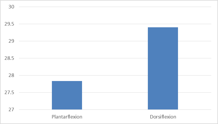
Figure 1: Comparison of Maximum Ankle Dorsiflexion Between Plantarflexion and Dorsiflexion Groups
The mean syndesmosis width in the plantarflexion group was 18.82 ± 2.18 mm under axial load alone, 18.96 ± 2.34 mm under axial load with 1 Nm torque, and 19.42 ± 2.41 mm under axial load with 5 Nm torque. In the dorsiflexion group, the corresponding values were 18.75 ± 1.42 mm, 18.88 ± 1.39 mm, and 19.51 ± 1.26 mm, respectively (Table 2, Figure 2).
Table 2: Comparison of Syndesmosis Width Under Different Loading Conditions Between Plantarflexion and Dorsiflexion Groups (n = 11 pairs)
|
Loading Condition |
Screw Insertion in Plantarflexion |
Screw Insertion in Dorsiflexion |
|
Axial load only |
18.82 (2.18) |
18.75 (1.42) |
|
Axial load + 1 Nm torque |
18.96 (2.34) |
18.88 (1.39) |
|
Axial load + 5 Nm torque |
19.42 (2.41) |
19.51 (1.26) |
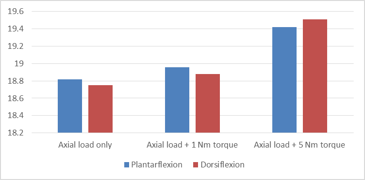
Figure 2: Comparison of Syndesmosis Width Under Different Loading Conditions Between Plantarflexion and Dorsiflexion Groups
The mean tibiotalar rotation in the plantarflexion group was 12.04 ± 6.44° under axial load alone, 12.68 ± 8.12° under axial load with 1 Nm torque, and 11.88 ± 4.36° under axial load with 5 Nm torque. In the dorsiflexion group, the corresponding values were 18.11 ± 5.08°, 16.52 ± 5.84°, and 12.34 ± 5.91°, respectively (Table 3, Figure 3).
Table 3: Comparison of Tibiotalar Rotation Under Different Loading Conditions Between Plantarflexion and Dorsiflexion Groups (n = 11 pairs)
|
Loading Condition |
Screw Insertion in Plantarflexion |
Screw Insertion in Dorsiflexion |
|
Axial load only |
12.04 (6.44)* |
18.11 (5.08)* |
|
Axial load + 1 Nm torque |
12.68 (8.12) |
16.52 (5.84) |
|
Axial load + 5 Nm torque |
11.88 (4.36) |
12.34 (5.91) |
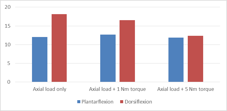
Figure 3: Comparison of Tibiotalar Rotation Under Different Loading Conditions Between Plantarflexion and Dorsiflexion Groups
The mean change in maximal ankle dorsiflexion relative to baseline for tricortical fixation was 0.88 ± 0.30° in the plantarflexion group and 1.18 ± 0.36° in the dorsiflexion group. For quadricortical fixation, the corresponding values were 0.96 ± 0.33° and 1.29 ± 0.39°, respectively. No statistically significant difference was observed between the groups for either fixation type (Table 4, Figure 4).
Table 4: Comparison of Change in Maximal Ankle Dorsiflexion Relative to Baseline Between Plantarflexion and Dorsiflexion Groups (n = 11 pairs)
|
Fixation Type |
Screw Insertion in Plantarflexion (deg) |
Screw Insertion in Dorsiflexion (deg) |
p-value |
|
3 Cortices |
0.88 ± 0.30 |
1.18 ± 0.36 |
0.091 |
|
4 Cortices |
0.96 ± 0.33 |
1.29 ± 0.39 |
0.087 |
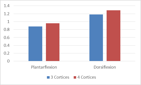
Figure 4: Comparison of Change in Maximal Ankle Dorsiflexion Relative to Baseline Between Plantarflexion and Dorsiflexion Groups
The change in syndesmosis width relative to baseline under axial load alone was −0.84 ± 0.22 mm and −0.79 ± 0.24 mm in the plantarflexion group for tricortical and quadricortical fixation, respectively. In the dorsiflexion group, the corresponding values were 0.46 ± 0.18 mm and 0.52 ± 0.20 mm. Under axial load with 1 Nm torque, the mean change ranged from −0.72 ± 0.19 mm to −0.69 ± 0.21 mm in the plantarflexion group and from 0.38 ± 0.16 mm to 0.41 ± 0.18 mm in the dorsiflexion group. With axial load and 5 Nm torque, the plantarflexion group demonstrated values of −0.18 ± 0.11 mm and −0.14 ± 0.12 mm for tricortical and quadricortical fixation, respectively, whereas the dorsiflexion group showed values of 0.82 ± 0.24 mm and 0.88 ± 0.26 mm. Statistically significant differences were observed between the groups under all loading conditions (Table 5, Figure 5).
Table 5: Comparison of Change in Syndesmosis Width Relative to Baseline Between Plantarflexion and Dorsiflexion Groups (n = 11 pairs)
|
Loading Condition |
3 Cortices – Plantarflexion (mm) |
3 Cortices – Dorsiflexion (mm) |
4 Cortices – Plantarflexion (mm) |
4 Cortices – Dorsiflexion (mm) |
p-value |
|
Axial load only |
-0.84 ± 0.22 |
0.46 ± 0.18 |
-0.79 ± 0.24 |
0.52 ± 0.20 |
0.036 |
|
Axial load + 1 Nm torque |
-0.72 ± 0.19 |
0.38 ± 0.16 |
-0.69 ± 0.21 |
0.41 ± 0.18 |
0.043 |
|
Axial load + 5 Nm torque |
-0.18 ± 0.11 |
0.82 ± 0.24 |
-0.14 ± 0.12 |
0.88 ± 0.26 |
0.025 |
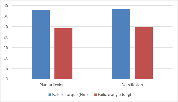
Figure 5: Comparison of Change in Syndesmosis Width Relative to Baseline Under Different Loading Conditions Between Plantarflexion and Dorsiflexion Groups
The change in tibiotalar rotation relative to baseline under axial load alone was 1.42 ± 0.38° and 1.57 ± 0.41° in the plantarflexion group for tricortical and quadricortical fixation, respectively. In the dorsiflexion group, the corresponding values were 1.86 ± 0.44° and 2.14 ± 0.48°. Under axial load with 1 Nm torque, the mean change ranged from 0.84 ± 0.27° to 0.91 ± 0.29° in the plantarflexion group and from 1.68 ± 0.39° to 1.92 ± 0.42° in the dorsiflexion group. With axial load and 5 Nm torque, the plantarflexion group demonstrated values of 0.62 ± 0.21° and 0.71 ± 0.24° for tricortical and quadricortical fixation, respectively, whereas the dorsiflexion group showed values of 1.34 ± 0.35° and 1.58 ± 0.37°. Statistically significant differences were observed between the groups under all loading conditions (Table 6, Figure).
Table 6: Comparison of Change in Tibiotalar Rotation Relative to Baseline Between Plantarflexion and Dorsiflexion Groups (n = 11 pairs)
|
Loading Condition |
3 Cortices – Plantarflexion (deg) |
3 Cortices – Dorsiflexion (deg) |
4 Cortices – Plantarflexion (deg) |
4 Cortices – Dorsiflexion (deg) |
p-value |
|
Axial load only |
1.42 ± 0.38 |
1.86 ± 0.44 |
1.57 ± 0.41 |
2.14 ± 0.48 |
0.021 |
|
Axial load + 1 Nm torque |
0.84 ± 0.27 |
1.68 ± 0.39 |
0.91 ± 0.29 |
1.92 ± 0.42 |
0.050 |
|
Axial load + 5 Nm torque |
0.62 ± 0.21 |
1.34 ± 0.35 |
0.71 ± 0.24 |
1.58 ± 0.37 |
0.028 |
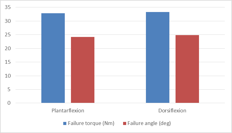
Figure 6: Comparison of Change in Tibiotalar Rotation Relative to Baseline Under Different Loading Conditions Between Plantarflexion and Dorsiflexion Groups
The mean failure torque in the plantarflexion group was 32.84 ± 4.62 Nm, compared with 33.27 ± 4.88 Nm in the dorsiflexion group. The mean failure angle was 24.18 ± 3.74° in the plantarflexion group and 24.91 ± 3.96° in the dorsiflexion group. No statistically significant difference was observed between the groups with respect to failure torque or failure angle (Table 7, Figure 7).
Table 7: Comparison of Failure Torque and Failure Angle Between Plantarflexion and Dorsiflexion Groups in 4-Cortex Fixation (n = 11 pairs)
|
Parameter |
Screw Insertion in Plantarflexion |
Screw Insertion in Dorsiflexion |
p-value |
|
Failure torque (Nm) |
32.84 ± 4.62 |
33.27 ± 4.88 |
0.613 |
|
Failure angle (deg) |
24.18 ± 3.74 |
24.91 ± 3.96 |
0.587 |
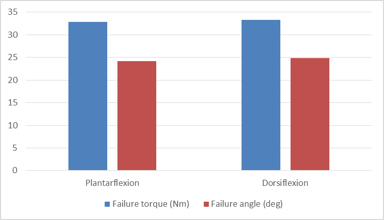
Figure 7: Comparison of Failure Torque and Failure Angle Between Plantarflexion and Dorsiflexion Groups in 4-Cortex Fixation
DISCUSSION:
In the current biomechanical cadaveric study, the effect of ankle joint position during syndesmotic screw placement (dorsiflexion vs. plantarflexion) and cortical engagement (tricortical vs. quadricortical) on syndesmotic width, tibiotalar rotation, ankle joint dorsiflexion range, and construct failure parameters was assessed in an experimental model of Weber C ankle injury. The main results of this investigation can be summed up as follows. Firstly, relative narrowing of the syndesmotic width occurred consistently in the case of plantarflexion insertion regardless of loading mode, while dorsiflexion insertion led to relative widening of syndesmosis width, with statistically significant intergroup differences recorded consistently (p < 0.05). Secondly, there was a trend toward higher tibiotalar rotation under isolated axial loading, which decreased as the level of rotational torque increased. Thirdly, the maximal dorsiflexion range of motion did not differ significantly between the two groups independently of cortical engagement. Fourthly, construct failure torque and failure angle were not statistically different between groups and types of fixation.
The lack of a significant difference in the post-operative range of motion of dorsiflexion between plantarflexion and dorsiflexion fixation is in agreement with the results obtained by Pallis et al. [6], who showed, using an anatomic pronation external rotation fracture model, that the positioning of the foot during the fixation procedure did not significantly affect post-operative range of motion at the ankle joint. Fixation in maximum dorsiflexion has been historically recommended to prevent excessive compression of the mortise through the use of the larger articular dome anteriorly. The current study, along with other biomechanical evidence, seems to imply that this may have little bearing on post-operative dorsiflexion. Li et al. [14] also noted that technical factors, including the level of screw placement, had no independent bearing on post-operative range of motion.
The fact that the effect of the ankle position on the syndesmosis width is always the same is one of the most clinically significant findings in the present study. Plantar flexion fixation led to a narrower syndesmosis width, while dorsiflexion led to a wider syndesmosis width when subjected to load. Malreduction in both ways is associated with poor clinical results and increased instability, leading to posttraumatic arthritis [5, 15]. According to Stenquist and Kwon, malreduction has persistently been observed at a high frequency in the literature, stressing the need for careful assessment during surgery [15]. Our results further support the findings by showing that fixation positioning during screw placement can consistently alter syndesmotic width after load.
Another significant outcome was the similarity of tibiotalar rotational values between the two groups at higher levels of rotational torque. While the dorsiflexion group showed increased rotational laxity at isolated axial loading, this became inconsequential during torsional loading, implying that physiological rotational loads might partially compensate for any positional discrepancies noted initially. According to Pang et al. [16], syndesmotic screw placement affects the point of contact pressure on the tibiotalar joint surface under torsional loading. It is therefore evident that the assessment of ankle mechanics should involve a combination loading model rather than a single static one. This point was further supported by Evers et al., who found that the rotational stiffness of ankle models was affected by posterior malleolar fixation rather than syndesmotic screw positioning alone [17].
With respect to cortical purchase, no significant differences were observed between tricortical and quadricortical fixation in terms of ankle dorsiflexion range, failure torque, or failure angle. These findings are consistent with previous clinical and biomechanical studies. Klepacki et al. reported that the number of cortices engaged did not significantly influence radiographic outcomes and suggested tricortical fixation as a practical alternative because of its easier hardware removal [9]. In a subsequent analysis from the same cohort, quadricortical fixation demonstrated reduced plantarflexion relative to the contralateral limb, indicating that increased construct rigidity may subtly influence ankle kinematics despite comparable overall functional outcomes [10]. Similarly, Li et al. [18], using finite element analysis, demonstrated that tricortical fixation more closely restored physiological ankle biomechanics, whereas quadricortical constructs generated greater stress concentration across the screw-bone interface. The comparable failure characteristics observed in the present study support the concept that increased cortical engagement may not necessarily confer superior biomechanical performance under physiological loading conditions.
The present study has several strengths. A standardised cadaveric Weber C injury model with controlled ligamentous sectioning was employed, and biomechanical testing was performed under progressively increasing loading conditions designed to simulate physiological ankle stresses. Furthermore, both ankle position during fixation and cortical purchase depth were evaluated simultaneously within a single experimental framework, allowing direct comparison of their biomechanical effects.
However, some of the limitations must also be addressed. First, the use of the cadaveric model itself precludes the possibility of making accurate predictions of biological behavior in vivo and the long-term functional outcomes. While the number of paired specimens used (eleven specimens) was similar to previous cadaveric experiments, it may have been too small to allow adequate statistical power to detect minor differences between the two groups. Second, there was no comminuted fracture, with intact fibulae being preserved in all specimens, while Weber C injuries in clinical practice involve comminution in most cases. The measured failure torque in our study was less compared to the results obtained by Lee et al. [19], most likely due to differences in quality of bones used, mineral density, and loading conditions. Lastly, variables like bone healing, loosening, and adaptation to implants cannot be mimicked in a cadaveric model
CONCLUSION:
In conclusion, ankle position during syndesmotic screw fixation influenced syndesmotic behaviour under loading, with plantarflexion tending to produce relative narrowing and dorsiflexion resulting in relative widening. However, these positional differences did not significantly affect ankle dorsiflexion or construct failure characteristics. The findings suggest that achieving an accurate syndesmotic reduction is likely more important than the specific ankle position or degree of cortical purchase used during fixation. Careful intraoperative assessment of syndesmotic alignment and ankle mortise congruity therefore remains essential irrespective of fixation technique. Further clinical studies are needed to determine whether these biomechanical differences have meaningful effects on long-term patient outcomes.
REFERENCES:
1. Tourné Y, Molinier F, Andrieu M, Porta J, Barbier G. Diagnosis and treatment of tibiofibular syndesmosis lesions. Orthop Traumatol Surg Res. 2019;105(8S):S275–S286. doi:10.1016/j.otsr.2019.09.014
2. Khambete P, Harlow E, Ina J, Miskovsky S. Biomechanics of the distal tibiofibular syndesmosis: a systematic review of cadaveric studies. Foot Ankle Orthop. 2021;6(2):24730114211012701. doi:10.1177/24730114211012701
3. Yammine K, Jalloul M, Assi C. Distal tibiofibular syndesmosis: a meta-analysis of cadaveric studies. Morphologie. 2022;106(355):241–251. doi:10.1016/j.morpho.2021.10.004
4. Liu J, Valentine D, Ebraheim NA. Management of syndesmosis injury: a narrative review. Orthop Res Rev. 2022;14:471–475. doi:10.2147/ORR.S340533
5. Akoh CC, Phisitkul P. Anatomic ligament repairs of syndesmotic injuries. Orthop Clin North Am. 2019;50(3):401–414. doi:10.1016/j.ocl.2019.02.004
6. Pallis MP, Pressman DN, Heida K, Nicholson T, Ishikawa S. Effect of ankle position on tibiotalar motion with screw fixation of the distal tibiofibular syndesmosis in a fracture model. Foot Ankle Int. 2018;39(6):746–750. doi:10.1177/1071100718759966
7. Weng Q, Lin C, Liu Y, Dai G, Lutchooman V, Hong J. Biomechanical analysis of a novel syndesmotic plate compared with traditional screw and suture button fixation. J Foot Ankle Surg. 2020;59(3):522–528. doi:10.1053/j.jfas.2019.07.025
8. Liu G, Chen L, Gong M, Xing F, Xiang Z. Clinical evidence for treatment of distal tibiofibular syndesmosis injury: a systematic review of clinical studies. J Foot Ankle Surg. 2019;58(6):1245–1250. doi:10.1053/j.jfas.2019.01.015
9. Klepacki K, Kowal I, Konieczny G, Tomczyk Ł, Miękisiak G, Morasiewicz P. Radiographic assessment of tibiofibular syndesmosis injury with different durations and types of fixation. J Clin Med. 2022;11(21):6331. doi:10.3390/jcm11216331
10. Klepacki K, Kowal I, Konieczny G, Tomczyk Ł, Miękisiak G, Kochańska-Bieri J, Morasiewicz P. Post-treatment functional outcomes of distal tibiofibular syndesmosis injuries with varying duration and method of stabilization. J Foot Ankle Surg. 2024;63(6):735–741. doi:10.1053/j.jfas.2024.07.011
11. Cartner JL, Hartsell ZM, Ricci WM, Tornetta P 3rd. Can we trust ex vivo mechanical testing of fresh--frozen cadaveric specimens? The effect of postfreezing delays. J Orthop Trauma. 2011 Aug;25(8):459-61. doi: 10.1097/BOT.0b013e318225b875. PMID: 21738060.
12. Sipahioglu S, Zehir S, Isikan E. Weber C ankle fractures with tibiofibular diastasis: syndesmosis-only fixation. Acta Ortop Bras. 2017;25(3):67-70. doi:10.1590/1413-785220172503151204.
13. D’Hooghe, P. and Salameh, M. (2020), Does the choice of syndesmotic screw versus suture button in ankle surgery has a silver lining? – a technical note. J EXP ORTOP, 7: 66. https://doi.org/10.1186/s40634-020-00279-x
14. Li JK, Yu Y, Wu YH, Wang J, Zeng XT, Zhao JG. Does the level of syndesmotic screw insertion affect clinical outcome after ankle fractures with syndesmotic instability? Orthop Surg. 2023;15(1):247–255. doi:10.1111/os.13569
15. Stenquist DS, Kwon JY. Strategies to avoid syndesmosis malreduction in ankle fractures. Foot Ankle Clin. 2020;25(4):613–630. doi:10.1016/j.fcl.2020.08.001
16. Pang EQ, Bedigrew K, Palanca A, Behn AW, Hunt KJ, Chou L. Ankle joint contact loads and displacement in syndesmosis injuries repaired with Tightropes compared to screw fixation in a static model. Injury. 2019;50(11):1901–1907. doi:10.1016/j.injury.2019.09.012
17. Evers J, Fischer M, Raschke M, Riesenbeck O, Milstrey A, Gehweiler D, et al. Leave it or fix it? How fixation of a small posterior malleolar fragment neutralizes rotational forces in trimalleolar fractures. Arch Orthop Trauma Surg. 2022;142(6):1031–1037. doi:10.1007/s00402-021-03772-9
18. Li H, Chen Y, Qiang M, Zhang K, Jiang Y, Zhang Y, et al. Computational biomechanical analysis of postoperative inferior tibiofibular syndesmosis: a modified modeling method. Comput Methods Biomech Biomed Engin. 2018;21(5):427–435. doi:10.1080/10255842.2018.1472770
19. Lee HS, Kim WJ, Young KW, Jeong GM, Yeo ED, Lee YK. Comparison of open anterior syndesmotic repair augmented with suture-tape and trans-syndesmotic screw fixation: a biomechanical study. J Foot Ankle Surg. 2021;60(2):339–344. doi:10.1053/j.jfas.2020.04.021.